Download presentation
Presentation is loading. Please wait.

1
Female Reproductive System
Kristine Krafts, M.D.

2
Female Reproductive System Outline
Cervix Uterus Ovaries Breast

3
Female Reproductive System Outline
Cervix Cervical carcinoma

4
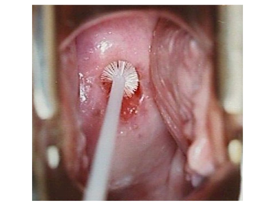
Cervical Carcinoma Once the most common cancer in women – now not even in top 10. Decrease due to Pap test! At the same time, precursor lesions are increasing (early detection)
")
5
Cervical Intraepithelial Neoplasia (CIN)
Precursor to carcinoma Almost all carcinomas arise in CIN; but not all cases of CIN progress to carcinoma! Three grades: CIN I: mild dysplasia (half regress, 20% progress) CIN II: moderate dysplasia CIN III: severe dysplasia (30% regress, 70% progress) The higher the grade, the more likely the lesion will progress to carcinoma
 CIN II: moderate dysplasia. CIN III: severe dysplasia (30% regress, 70% progress) The higher the grade, the more likely the lesion will progress to carcinoma.")
6
Cervical Carcinoma Risk Factors
Early age at first intercourse Multiple sexual partners A male partner with multiple previous partners Persistent infection with “high-risk” HPV Smoking Immunodeficiency

7
Cervical Carcinoma and HPV
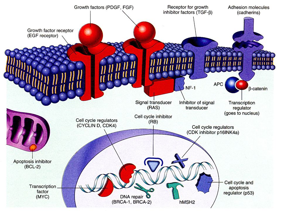
HPV is detectable in almost all CIN and cancer. “High-risk” types: 16, 18, 45, 31 Found in carcinomas Integrate into genome, inactivate p53, RB “Low-risk” types: 6, 11 Found in condylomas (benign lesions) Do not integrate into genome
 Do not integrate into genome.")
9
Development of transformation zone

10
Normal cervix, young adult
Transformation zone Normal cervix, young adult

11
Transformation zone

12
Spectrum of cervical intraepithelial neoplasia (CIN)
")
13
Normal turning into CIN

15
Cytology of CIN (Pap smear)
normal CIN I CIN II CIN III Cytology of CIN (Pap smear)
")
16
Cytology of CIN (Pap smear)
normal CIN I CIN II CIN III “High-grade dysplasia” “Low-grade dysplasia” Cytology of CIN (Pap smear)
")
17
Invasive Cervical Carcinoma
Most cases are squamous, arising from CIN Small number are adenocarcinomas Peak age: 45 (10-15 years after CIN develops!) Spreads slowly Most cases are diagnosed early Mortality is related to stage Stage 0 (preinvasive): 100% 5 year survival Stage 4: 10% 5 year survival
 Spreads slowly. Most cases are diagnosed early. Mortality is related to stage. Stage 0 (preinvasive): 100% 5 year survival. Stage 4: 10% 5 year survival.")
18
Cervical carcinoma

19
Cervical carcinoma

20
Female Reproductive System Outline
Cervix Uterus Endometriosis Endometrial hyperplasia Tumors

21
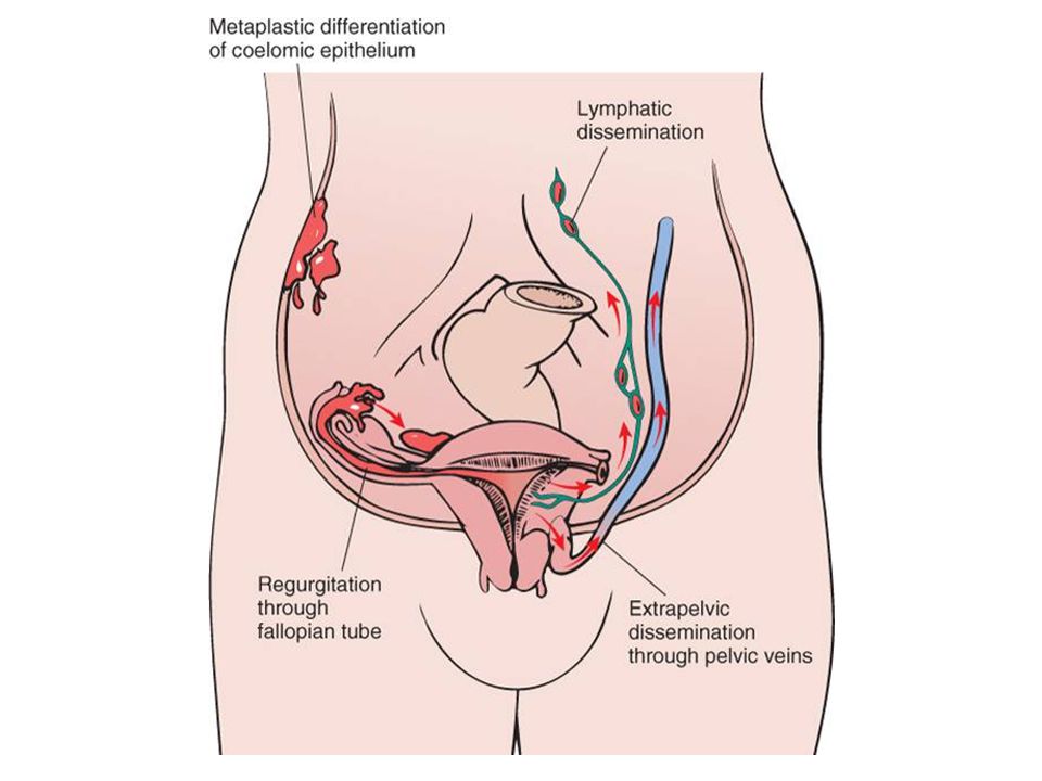
Endometriosis Location of endometrial glands outside uterus
Usually peritoneum, rarely lymph nodes Endometrium undergoes cyclic bleeding Causes scarring, pain, sometimes sterility How does endometrium get out?

23
Endometriosis in ovary (“chocolate cyst”)
")
24
Endometrial Hyperplasia
Proliferation of endometrium due to estrogen excess Risk factors: anovulatory cycles, obesity, estrogen-producing ovarian tumors, exogenous hormone use Three categories: simple, complex, and atypical The more severe the hyperplasia, the greater the chance that it will evolve into carcinoma

25
Normal endometrium

26
Endometrial hyperplasia
Simple Complex Atypical Endometrial hyperplasia

27
Leiomyoma “Fibroid” Benign tumor of smooth muscle Common!
Stimulated by estrogen Menorrhagia, metrorrhagia, or asymptomatic

28
Leiomyosarcoma Malignant tumor of smooth muscle
Necrotic, with atypical cells and lots of mitoses Often recur after surgery Many metastasize, especially to lungs 5 year survival = 40%

29
Leiomyoma Leiomyosarcoma

30
Leiomyoma Leiomyosarcoma

31
Endometrial Carcinoma
Peak age: (not before 40) Frequently arises in endometrial hyperplasia Risk factors: obesity, nulliparity, estrogen replacement Symptoms: leukorrhea, irregular bleeding Metastasizes late
 Frequently arises in endometrial hyperplasia. Risk factors: obesity, nulliparity, estrogen replacement. Symptoms: leukorrhea, irregular bleeding. Metastasizes late.")
32
Endometrial adenocarcinoma

33
Female Reproductive System Outline
Cervix Uterus Ovaries Tumors

34
Origin of Ovarian Tumors
Surface epithelial tumors Germ cell tumors Sex cord-stromal tumors Cystadenoma Cystadenocarcinoma Teratoma Dysgerminoma Yolk sac tumor Choriocarcinoma Granulosa-theca cell tumor Sertoli-Leydig cell tumor

35
Cystadenoma Benign tumor derived from surface epithelium
Repeated ovulation, scarring, infolding of epithelium leads to cysts, which can undergo neoplastic transformation Typically large, occasionally bilateral

36
Patient with ovarian cystadenoma

37
Patient with ovarian cystadenoma

38
Ovarian cystadenoma

39
Ovarian cystadenoma

40
Ovarian cystadenoma

41
Teratoma Benign tumor with differentiation along all three germ cell layers (ectoderm, endoderm, mesoderm) Usually cystic, with skin inside (“dermoid cyst”) Sebaceous material, matted hair, teeth, bone… Malignant variant has immature tissues
 Sebaceous material, matted hair, teeth, bone… Malignant variant has immature tissues.")
42
Teratoma

43
Teratoma

44
Ovarian Cancer 22,000 new cases / 14,000 deaths predicted in 2014
5th commonest, 5th most deadly cancer in women Danger: no definitive signs until advanced Peak age: 50 Most are cystadenocarcinomas

45
Papillary cystadenocarcinoma

46
Papillary cystadenocarcinoma

47
Ovarian Cancer Symptoms Feeling of fullness or bloating Pelvic pain
Back pain Abnormal menses Risk factors Nulliparity Family history (BRCA gene mutation) NOT using oral contraceptives!
 NOT using oral contraceptives!")
48
Ovarian Cancer Treatment: surgery, radiation, chemotherapy
Prognosis depends on stage Cancer confined to the ovary: 5y survival 70% Cancer through ovarian capsule: 5y survival 13%

49
Female Reproductive System Outline
Cervix Uterus Ovaries Breast Fibrocystic change Tumors

50
Breast Many breast diseases present as lumps
Most lumps represent benign things… …but a lump always needs to be evaluated Ultrasound, mammography, fine needle aspiration, and biopsy are the usual methods

51
Most breast lumps are benign

52
Fibrocystic Change Two kinds: nonproliferative and proliferative change Cause: exaggeration of normal breast cycles Rarely associated with increased cancer risk Very common (present in most women at autopsy) Called fibrocystic change, not fibrocystic disease
 Called fibrocystic change, not fibrocystic disease.")
53
Fibrocystic Change Nonproliferative fibrocystic change
Increased stroma Dilation of ducts, formation of cysts Proliferative fibrocystic change Hyperplasia of breast epithelium If epithelium shows atypia, 5x ↑ risk of cancer

54
Fibrocystic change

55
Normal breast

56
Nonproliferative fibrocystic change

57
Proliferative fibrocystic change

58
Fibroadenoma Most common benign breast tumor Stimulated by estrogen
Peak incidence in 20s Solitary, discrete, moveable mass Fibrous tissue with compressed ducts and lobules

59
Fibroadenoma

60
Fibroadenoma

61
Breast Carcinoma 233,000 new cases / 40,000 deaths predicted in 2014
Most common, 2nd deadliest cancer in women Lifetime risk: 1 in 8 75% of patients are >50 Rate was increasing but now stable

62
Breast Carcinoma Risk Factors
Age Family history Increased estrogen exposure Obesity Alcohol consumption High-fat diet

63
Breast Carcinoma Family History
5-10% of all cases are hereditary Worry if first degree relative with breast cancer Most have BRCA-1 or BRCA-2 mutations Tumor suppressor genes; help repair DNA Genetic testing difficult Most carriers get cancer by age 70

64
Breast Carcinoma Clinical Findings
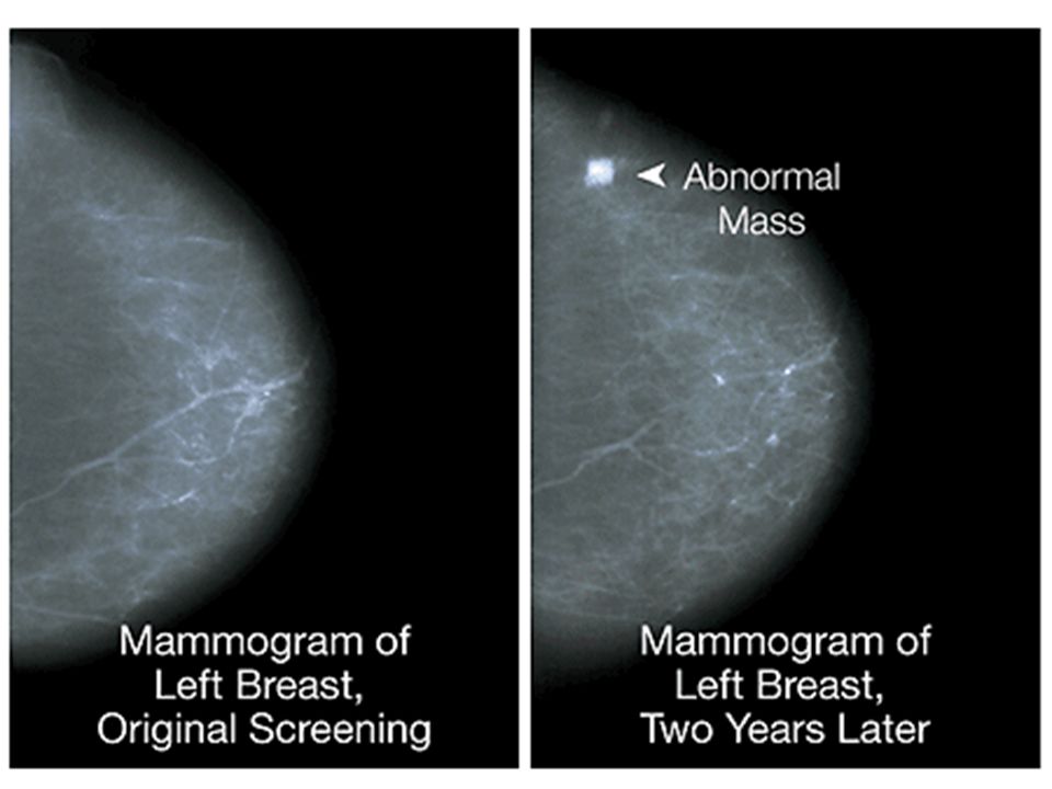
If discovered by palpation Solitary, painless, moveable mass 2-3 cm in diameter Axillary nodes positive in 50% of patients If discovered by mammography 1 cm in size Axillary nodes positive in only 15% of patients As disease progresses Fixation to chest wall Adherence to overlying skin Peau d’orange

66
Advanced breast carcinoma: fixation to skin

67
Peau d’orange

68
Inflammatory breast carcinoma

69
Breast Carcinoma Histologic Types
Non-invasive Ductal carcinoma in situ (DCIS) Lobular carcinoma in situ (LCIS) Invasive Ductal Lobular Inflammatory Others
 Lobular carcinoma in situ (LCIS) Invasive. Ductal. Lobular. Inflammatory. Others.")
70
Normal breast

71
Lobular carcinoma in situ

72
Invasive breast carcinoma

73
Low-grade invasive ductal carcinoma

74
High-grade invasive ductal carcinoma

75
Inflammatory breast carcinoma

76
Breast Carcinoma Prognostic Factors
Size of tumor Lymph node involvement Distant metastases Grade of tumor Histologic type of tumor

77
Sentinel node biopsy

78
TNM* staging system for breast cancer
Stage T N M 5-year survival Stage 0 Stage I Stage II Stage III Stage IV DCIS <2 cm <5 cm >5 cm Any T <3 4+ 1+ 10+ Any N M0 skin or chest wall M1 92% 87% 75% 46% 13% * Tumor (size), nodes (# positive), metastases
, nodes (# positive), metastases.")
Similar presentations

 Consultant Obstetrician & Gynaecologist Infertility Specialist.>")

>")


