Download presentation
Presentation is loading. Please wait.

1
Intracranial Hemorrhage
Nikdokht Farid, M.D. Assistant Professor UCSD Department of Radiology

2
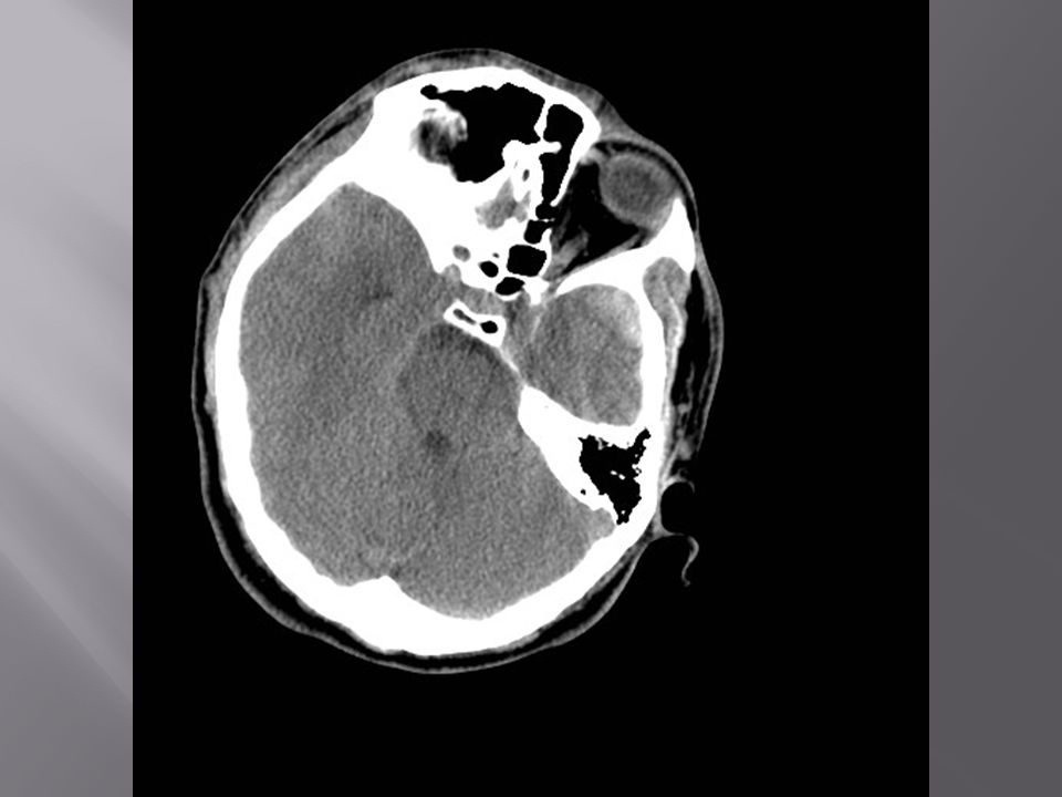
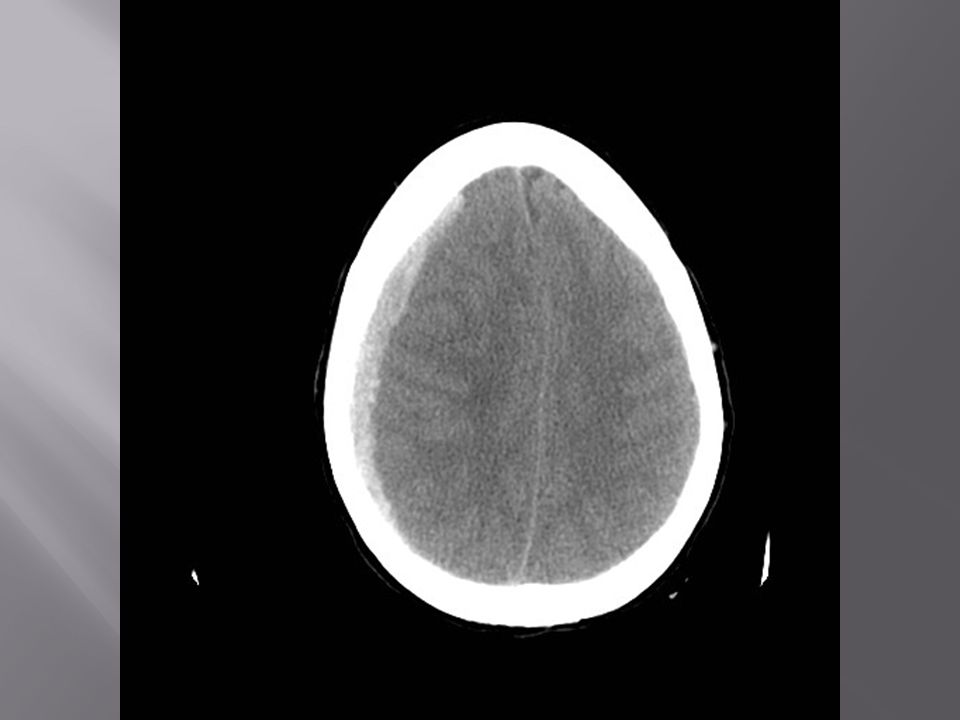
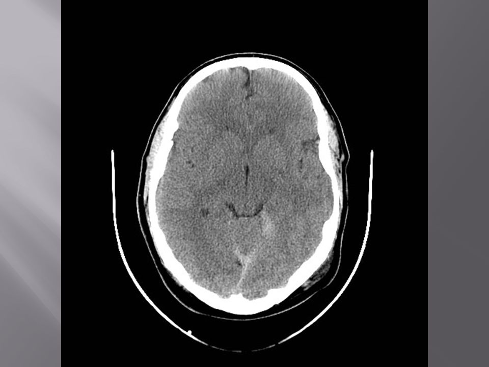
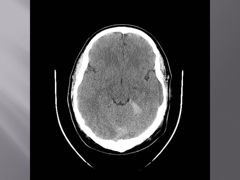
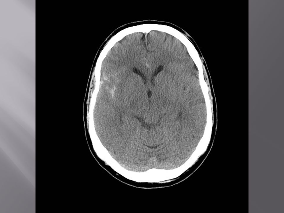
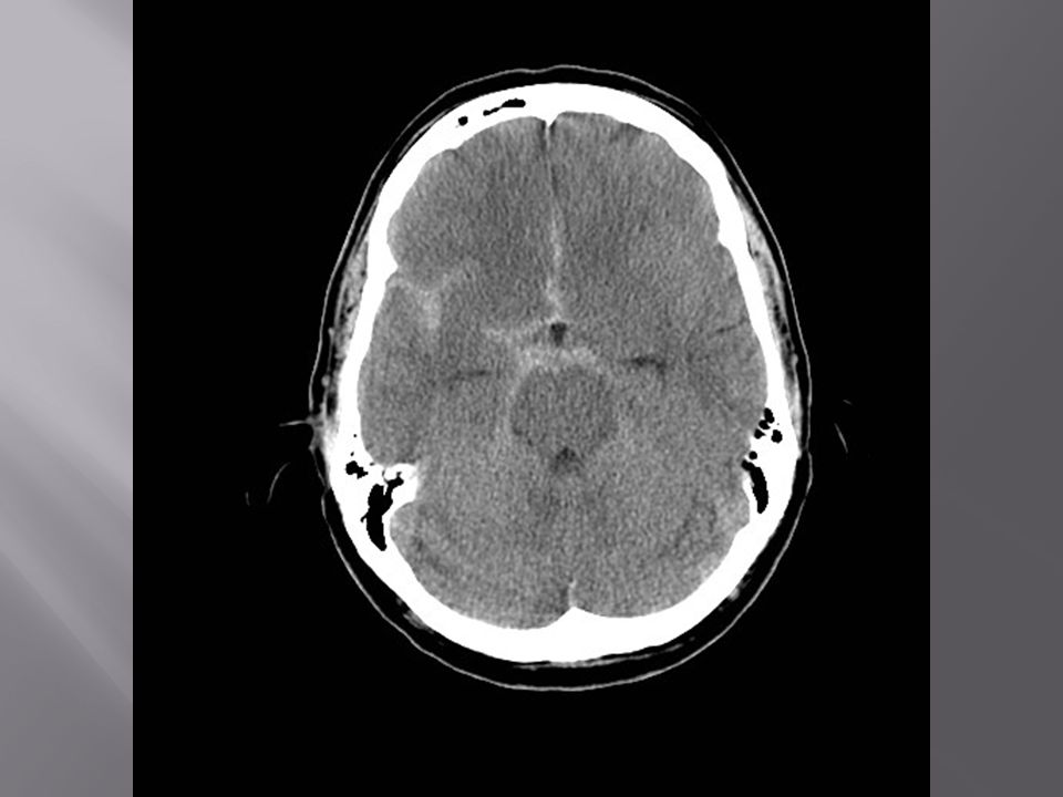
25 year old male status post severe head trauma

4
Repeat CT few hours later…

9
Cerebral Contusion Patchy multifocal superficial hemorrhages surrounded by edema Anterior inferior frontal and temporal lobes are most common site (“frontal and temporal poles”) Coup and contracoup injury sites Often associated with subdural hematoma (SDH), traumatic subarachnoid hemorrhage (SAH), intraventricular hemorrhage (IVH) “Blossoming” of contusions may occur (ongoing hemorrhage or worsening edema)
 Coup and contracoup injury sites. Often associated with subdural hematoma (SDH), traumatic subarachnoid hemorrhage (SAH), intraventricular hemorrhage (IVH) Blossoming of contusions may occur (ongoing hemorrhage or worsening edema)")
10
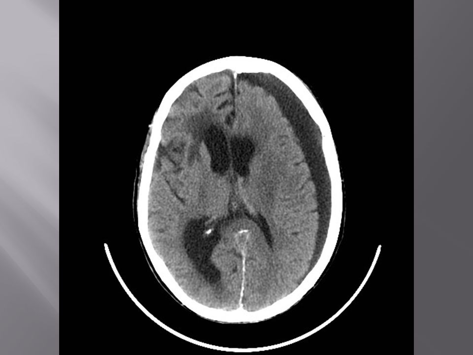
20 year old female status post head trauma, “lucid interval” prior to ALOC

15
Epidural Hematoma-EDH
Biconvex or lentiform extra-axial collection—usually hyperdense (low density “swirl sign”) Does not cross suture lines 90% arterial, 10% venous Arterial EDH often associated with fracture near middle meningeal artery groove Venous EDH often associated with fracture near dural sinus attachment Mass effect on underlying brain and subarachnoid space
 Does not cross suture lines. 90% arterial, 10% venous. Arterial EDH often associated with fracture near middle meningeal artery groove. Venous EDH often associated with fracture near dural sinus attachment. Mass effect on underlying brain and subarachnoid space.")
16
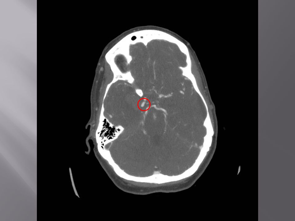
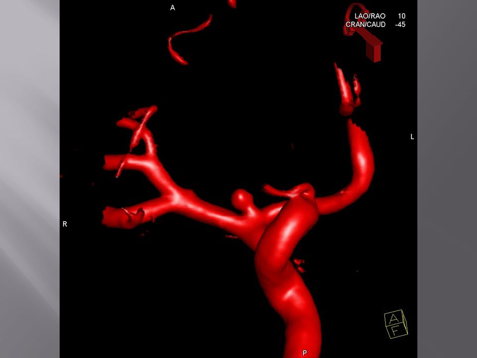
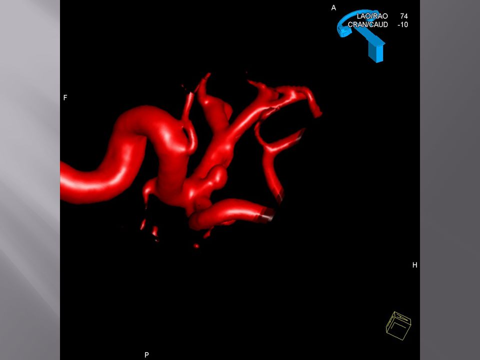
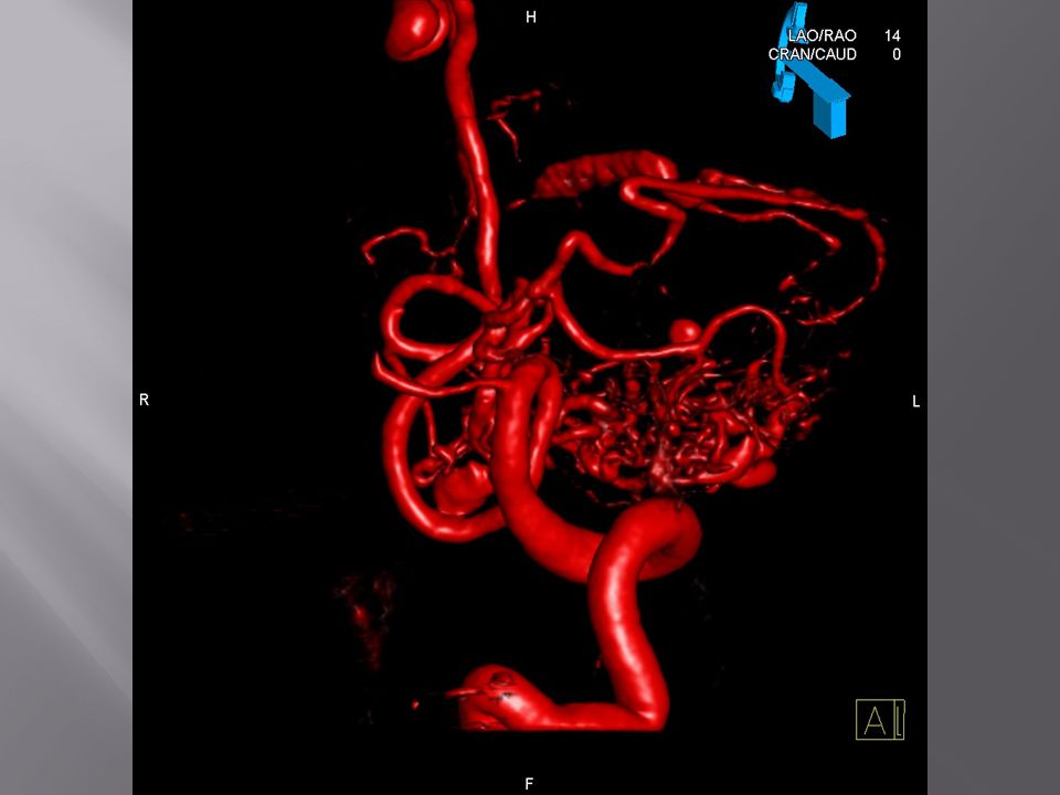
30 year old female with sudden onset severe headache and altered mental status

21
CT Angiogram— Ruptured right PCOM aneurysm

25
Subdural Hematoma-SDH
Crescentic extra-axial collection (acute—hyperdense, subacute—isodense, chronic—hypodense) Crosses suture lines, spreading diffusely over the convexity (does not cross dural attachment) May also extend along falx and tentorium Etiology: TRAUMA (most common)—tearing of bridging cortical veins Aneurysm rupture Vascular malformation (dural AVF, AVM)
 Crosses suture lines, spreading diffusely over the convexity (does not cross dural attachment) May also extend along falx and tentorium. Etiology: TRAUMA (most common)—tearing of bridging cortical veins. Aneurysm rupture. Vascular malformation (dural AVF, AVM)")
26
75 year old female with repeated falls, altered mental status

29
Chronic SDH with marked mass effect on left cerebral hemisphere resulting in “trapping” of the right lateral ventricle with transependymal flow of CSF

30
35 year old male status post trauma

35
Subdural hematoma layering along the tentorium

36
60 year old male status post trauma

41
Subarachnoid Hemorrhage-SAH
High density within the sulci and cisterns Etiology: TRAUMA (most common) Rupture aneurysm (2nd most common) Others (much less common)—AVM, perimesencephalic venous hemorrhage, etc. Traumatic SAH often associated with other forms of ICH (contusions, SDH, etc.)
 Rupture aneurysm (2nd most common) Others (much less common)—AVM, perimesencephalic venous hemorrhage, etc. Traumatic SAH often associated with other forms of ICH (contusions, SDH, etc.)")
42
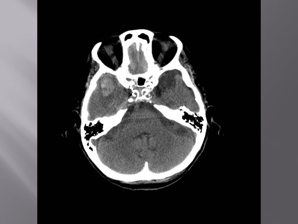
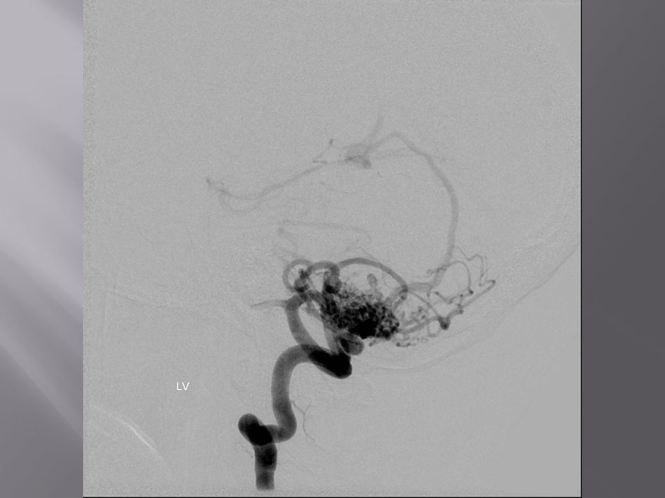
40 year old female presenting with “worst headache of life”

49
Cerebral Angiogram- Ruptured right ICA terminus aneurysm

56
40 year old male status post trauma

62
Combination ICH: SDH, SAH, IVH, contusions

63
45 year old female with sudden onset ataxia, nausea, vomiting

70
Large left cerebellar AVM with bleed

71
Diffuse axonal injury Traumatic axonal stretch injury
Punctate microhemorrhages at grey/white matter junction, corpus callosum, upper brainstem MR much more sensitive than CT—specifically T2* gradient echo sequence (GRE) CT is often normal (50-80%)
 CT is often normal (50-80%)")
72
T2* GRE Image from UCSD Neuroradiology Teaching File Database--spinwarp.ucsd.edu/neuroweb/

Similar presentations


>")


>")

