Download presentation
Presentation is loading. Please wait.

3
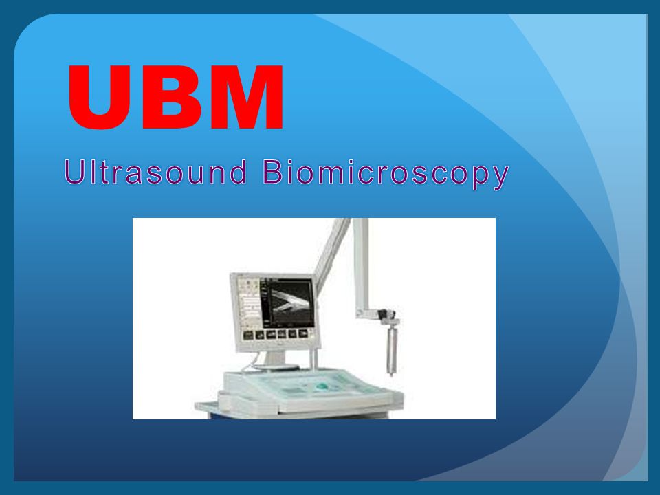
Ultrasound biomicroscopy is a new imaging technique that uses high frequency ultrasound to produce images of the eye at near microscopic resolution

4
Basic of ultrasound biomicroscopy Is based on 40 to 100 MHZ transduceres incorporated into a B- mode clinical scanner Image resolution is approximately 50 μm Reduced depth of penetration to approximately 5 mm

6
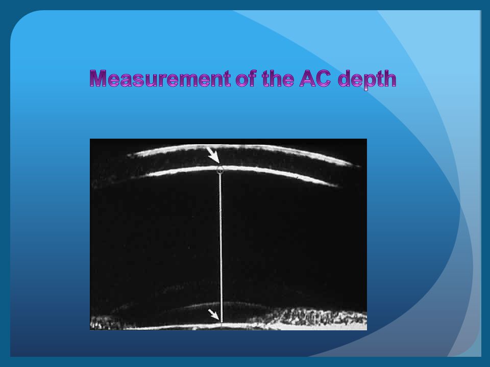
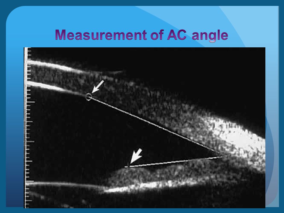
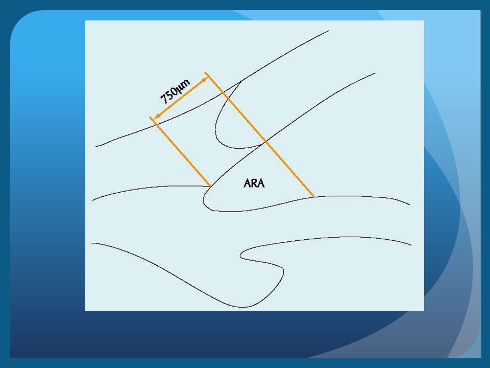
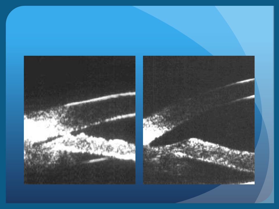
Quantitative studies includes Simple measurements methods such as a) Distance b) Angle measurement
 Distance b) Angle measurement")
9
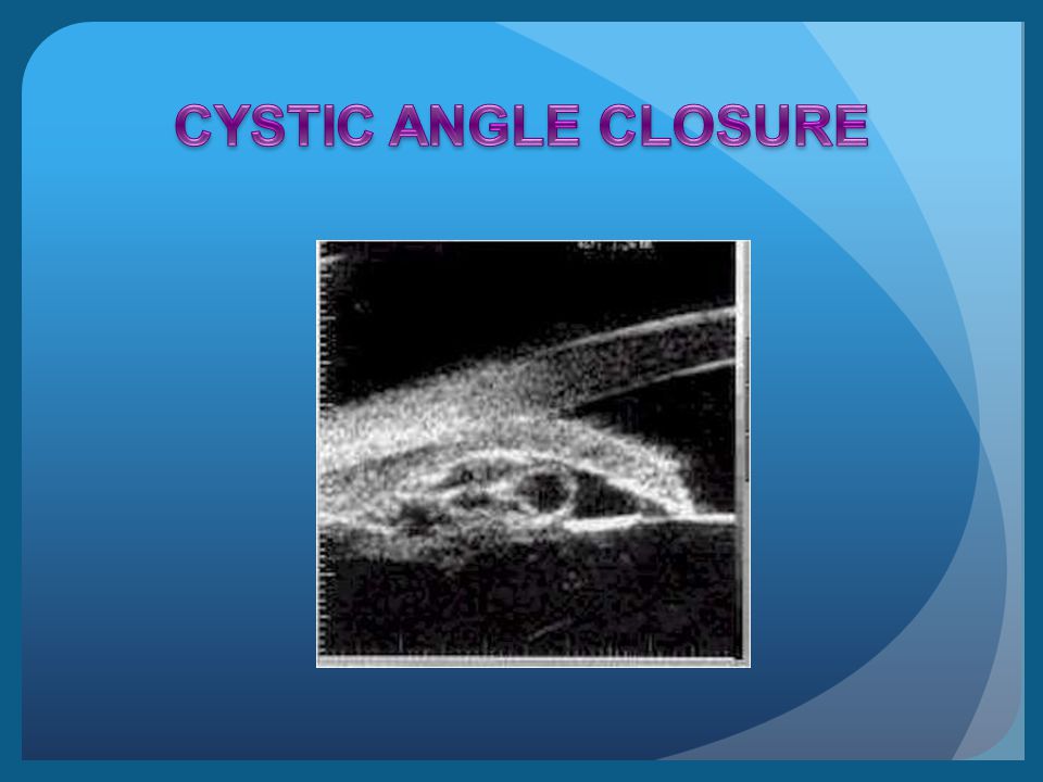
Qualitative analysis include : 1) Pathophysiology of anterior segment disorders 2) Mechanism of appositional angle closure
 Pathophysiology of anterior segment disorders 2) Mechanism of appositional angle closure")
10
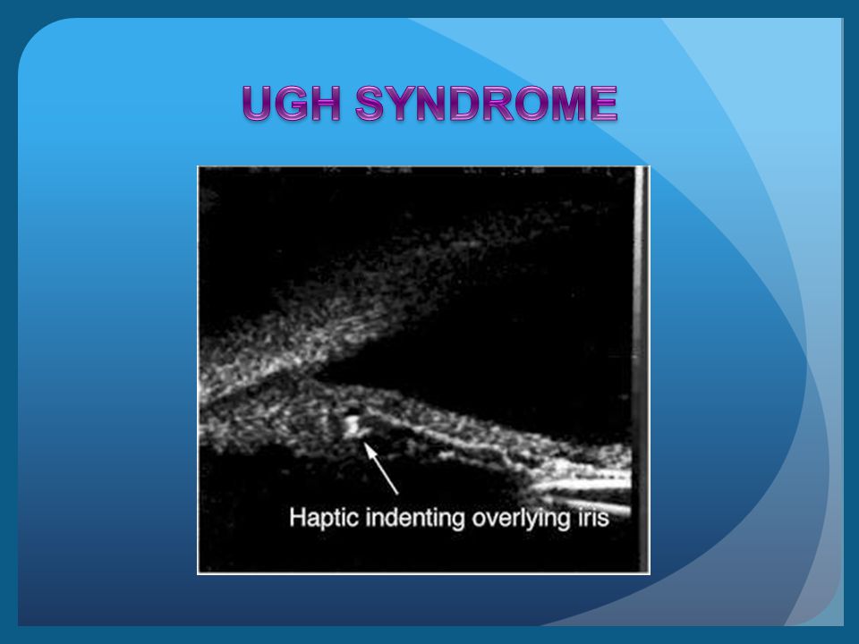
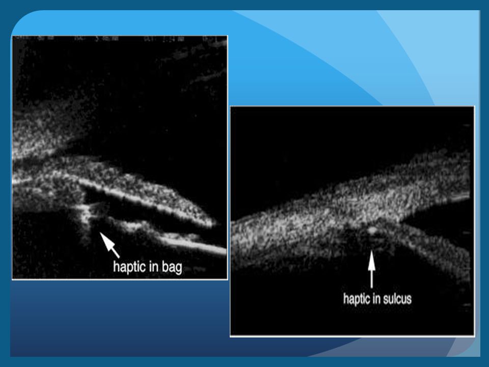
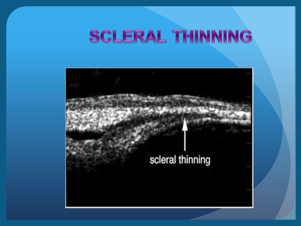
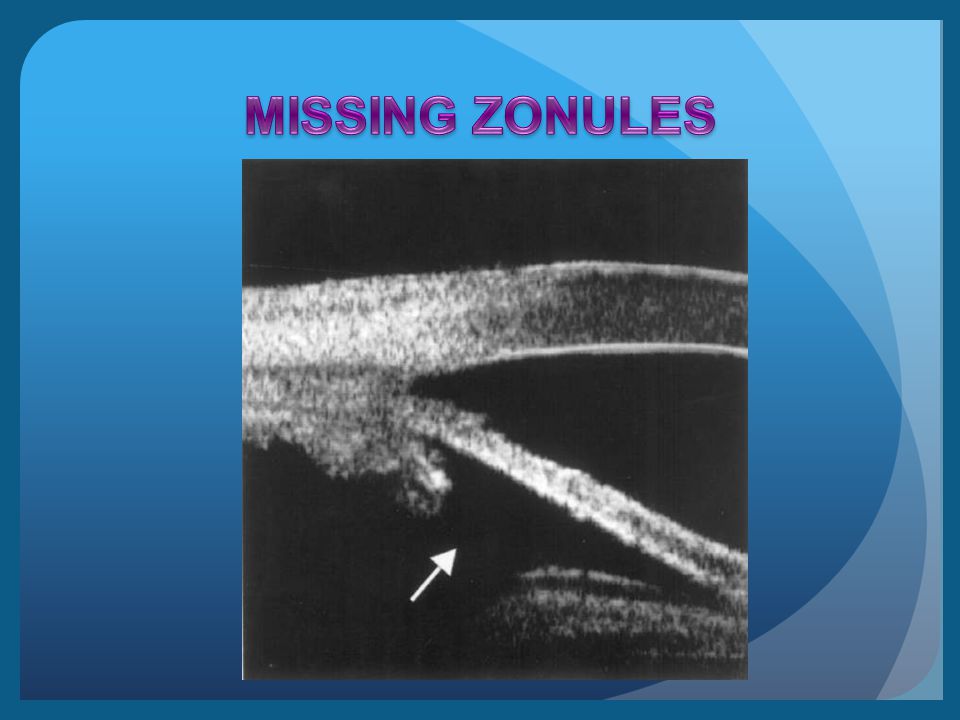
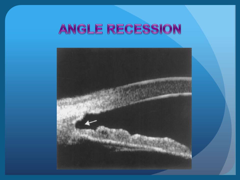
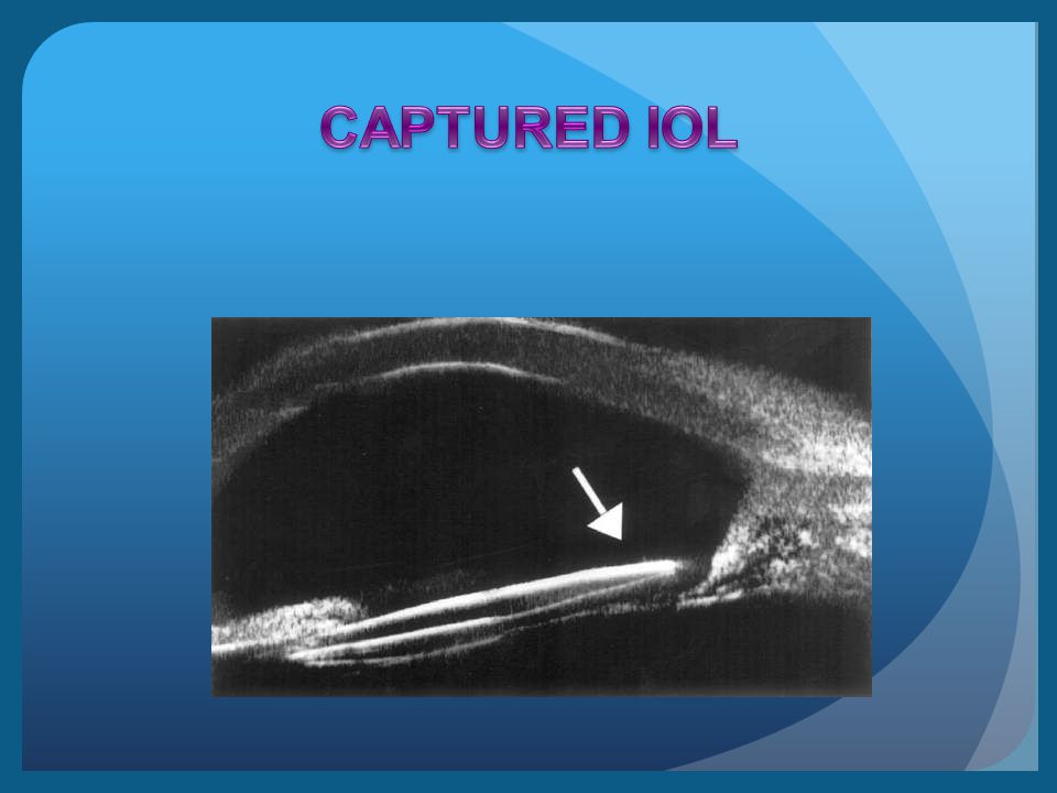
The cornea Glaucoma Tumors IOLs Ocular adnexa The sclera Trauma

13
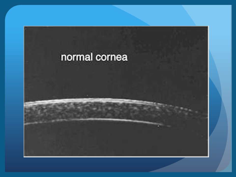
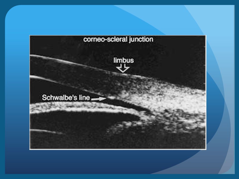
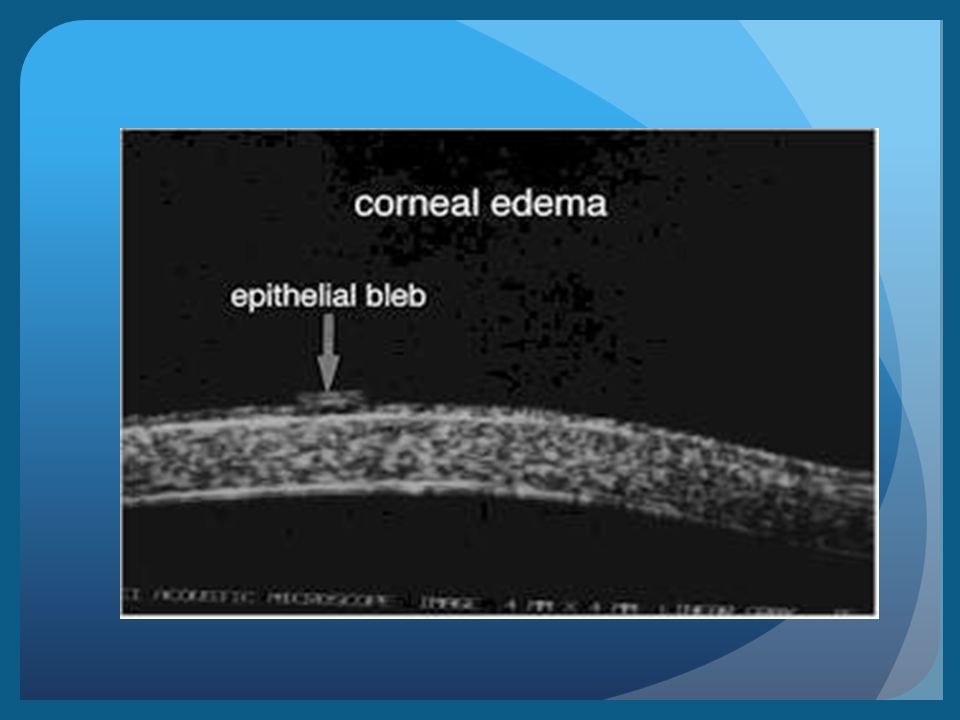
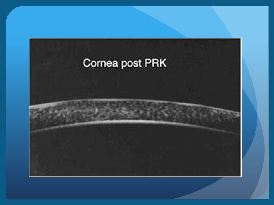
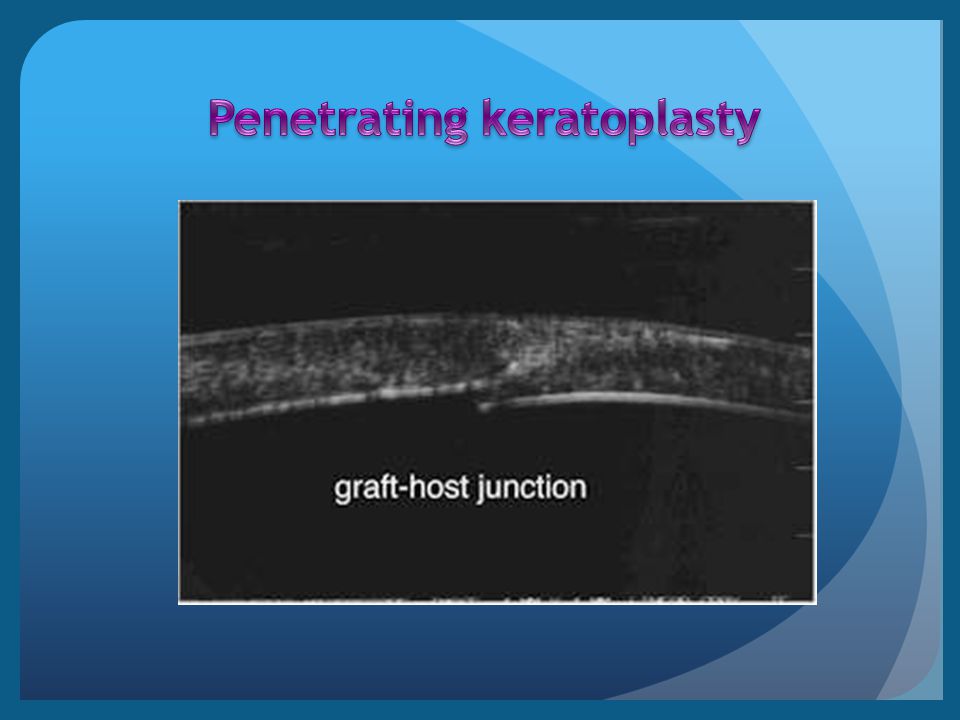
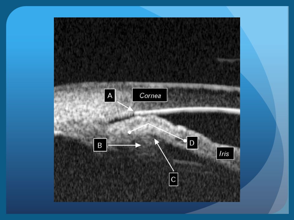
CORNEA: Internal corneal details the depth of abnormalities

26
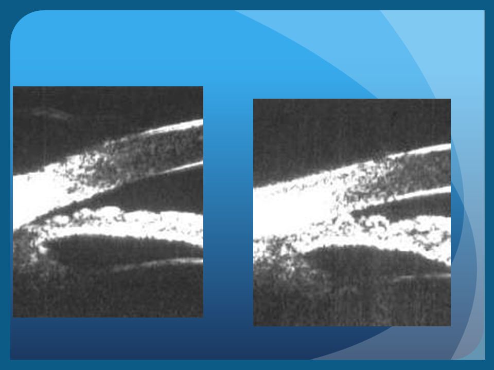
Plateau iris: Anatomically anterior position of the ciliary processes Large ciliary process Medium depth of AC Slightly convex of iris surface

29
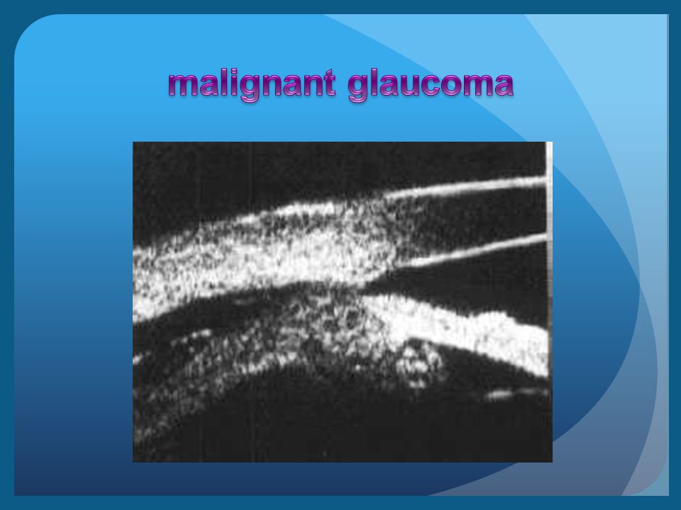
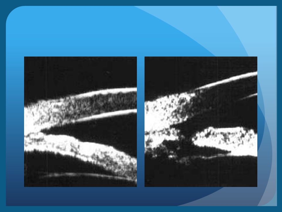
MALIGNANT GLAUCOMA: Shallow supraciliary detachmen Anterior rotation of ciliary body Aqueous humor is secreted posterior to the lens

31
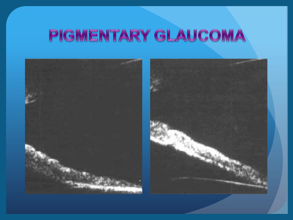
Reverse pupillary block (pigmentary glaucoma) Concave configuration of midperipheral iris Large iris Posteriorly insertion of iris
 Concave configuration of midperipheral iris Large iris Posteriorly insertion of iris")
38
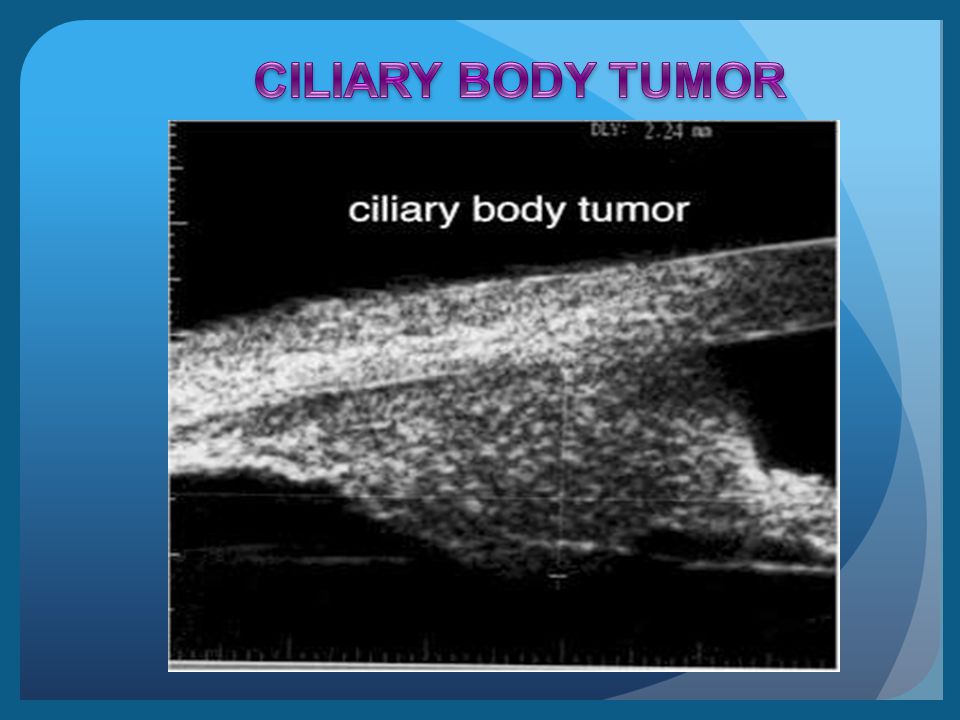
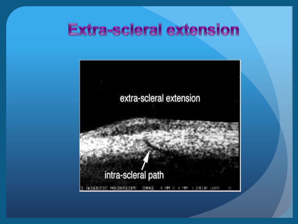
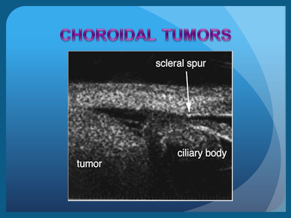
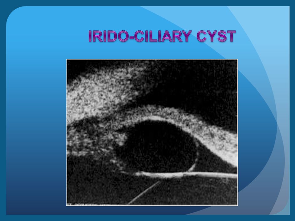
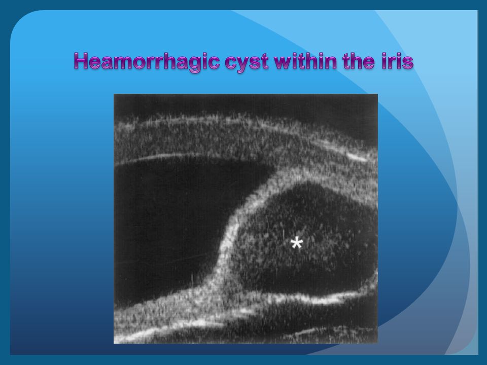
Iris tumors, ciliary body tumors and anterior choroidal tumors can be imaged Borders of the tumor are usually detectable by the change in reflectivity from surrounding structures

74
Ultrasound biomicroscopy is a new imaging technique that uses high frequency ultrasound to produce images of the eye at near microscopic resolution

Similar presentations


 What is the difference between a mirror an a lens? 2) Why do you think we have a lens in our eye instead of a mirror?>")


 17 April 2017 Biology Matters textbook page 281 Concept Map.>")


