Download presentation
Presentation is loading. Please wait.

1
Vision and Eye Problems:
How to recognize them and what we can do about them Nicholas J. Volpe, MD Tarry Professor and Chairman Department of Ophthalmology Feinberg School of Medicine Northwestern University

2
Goals common visual symptoms in elderly people
followed by a discussion of what we do to make the diagnosis how we treat common problems including dry eye, glaucoma, cataract, eyelid problems, diabetic retinopathy, macular degeneration and double vision"

3
Strategies to Preserve Your Vision
Prevention is our most potent tool in the quest to reduce disease (and healthcare costs) Choose your parents well and stop aging!!! OR Don’t Smoke Wear Glasses that are UV protective Safety glasses for high risk activities Pay Attention to Nutrition and Vitamins Don’t Ignore Symptoms Get Regular Eye Examinations
 Choose your parents well and stop aging!!! OR. Don’t Smoke. Wear Glasses that are UV protective. Safety glasses for high risk activities. Pay Attention to Nutrition and Vitamins. Don’t Ignore Symptoms. Get Regular Eye Examinations.")
4
Major Causes of Chronic Visual Loss Preventable and Treatable
Cataracts Glaucoma Macular degeneration Diabetic retinopathy Other Issues Dry eye Double Vision Eyelid Abnormalities Presbyopia near vision blurring

5
Stop Smoking Clearly a risk factor for cataracts 3X the risk
Clearly a risk factor for macular degeneration and its response to treatment

6
Nutrition Healthy tear film Macular degeneration
Fruits and Green Leafy Vegetables Carotenoid pigments (lutein) accumulate in macula and prevent light damage Omega fatty acids Lutein and Zeaxanthin Studied in AREDS 2 Vitamins A,C, E
 accumulate in macula and prevent light damage. Omega fatty acids. Lutein and Zeaxanthin. Studied in AREDS 2. Vitamins A,C, E.")
7
Regular Check Ups Many diseases can be detected
Every 2-3 years from age 40-65 Every 1-2 years after age 65 More frequently with diabetes or family history of glaucoma or macula degeneration Young adults, in the absence of symptoms, do not require routine examinations

8
Common Eye Symptoms Foreign body sensation Itching and burning
Blurred vision flashes and floaters Distortion of shape Blind spots Loss of peripheral vision Double vision Eyelid position changes

9
Foreign Body Sensation,Itching and Burning
Dry eye Blepharitis Allergy Eyelid malposition

10
Dry Eye Tears Cyclosporine Punctal plugs

11
Ptosis, Entropion, Ectropion

12
Flashes and Floaters (ahujaeyecenter.com)
")
13
Cataracts Symptoms Age Steroids (PSC) Trauma Inflammation Diabetes
Expected if ≥ 60 years old 50% years old 70% > 75 years old Most common cause of decreased vision Symptoms Loss of acuity Difficulty with colors Glare at night Trouble reading small print Age Steroids (PSC) Trauma Inflammation Diabetes Other drugs
 Trauma. Inflammation. Diabetes. Other drugs.")
14
Subcapsular cataract Anterior Posterior

15
Nuclear cataract Progression Exaggeration of normal nuclear
ageing change Increasing nuclear opacification Causes increasing myopia Initially yellow then brown

16
Cataract Surgery Outpatient Very successful > 95%
Almost all with intraocular lenses Most common surgical procedure in U.S. >1.4 million/year Most successful surgical intervention Complications uncommon sight threatening IOL technology continues to evolve for astigmatic correction and presbyopia Newest modality is femtosecond laser

17
Cataract Prevention Smoking cessation Reduces Vitamin C in the eye
Vitamin C levels are high in the eye and this helps remove prooxidants Ultraviolet light Fruits and vegetables 5 fold decrease at 3-4 servings per day Regular alcohol consumption increases risk of cataract Steroids and inflammatory conditions are risks for cataracts

18
Age- Related Macular Degeneration
Age-related macular degeneration (AMD) is the most common cause of severe, irreversible vision loss in older Americans and Europeans Worldwide, AMD disease affects million people. Etiology is complex and poorly understood Free-radical mediated damage to the photoreceptors and the RPE Angiogenesis is a feature of neovascular AMD AMD may be associated with a systemic vascular disorder Genetic and environmental factors Variation in the complement factor H gene
 is the most common cause of severe, irreversible vision loss in older Americans and Europeans. Worldwide, AMD disease affects million people. Etiology is complex and poorly understood. Free-radical mediated damage to the photoreceptors and the RPE. Angiogenesis is a feature of neovascular AMD. AMD may be associated with a systemic vascular disorder. Genetic and environmental factors. Variation in the complement factor H gene.")
19
AMD Risk Factors Gender ♀ > ♂ Race/Ethnicity Smoking Family History
Symptoms early = None, mild distortion late = acute loss of vision Gender ♀ > ♂ Race/Ethnicity Smoking Family History Atherosclerosis Hypertension

20
Atrophic AMD Progression Initially drusen and non-specific RPE changes
Late RPE (geographic) atrophy
 atrophy.")
21
Atrophic AMD Fluorescein angiogram Management
Hyperfluorescence from RPE window defect Low-vision aids if appropriate

22
Choroidal Neovascularization (CNV)
Less common than atrophic AMD but more serious Metamorphopsia is initial symptom Many lesions are not visible clinically Suspicious clinical signs Subretinal blood or lipid Gray-yellow subretinal lesion with fluid

23
Current Status of Therapies for CNV
Antiangiogenic therapy Lucentis, Avastin, Eylea CATT trial (Avastin vs Lucentis) Photodynamic therapy with verteporfin Steroids Thermal Laser Choroidal neovascularization (CNV) has proved difficult to treat. Laser photocoagulation reduces vision loss in some forms of CNV in the long term, but the technique has many limitations. It is suitable for only a minority of patients, and around half of all treated eyes have persistent or recurrent CNV within 2 years.[18] Most other approaches to treatment of CNV – such as radiation,[32][93] antiangiogenic drugs[33] including steroids [83], transpupillary thermotherapy [97] and surgery [34][89] – are under investigation. Progress in patient selection and development of new drugs and operating techniques may bring about benefits in other therapies in the future.[32][33][34] At present, however, only photodynamic therapy (PDT) with verteporfin has been proven effective for treatment of CNV in clinical trials, [1][2][3][94] and may be suitable for a larger proportion of patients than laser photocoagulation.[1][2][3][94]
 Photodynamic therapy with verteporfin. Steroids. Thermal Laser. Choroidal neovascularization (CNV) has proved difficult to treat. Laser photocoagulation reduces vision loss in some forms of CNV in the long term, but the technique has many limitations. It is suitable for only a minority of patients, and around half of all treated eyes have persistent or recurrent CNV within 2 years.[18] Most other approaches to treatment of CNV – such as radiation,[32][93] antiangiogenic drugs[33] including steroids [83], transpupillary thermotherapy [97] and surgery [34][89] – are under investigation. Progress in patient selection and development of new drugs and operating techniques may bring about benefits in other therapies in the future.[32][33][34] At present, however, only photodynamic therapy (PDT) with verteporfin has been proven effective for treatment of CNV in clinical trials, [1][2][3][94] and may be suitable for a larger proportion of patients than laser photocoagulation.[1][2][3][94]")
24
Treatment w/Anti VEGF

26
Treatment for Dry AMD -Age-related Eye Disease Study (AREDS) –role of antioxidants vitamin E, 400 IU vitamin C, 500 mg beta carotene, 15 mg (approximately 25,000 IU Vitamin A) zinc 80 mg as zinc oxide copper, 2 mg, as cupric oxide Copper should be taken with zinc, because high-dose zinc is associated with copper deficiency. (Category 3: extensive intermediate size drusen, or at least 1 large druse (> 125 ?m), or noncentral geographic atrophy in 1 or both eyes) (Category 4: advanced or neovascular AMD in 1 eye or vision loss due to AMD in 1 eye) should consider taking high-dose anti-oxidants plus zinc on a daily basis.
 zinc 80 mg as zinc oxide. copper, 2 mg, as cupric oxide. Copper should be taken with zinc, because high-dose zinc is associated with copper deficiency. (Category 3: extensive intermediate size drusen, or at least 1 large druse (> 125 m), or noncentral geographic atrophy in 1 or both eyes) (Category 4: advanced or neovascular AMD in 1 eye or vision loss due to AMD in 1 eye) should consider taking high-dose anti-oxidants plus zinc on a daily basis.")
27
Established Age Related Macular Degeneration
Use Amsler Grid to monitor central vision AREDS-Occuvite Preservision B carotene vs. Lutein and Zeaxanthin (AREDS 2) Vitamin C Vitamin E Zinc Oxide (?necessary and ? Stomach upset) Copper NB: No beta carotene for smokers and others at risk for lung cancer Others??? Lutein Eyes, PhoVision, Perspective, Ocu-force
 Vitamin C. Vitamin E. Zinc Oxide ( necessary and Stomach upset) Copper. NB: No beta carotene for smokers and others at risk for lung cancer. Others Lutein Eyes, PhoVision, Perspective, Ocu-force.")
28
AREDS Results Recommendations
Evaluation: Persons over 55 years old receive a dilated eye exam to assess risk of advanced AMD. Contraindications to Treatment: Smokers and ex-smokers should not use beta carotene, because previous studies have suggested an association with lung cancer and beta carotene in smokers. There were no benefits from treatment shown in the AREDS for patients with no AMD (Category 1) and early AMD (Category 2).
 and early AMD (Category 2).")
29
AREDS 2 Adding omega 3’s did not help
Taking away B Carotene did not hurt and lutein and zexanthine may have been a bit more protective Reducing zinc dose did not hurt and less side effects No prevention of cataracts

30
Diabetic Retinopathy most common cause of
new blindness among adults yo Blindness in working adults affects over 5.3 million Americans age >18 (2.5% of this population) Prevention- worse in HTN, obesity, renal failure, hyperlipidema, smoking, anemia, pregnancy and POOR glycemic control Diabetic retinopathy develops, to some degree, in nearly all patients with diabetes, and is the most common cause of new cases of blindness among adults. The most predominant causes of vision loss are clinically significant macular edema (CSME) and proliferative diabetic retinopathy (PDR). Vision loss can be avoided or minimized with proper ophthalmic care and examination to identify retinopathy in its early stages.
 Prevention- worse in HTN, obesity, renal failure, hyperlipidema, smoking, anemia, pregnancy and POOR glycemic control. Diabetic retinopathy develops, to some degree, in nearly all patients with diabetes, and is the most common cause of new cases of blindness among adults. The most predominant causes of vision loss are clinically significant macular edema (CSME) and proliferative diabetic retinopathy (PDR). Vision loss can be avoided or minimized with proper ophthalmic care and examination to identify retinopathy in its early stages.")
31
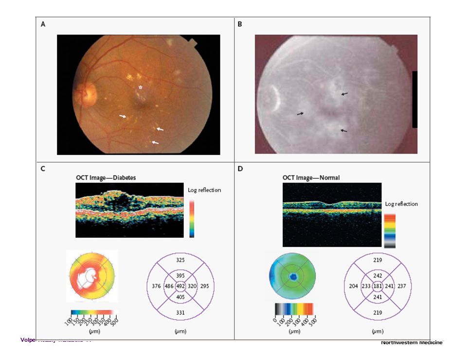
Clinical Findings in NPDR
Microaneurysms Earliest clinical sign of diabetic retinopathy Appear as small red dots in the superficial retinal layers Rupture produces blot/flame hemorrhages

32
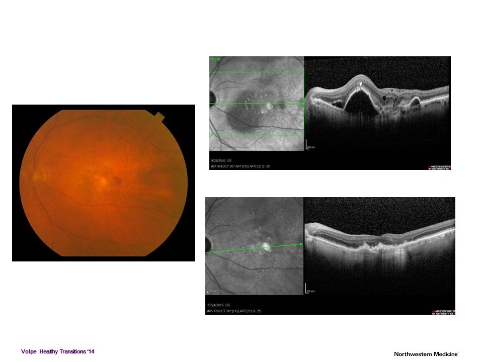
Macular Edema (CSME) Leading cause of visual impairment in patients with diabetes
 Leading cause of visual impairment in patients with diabetes")
34
Macular Edema Treatments
ETDRS focal laser surgery for CSME reduces the incidence of moderate visual loss (doubling of visual angle or roughly a 2-line visual loss) from 30% to 15% over a 3-year period Steroids -peri-ocular -intraocular Anti-VEGF agents Favorable prognostic factors Circinate exudates of recent onset Well-defined leakage Good perifoveal perfusion Unfavorable prognostic factors Diffuse edema/multiple leaks Lipid deposition in the fovea Macular ischemia Cystoid macular edema Preoperative vision of less than 20/200 Hypertension If specific microaneurysms leak- they are treated directly with focal laser photocoagulation. Diffuse leakage - grid pattern of laser Medium intensity burns ( µm) are placed at least 1 burn size apart Intravitreal or sub-tenons steroid injections- when edema persists after multiple focal laser treatments. CSME should be treated and observed closely (every 2-3 mo)
 from 30% to 15% over a 3-year period. Steroids. -peri-ocular. -intraocular. Anti-VEGF agents. Favorable prognostic factors. Circinate exudates of recent onset. Well-defined leakage. Good perifoveal perfusion. Unfavorable prognostic factors. Diffuse edema/multiple leaks. Lipid deposition in the fovea. Macular ischemia. Cystoid macular edema. Preoperative vision of less than 20/200. Hypertension. If specific microaneurysms leak- they are treated directly with focal laser photocoagulation. Diffuse leakage - grid pattern of laser. Medium intensity burns ( µm) are placed at least 1 burn size apart. Intravitreal or sub-tenons steroid injections- when edema persists after multiple focal laser treatments. CSME should be treated and observed closely (every 2-3 mo)")
35
Ischemic diabetic maculopathy
Macula appears relatively normal Capillary non-perfusion on FA Poor visual acuity Treatment not appropriate

36
PDR Proliferation of new blood vessels due to ischemia NVD Disc
NVE Elsewhere NVI Iris NVA Angle When proliferation of new blood vessels occurs, the patient is diagnosed as having PDR. New blood vessel formation is caused by retinal ischemia. Neovascularization can occur at the optic disk (NVD) or elsewhere in the retina (NVE). These new vessels are weak and, when they break, can cause vitreous hemorrhage. They can also cause retinal traction, retinal tears, and retinal detachment over time.
 or elsewhere in the retina (NVE). These new vessels are weak and, when they break, can cause vitreous hemorrhage. They can also cause retinal traction, retinal tears, and retinal detachment over time.")
37
PDR - cont. Treatment Options Pan-retinal photocoagulation
Peripheral Retinal Cryotherapy Vitrectomy Anti-VEGF DRS proved that xenon and argon laser decreases to chances of visual loss in eye with high risk characteristics (HRC) High risk characteristics (HRC) was defined as: NVD > ½ the disc area Any NVD and VH NVE > ½ the disc area and VH or pre-retinal hemorrhage Peripheral Retinal Cryotherapy Employed when HRC is present but the media are too hazy for laser Major complication is increased risk of traction RD, seen in 25-38% Vitrectomy Major indications: non-clearing vitreous hemorrhage, macular involving or threatening TRD, or combined traction-rhegmatogenous RD Other indications: ME with a thickened and taut posterior hyaloid, macular heterotopia, ERM, severe pre-retinal hemorrhage, and NVG with cloudy media
 High risk characteristics (HRC) was defined as: NVD > ½ the disc area. Any NVD and VH. NVE > ½ the disc area and VH or pre-retinal hemorrhage. Peripheral Retinal Cryotherapy. Employed when HRC is present but the media are too hazy for laser. Major complication is increased risk of traction RD, seen in 25-38% Vitrectomy. Major indications: non-clearing vitreous hemorrhage, macular involving or threatening TRD, or combined traction-rhegmatogenous RD. Other indications: ME with a thickened and taut posterior hyaloid, macular heterotopia, ERM, severe pre-retinal hemorrhage, and NVG with cloudy media.")
38
Retinopathy Screening
Type 1 diabetes - screen within 3-5 years of diagnosis after age 101 Type 2 diabetes - screen at time of diagnosis1 Pregnancy - women with preexisting diabetes should be screened prior to conception and during first trimester1 Follow-up depends on severity of disease A comprehensive eye exam should be given to all patients with diabetes. The exam should include all of the components recommended by the American Academy of Ophthalmology and the American Optometric Association for non-diabetic patients, however additional attention should be given to assess abnormalities of the retina and vitreous fluid. Dilated indirect ophthalmoscopy, slitlamp biomicroscopy, and fundus examination should be used to identify retinal or vitreous hemorrhages, microaneurysms, retinal tears or detachment, intraretinal microvascular abnormality (IRMA), neovascularization at the optic disk (NVD) and elsewhere in the retina (NVE). Patients with type 1 diabetes should be screened for NPDR (non-proliferative diabetic retinopathy) within three to five years of diagnosis after age 10. Retinopathy leading to vision loss rarely develops in children prior to puberty, regardless of how long the child has had diabetes. Patients with type 2 diabetes should be screened at time of diagnosis. Finally, women with preexisting diabetes should be screened prior to conception and during the first trimester. Follow-up exams should be scheduled at the physicians discretion based on the results of the first trimester exam. Note that follow-up exams should be conducted yearly, however new ADA Clinical Practice Recommendations state that less frequent exams every 2-3 years can be considered in the setting of a normal eye exam upon the advice of an eye care professional.
, neovascularization at the optic disk (NVD) and elsewhere in the retina (NVE). Patients with type 1 diabetes should be screened for NPDR (non-proliferative diabetic retinopathy) within three to five years of diagnosis after age 10. Retinopathy leading to vision loss rarely develops in children prior to puberty, regardless of how long the child has had diabetes. Patients with type 2 diabetes should be screened at time of diagnosis. Finally, women with preexisting diabetes should be screened prior to conception and during the first trimester. Follow-up exams should be scheduled at the physicians discretion based on the results of the first trimester exam. Note that follow-up exams should be conducted yearly, however new ADA Clinical Practice Recommendations state that less frequent exams every 2-3 years can be considered in the setting of a normal eye exam upon the advice of an eye care professional.")
39
Diabetic Eye Care Like glaucoma, you will NOT HAVE SYMPTOMS UNTIL IT IS TOO LATE! 95-100% treatable with early detection Regular eye exams at 6 or 12 month interval depending on what MD sees Bleeding, swelling and growth of blood vessels Diabetes control (Hemoglobin A1c) is the most important way to reduce your risk High blood pressure is a risk Diet and exercise
 is the most important way to reduce your risk. High blood pressure is a risk. Diet and exercise.")
40
Glaucoma Optic nerve 1.2 million nerve fibers Ganglion cells in retina
exit to brain as optic nerve

41
Definition of Glaucoma
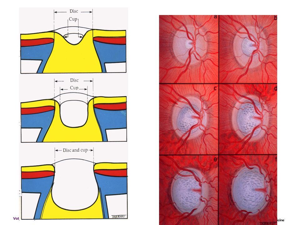
A group of optic neuropathies in which retinal ganglion cells die by apoptosis with resultant optic disc cupping and characteristic visual field deficits Optic neuropathy Retinal ganglion cell apoptosis Optic disc cupping or excavation Loss of visual function -IOP is too high for the nerve??? Most common cause blindness: African-Americans COMPLETE/TOTAL BLINDNESS

44
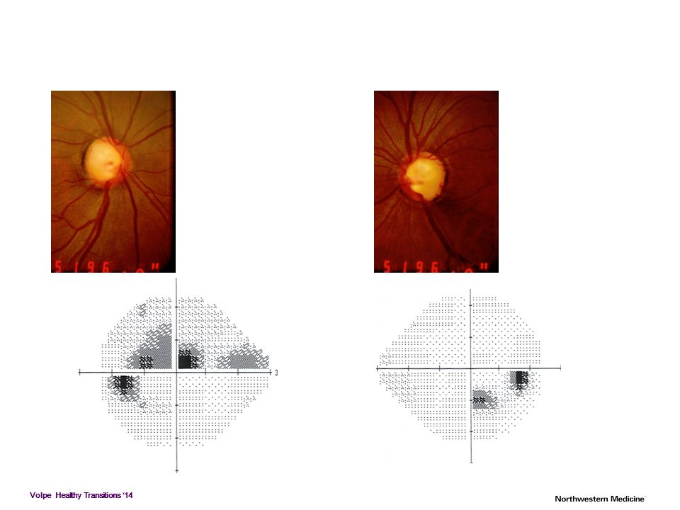
Glaucoma Loss of visual field Site of visual field loss
corresponds to area of damage on optic disc, e.g., “cupping”

45
Angle Closure Glaucoma
Acute pain, redness, tearing Associated with dilation of pupil Natural (e.g., movie theater) Pharmacologic Nausea & vomiting often in ER with “acute abdomen” Risk factor of narrow angle can be detected on screening exam (esp hyperope) and prophylactic iridotomy is preventative of attack in 100%
 Pharmacologic. Nausea & vomiting. often in ER with acute abdomen Risk factor of narrow angle can be detected on screening exam (esp hyperope) and prophylactic iridotomy is preventative of attack in 100%")
46
Acute angle-closure glaucoma
Signs Ciliary injection Complete angle closure Severe corneal edema Dilated, unreactive, vertically oval pupil Shallow anterior chamber Medical rx to lower IOP, followed by laser (Yag) iridotomy
 iridotomy.")
47
Primary Open-Angle Glaucoma
The most prevalent type of glaucoma in the United States Elevated intraocular pressure is not part of the diagnostic criteria 25% of patients with primary open-angle glaucoma in the US have normal intraocular pressure Asymptomatic Some loss of visual field Most common type Familial, bilateral “Sneak thief of sight”

48
Primary Open-Angle Glaucoma
Evidence that IOP reduction is beneficial Collaborative Normal-Tension Glaucoma Study (CNTGS) Advanced Glaucoma Intervention Study (AGIS) Early Manifest Glaucoma Study (EMGT) 25% IOP reduction RoP 62% to 45% at a median of 6 years. Ocular Hypertension Treatment Study (OHTS)
 Advanced Glaucoma Intervention Study (AGIS) Early Manifest Glaucoma Study (EMGT) 25% IOP reduction RoP 62% to 45% at a median of 6 years. Ocular Hypertension Treatment Study (OHTS)")
49
Treatment for POAG Lower the IOP Medical therapy
Prostaglandin , B-blockers,Sypathomimetics, Carbonic-anyhrase inhibitors Laser surgery (ALT, SLT) Incisional surgery (Trab, shunt)
 Incisional surgery (Trab, shunt)")
50
Tube Shunts

51
Double Vision Monocular vs binocular Eye misalignment
Stroke, tumor, cranial nerve palsy However most are benign Thyroid Eye Disease Patch, prism, surgery Utsavaeyelinic.com

52
Summary FBS, itching burning Floaters and flashes Diabetic Retinopathy
Prevention, treatment Cataract Surgical treatment continues to improve Double Vision Most benign-eye misalignment Must be evaluated Glaucoma Silent blindness, family history Medical and surgical rx ARMD New age of available prevention strategies and treatments exudative varietyFBS

53
Strategies to Preserve Your Vision
Prevention is our most potent tool in the quest to reduce disease (and healthcare costs) Choose your parents well and stop aging!!! OR Don’t Smoke Wear Glasses that are UV protective Safety glasses for high risk activities Pay Attention to Nutrition and Vitamins Don’t Ignore Symptoms Get Regular Eye Examinations
 Choose your parents well and stop aging!!! OR. Don’t Smoke. Wear Glasses that are UV protective. Safety glasses for high risk activities. Pay Attention to Nutrition and Vitamins. Don’t Ignore Symptoms. Get Regular Eye Examinations.")
Similar presentations


 Waxman MD PhD>")


Observation occult CNV also requires close follow – up.every 3 month 2) Laser coagulation 3) Surgery.>")


