Download presentation
Presentation is loading. Please wait.

1
Jed Hummel RAD 4001 CASE PRESENTATION

3
CT HALO SIGN Ground glass attenuation surrounding a pulmonary nodule Causes of the ground glass halo can be divided into three categories hemorrhage tumor infiltration inflammatory processes Initially proposed as an early, specific finding of invasive pulmonary aspergillosis Lee, Y. R., Y. W. Choi, et al. (2005). "CT halo sign: the spectrum of pulmonary diseases." Br J Radiol 78(933): 862-5.
. CT halo sign: the spectrum of pulmonary diseases. Br J Radiol 78(933):")
4
HALO SIGN DIFFERENTIAL DIAGNOSIS Infectious diseases Neoplastic diseases Non-neoplastic, non-infectious, inflammatory diseases

5
HALO SIGN DIFFERENTIAL DIAGNOSIS Infectious diseases Fungus Aspergillosis Mucormycosis Candidiasis Coccidioidomycosis cryptococcosis Septic embolism Mycobacteria: TB, MAC Rickettsia: Coxiella burnetti Virus: HSV, VZV, cytomegalovirus Aspergillosis is most common cause in immunocompromised patient Lee, Y. R., Y. W. Choi, et al. (2005). "CT halo sign: the spectrum of pulmonary diseases." Br J Radiol 78(933): 862-5.
. CT halo sign: the spectrum of pulmonary diseases. Br J Radiol 78(933):")
6
HALO SIGN DIFFERENTIAL DIAGNOSIS Neoplastic diseases Primary tumors: bronchioloalveolar carcinoma squamous cell Kaposi sarcoma adenocarcinoma Metastatic tumors: Choriocarcinoma Osteosarcoma Melanoma hydatidiform mole GI malignancies Lymphoproliferative disorders Bronchioloalveolar carcinoma is most common cause in immunocompetent patients Lee, Y. R., Y. W. Choi, et al. (2005). "CT halo sign: the spectrum of pulmonary diseases." Br J Radiol 78(933): 862-5.
. CT halo sign: the spectrum of pulmonary diseases. Br J Radiol 78(933):")
7
HALO SIGN DIFFERENTIAL DIAGNOSIS Non-neoplastic, non-infectious, inflammatory diseases Wegener’s granulomatosis Eosinophilic lung disease parasitic infestation (schistosomiasis) simple pulmonary eosinophilia hypereosinophilic syndrome Pulmonary endometriosis Organizing pneumonia Hypersensitivity pneumonitis Iatrogenic injury: transbronchial lung biopsy catheter-induced pulmonary pseudoaneurysm Lee, Y. R., Y. W. Choi, et al. (2005). "CT halo sign: the spectrum of pulmonary diseases." Br J Radiol 78(933): 862-5.
. CT halo sign: the spectrum of pulmonary diseases. Br J Radiol 78(933):")
8
HISTOPATHOLOGICAL CAUSES OF HALO Hemorrhage into lung tissue Vasculitis (Wegener’s) Metastases from hypervascular tumors (angiosarcoma, choriocarcinoma, osteosarcoma and melanoma) hemorrhage secondary to the fragility of neovascular tissue. Vascular invasion of aspergillus organisms causes thrombosis of small- to medium-sized vessels and ischemic necrosis of the lung parenchyma. In a group of 25 patients the frequency of halo sign ranged from 96% at day 0 to 19% at day 14. One study showed that CT is more sensitive in diagnosis of early invasive aspergillus than serologic testing. Thus, in severely neutropenic patients with a fever that does not respond to antibiotics, the CT halo sign is an early indicator for diagnosing invasive aspergillosis even before serological tests become positive, and it warrants administration of systemic antifungal therapy. Lee, Y. R., Y. W. Choi, et al. (2005). "CT halo sign: the spectrum of pulmonary diseases." Br J Radiol 78(933): 862-5.
. CT halo sign: the spectrum of pulmonary diseases. Br J Radiol 78(933):")
9
HISTOPATHOLOGICAL CAUSES OF HALO Inflammatory cell infiltration Infections such as: cryptococcosis and cytomegalovirus pneumonia. In eosinophilic lung diseases the halo sign is caused by pulmonary infiltrations of eosinophils and other inflammatory cells Lee, Y. R., Y. W. Choi, et al. (2005). "CT halo sign: the spectrum of pulmonary diseases." Br J Radiol 78(933): 862-5.
. CT halo sign: the spectrum of pulmonary diseases. Br J Radiol 78(933):")
10
HISTOPATHOLOGICAL CAUSES OF HALO Tumor cell infiltration In bronchioloalveolar carcinoma the halo reflects a unique lepidic growth pattern in which tumor spreads into distal air spaces by using the alveolar septa as a stroma with relative lack of acinar filling. Lee, Y. R., Y. W. Choi, et al. (2005). "CT halo sign: the spectrum of pulmonary diseases." Br J Radiol 78(933): 862-5.
. CT halo sign: the spectrum of pulmonary diseases. Br J Radiol 78(933):")
11
HALO SIGN Invasive pulmonary aspergillosis in a 39-year-old man with AML and neutropenia. Lee, Y. R., Y. W. Choi, et al. (2005). "CT halo sign: the spectrum of pulmonary diseases." Br J Radiol 78(933): 862-5.
. CT halo sign: the spectrum of pulmonary diseases. Br J Radiol 78(933):")
12
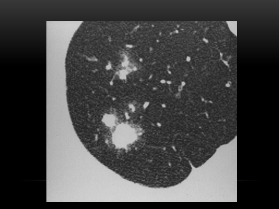
HALO SIGN Wegener's granulomatosis in a 70-year-old woman. Pulmonary nodule with the CT halo sign in the left lower lobe (thick arrow). Nodule in the right middle lobe without surrounding ground glass opacity (thin arrow) Lee, Y. R., Y. W. Choi, et al. (2005). "CT halo sign: the spectrum of pulmonary diseases." Br J Radiol 78(933): 862-5.
. Nodule in the right middle lobe without surrounding ground glass opacity (thin arrow) Lee, Y. R., Y. W. Choi, et al. (2005). CT halo sign: the spectrum of pulmonary diseases. Br J Radiol 78(933):")
13
HALO SIGN Cytomegalovirus pneumonia in a 45-year-old woman Multiple tiny nodules with the CT halo sign in the right lower lobe. Lee, Y. R., Y. W. Choi, et al. (2005). "CT halo sign: the spectrum of pulmonary diseases." Br J Radiol 78(933): 862-5.
. CT halo sign: the spectrum of pulmonary diseases. Br J Radiol 78(933):")
14
HALO SIGN Bronchioloalveolar carcinoma in a 60-year-old woman. Lee, Y. R., Y. W. Choi, et al. (2005). "CT halo sign: the spectrum of pulmonary diseases." Br J Radiol 78(933): 862-5.
. CT halo sign: the spectrum of pulmonary diseases. Br J Radiol 78(933):")
15
HALO SIGN Simple pulmonary eosinophilia (Loeffler syndrome) in a 42-year- old man Lee, Y. R., Y. W. Choi, et al. (2005). "CT halo sign: the spectrum of pulmonary diseases." Br J Radiol 78(933): 862-5.
. CT halo sign: the spectrum of pulmonary diseases. Br J Radiol 78(933):")
17
H&P 50 y/o male with pmh of sinusitis and severe headaches for 4 years. Treated with different antibiotics as outpatient with little improvement. Presents with cough with blood streaked sputum for the last two days. ROS positive for fatigue, subjective weight loss, night sweats, arthralgias. PE: Purpuric rash on both of his lower extremities Labs: ESR 55 mm/hr, CRP 22.4 mg/dl, RF 1175.7 IU/ml UA: 100 red blood cells per high-power field

18
WEGENER'S GRANULOMATOSIS Necrotizing medium-sized and small vessel vasculitis involving upper respiratory tract, lung, renal vessels Necrotizing granulomas in upper respiratory tract (saddle nose deformity), kidneys (crescentic glomerulonephritis) Patients often present with chronic sinusitis (67%). Other symptoms: fever, weight loss, hematuria, hearing disturbances, palpable purpura, arthritis, cough, cavitary pulmonary lesions -> hemoptysis. c-ANCA antibodies (>90% of cases) Treatment: High-dose corticosteroids, cyclophosphamide. Patients tend to have frequent relapses. Shafiei K, Luther E, Archie M, Gulick J, Fowler MR. Wegener granulomatosis: case report and brief literature review. J Am Board Fam Pract. Nov-Dec 2003;16(6):555-559.
 Treatment: High-dose corticosteroids, cyclophosphamide. Patients tend to have frequent relapses. Shafiei K, Luther E, Archie M, Gulick J, Fowler MR. Wegener granulomatosis: case report and brief literature review. J Am Board Fam Pract. Nov-Dec 2003;16(6):")
19
WEGENER'S GRANULOMATOSIS PA chest radiograph shows cavitating masses with fluid levels in both lungs of intermediate wall thickness. Typically finding: multiple pulmonary nodules that cavitate in one-third to one- half of patients. The cavities tend to be thick walled with irregular inner lining. There is no predilection for any lung zone. The cavities may resolve spontaneously.

20
WEGENER'S GRANULOMATOSIS Coronal CT image shows the absence of the midline nasal structures and the medial wall of the maxillary sinuses. Valencia MP, Castillo M. Congenital and acquired lesions of the nasal septum: a practical guide for differential diagnosis. Radiographics. Jan-Feb 2008;28(1):205-224; quiz 326.
: ; quiz")
21
WEGENER'S GRANULOMATOSIS Axial CT image shows a midline intranasal mass that has eroded the septum. The maxillary sinuses are small, and their lateral and anterior walls have a thickened, ground-glass appearance. Valencia MP, Castillo M. Congenital and acquired lesions of the nasal septum: a practical guide for differential diagnosis. Radiographics. Jan-Feb 2008;28(1):205-224; quiz 326.
: ; quiz")
22
WEGENER'S GRANULOMATOSIS

23
SOURCES 1.Pinto PS. The CT Halo Sign. Radiology. Jan 2004;230(1):109-110. 2.Lee YR, Choi YW, Lee KJ, Jeon SC, Park CK, Heo JN. CT halo sign: the spectrum of pulmonary diseases. Br J Radiol. Sep 2005;78(933):862-865. 3.Prince JS, Duhamel DR, Levin DL, Harrell JH, Friedman PJ. Nonneoplastic lesions of the tracheobronchial wall: radiologic findings with bronchoscopic correlation. Radiographics. Oct 2002;22 Spec No:S215-230. 4.Shafiei K, Luther E, Archie M, Gulick J, Fowler MR. Wegener granulomatosis: case report and brief literature review. J Am Board Fam Pract. Nov-Dec 2003;16(6):555-559. 5.Valencia MP, Castillo M. Congenital and acquired lesions of the nasal septum: a practical guide for differential diagnosis. Radiographics. Jan-Feb 2008;28(1):205-224; quiz 326.
: Lee YR, Choi YW, Lee KJ, Jeon SC, Park CK, Heo JN. CT halo sign: the spectrum of pulmonary diseases. Br J Radiol. Sep 2005;78(933): Prince JS, Duhamel DR, Levin DL, Harrell JH, Friedman PJ. Nonneoplastic lesions of the tracheobronchial wall: radiologic findings with bronchoscopic correlation. Radiographics. Oct 2002;22 Spec No:S Shafiei K, Luther E, Archie M, Gulick J, Fowler MR. Wegener granulomatosis: case report and brief literature review. J Am Board Fam Pract. Nov-Dec 2003;16(6): Valencia MP, Castillo M. Congenital and acquired lesions of the nasal septum: a practical guide for differential diagnosis. Radiographics. Jan-Feb 2008;28(1): ; quiz")
Similar presentations

 Active TB Routine; FBE WCC (Infection) Hb (Anaemic of chronic disease) U&Es (baseline) LFTs (baseline) ESR/CRP (inflammation/infection)>")


 SHEN JIN The First Affiliated Hospital of Kunming Medical College.>")

