Download presentation
Presentation is loading. Please wait.

1
Limbic System Amydala & Hippocampus
Done By: Maddi Gervasio, Kelly Camardo, Catherine Murphy, Andrea Alcala, Riham Majeed, and Ro-Anne Thomas

2
History Paul Broca (1878) first referred to the curved rim of the cortex that borders the cerebral hemispheres and brainstem as the great limbic lobe (from Latin Limbus for border) Paul D. MacLean (1952) coined term “Limbic System” while introducing Triune Brain Theory and attributed this system to Broca’s limbic lobe and related subcortical structures as the collective entity for emotional and motivational regulation. Since then, disagreements have arisen on whether or not one collective entity was purely responsible for emotional and motivational regulation. Some neuroscientists no longer believe that the concept of a unified "limbic system" is valid; however the term “Limbic System” is still used. The limbic system can now be attributed to one of the parts of the brain that control visceral, autonomic processes; as well as some structures playing a part in memory.
 first referred to the curved rim of the cortex that borders the cerebral hemispheres and brainstem as the great limbic lobe (from Latin Limbus for border) Paul D. MacLean (1952) coined term Limbic System while introducing Triune Brain Theory and attributed this system to Broca’s limbic lobe and related subcortical structures as the collective entity for emotional and motivational regulation. Since then, disagreements have arisen on whether or not one collective entity was purely responsible for emotional and motivational regulation. Some neuroscientists no longer believe that the concept of a unified limbic system is valid; however the term Limbic System is still used. The limbic system can now be attributed to one of the parts of the brain that control visceral, autonomic processes; as well as some structures playing a part in memory.")
3
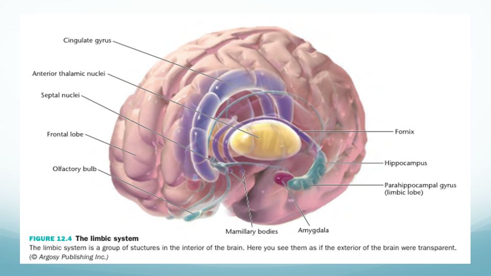
General Structure The limbic system is a set of several functionally and anatomically interconnected nuclei and cortical structures that are located in the telencephalon, or cerebral cortex, and diencephalon, or subcortical regions. Despite a lack of universal agreement on the limbic system structure, the following have been attributed as a part of the Limbic System:

4
General Structure (continued)
Cortical areas: Limbic lobe Orbitofrontal cortex, a region in the frontal lobe. Piriform cortex, part of the olfactory system. Entorhinal cortex (ento = interior, rhino = nose, entorhinal = interior to the rhinal sulcus) Hippocampus (and associated structures), a bilateral structure located under the cerebral cortex. Fornix, a white matter structure connecting the hippocampus with other brain structures, particularly the mammillary bodies and septal nuclei Subcortical areas: Septal nuclei, a set of structures that lie in front of the lamina terminalis, considered a pleasure zone. Amygdala, located deep within the temporal lobes and related with a number of emotional processes. Nucleus accumbens, a region in the basal forebrain. Diencephalic structures: Hypothalamus: a center for the limbic system, connected with the frontal lobes, septal nuclei and the brain stem reticular formation via the medial forebrain bundle, with the hippocampus via the fornix, and with the thalamus via the mammillothalamic fasciculus. It regulates a great number of autonomic processes. Mammilary bodies, part of the hypothalamus that receives signals from the hippocampus via the fornix and projects them to the thalamus. Anterior nuclei of thalamus receive input from the mammillary bodies. Involved in memory processing
 Hippocampus (and associated structures), a bilateral structure located under the cerebral cortex. Fornix, a white matter structure connecting the hippocampus with other brain structures, particularly the mammillary bodies and septal nuclei. Subcortical areas: Septal nuclei, a set of structures that lie in front of the lamina terminalis, considered a pleasure zone. Amygdala, located deep within the temporal lobes and related with a number of emotional processes. Nucleus accumbens, a region in the basal forebrain. Diencephalic structures: Hypothalamus: a center for the limbic system, connected with the frontal lobes, septal nuclei and the brain stem reticular formation via the medial forebrain bundle, with the hippocampus via the fornix, and with the thalamus via the mammillothalamic fasciculus. It regulates a great number of autonomic processes. Mammilary bodies, part of the hypothalamus that receives signals from the hippocampus via the fornix and projects them to the thalamus. Anterior nuclei of thalamus receive input from the mammillary bodies. Involved in memory processing.")
6
Hippocampus (structure)
Means “seahorse” in Greek because of seahorse shape A bilateral structure, it is located in medial aspect of temporal lobe, forming the medial wall of the lateral ventricle in this area Part of the Hippocampal Formation which also includes the denate gyrus and subiculum The Hippocampus is essentially a curved area of cortex that is divided into four regions called the CA (Cornu Ammonis) fields. These are designated as CA1, CA2, CA3 and CA4 and contain prominent pyramidal cells. CA4 is embedded into a backward-facing, V-shaped cortex, the dentate gyrus The denate gyrus blends into the adjacent subiculum, which, in turn, is connected to the entorhinal cortex (EC) on the parahippocampal gyrus of the temporal lobe.
 fields. These are designated as CA1, CA2, CA3 and CA4 and contain prominent pyramidal cells. CA4 is embedded into a backward-facing, V-shaped cortex, the dentate gyrus. The denate gyrus blends into the adjacent subiculum, which, in turn, is connected to the entorhinal cortex (EC) on the parahippocampal gyrus of the temporal lobe.")
7
(EC)
")
8
Hippocampal Pathways Afferents and efferents of the hippocampus are bundled together in the same paths. Thus, by knowing the output paths, for example, you will also know the input paths, or vice versa. Two major pathways into and out of the hippocampus are the fornix and entorhinal cortex (EC) (via the cingulate cortex). The precommissural branch of the fornix connects to the septal nuclei, preoptic nuclei, ventral striatum, orbital cortex and anterior cingulate cortex. The postcommissural branch of the fornix connects to the anterior nucleus of the thalamus and the mammillary bodies of the hypothalamus. Mammillothalamic tract also goes to the anterior thalamic nucleus. The anterior thalamic nuclei in turn connects to the cingulate cortex. The cingulate cortex projects back to the entorhinal cortex of parahippocampal gyrus, completing a “great” loop called the Papez circuit.
 (via the cingulate cortex). The precommissural branch of the fornix connects to the septal nuclei, preoptic nuclei, ventral striatum, orbital cortex and anterior cingulate cortex. The postcommissural branch of the fornix connects to the anterior nucleus of the thalamus and the mammillary bodies of the hypothalamus. Mammillothalamic tract also goes to the anterior thalamic nucleus. The anterior thalamic nuclei in turn connects to the cingulate cortex. The cingulate cortex projects back to the entorhinal cortex of parahippocampal gyrus, completing a great loop called the Papez circuit.")
9
Amygdala (structure) Identified by Karl Friedrich Burdach in the early 19th century, the amygdala, an almond-shaped structure deep within the temporal lobe, is a collection of nuclei lying beneath the uncus. Lying at the anterior end of the hippocampal formation and the anterior tip of the inferior horn of the lateral ventricle, it merges with the periamygdaloid cortex, which forms part of the surface of the uncus. The amygdaloid complex is structurally diverse and comprises of approximately 13 nuclei. These are further divided into subdivisions that have extensive internuclear and intranuclear connections. The major groups are: Basolateral nuclei Cortical-like nuclei Centromedial nuclei Other amygdaloid nuclei Extended Amygdala (centromedial amygdala, sublenticular substantia innominata and bed nucleus of the stria terminalis) Indian J Psychiatry Apr-Jun; 49(2): 132–139
 Indian J Psychiatry Apr-Jun; 49(2): 132–139.")
10
Amygdala Pathways Besides projecting back to the cortex, (mostly the orbitofrontal cortex) the amygdala makes reciprocal connections with brain regions including the thalamus, hypothalamus, septal nuclei, orbital frontal cortex, cingulate gyrus, hippocampus, parahippocampal gyrus, and brain stem. The Olfactory bulb is only structure that does not receive reciprocal projections from the amygdala. The Amygdala serves to integrate information processing between prefrontal / temporal association cortices and the hypothalamus. The amygdala has two major output pathways: Dorsal route via stria terminalis projects to the septal area and hypothalamus. Ventral route via the ventral amygdalofugal pathway terminates in the septal area, hypothalamus and the medial dorsal thalamic nucleus.
 the amygdala makes reciprocal connections with brain regions including the thalamus, hypothalamus, septal nuclei, orbital frontal cortex, cingulate gyrus, hippocampus, parahippocampal gyrus, and brain stem. The Olfactory bulb is only structure that does not receive reciprocal projections from the amygdala. The Amygdala serves to integrate information processing between prefrontal / temporal association cortices and the hypothalamus. The amygdala has two major output pathways: Dorsal route via stria terminalis projects to the septal area and hypothalamus. Ventral route via the ventral amygdalofugal pathway terminates in the septal area, hypothalamus and the medial dorsal thalamic nucleus.")
11
Overview of Function The “feeling and reacting brain”
Most of the functions are necessary for evolutionary survival (species preservation) and self-preservation Regulates the autonomic and endocrine function, particularly in response to emotional stimuli Sets the level of arousal and involved in motivation and reinforcing behaviors Certain areas are critical for types of memory (spatial, episodic-autobiographical memory) Other functions include: sensory processing, time perception, attention, consciousness, and instincts Some areas are closely connected to: olfaction, adrenaline flow, emotions, formation and retention of memories, motivation and especially emotions Swenson (Ed.). (2006, January 1). Chapter 9 - Limbic System. Retrieved April 23, 2015, from
 and self-preservation. Regulates the autonomic and endocrine function, particularly in response to emotional stimuli. Sets the level of arousal and involved in motivation and reinforcing behaviors. Certain areas are critical for types of memory (spatial, episodic-autobiographical memory) Other functions include: sensory processing, time perception, attention, consciousness, and instincts. Some areas are closely connected to: olfaction, adrenaline flow, emotions, formation and retention of memories, motivation and especially emotions. Swenson (Ed.). (2006, January 1). Chapter 9 - Limbic System. Retrieved April 23, 2015, from")
12
Two Major Areas Amygdala and Hippocampus:
involved with sensory input and processing motivation, emotion, learning and memory

13
Amygdala Encodes, stores and retrieves episodic-autobiographical memories (EAM) Memories from personal experiences or events involving a specific time, place, emotions and factual knowledge (who, want, when, where, why) General facts and knowledge about the world Main function: cues so mnemonic events of emotional significance can be found within the appropriate neural pathway and re-activated or remembered Attentional and emotional processes: Helps one define an organism or object by focusing on it and blocking out surrounding stimuli Helps one respond appropriately to one’s surroundings Social processing: Helps define a stimulus and respond appropriately Plays key role in the evaluation of face Evaluating trustworthiness of people and first impressions of faces
 General facts and knowledge about the world. Main function: cues so mnemonic events of emotional significance can be found within the appropriate neural pathway and re-activated or remembered. Attentional and emotional processes: Helps one define an organism or object by focusing on it and blocking out surrounding stimuli. Helps one respond appropriately to one’s surroundings. Social processing: Helps define a stimulus and respond appropriately. Plays key role in the evaluation of face. Evaluating trustworthiness of people and first impressions of faces.")
14
Hippocampus Key role in processes of cognition
Formation and recall of spatial memory: memory on information about one’s environment or surroundings Left Hippocampus is involved in the recall of the spatial memory General area for binding together pieces of memory from the hippocampus and other areas of the brain “critical for effectively combining the ‘what’, ‘when,’ and ‘where’ qualities of each experience to compose the retrieved memory” (study done by Eichenbaum and his team, hippocampal lesions in rats) Dorsal Hippocampus is important for the formation of new neurons called adult-born granules cells (GC) during adolescence and adulthood and for spatial memory formation These adult-born granules cells have a key role in the improvement of learning new tasks These have to do with the pattern separation in spatial memory, increasing the firing in cell networks and causing stronger memory formations Boeree, G. (n.d.). The limbic system: The emotional nervous system. Retrieved April 23, 2015, from
 Dorsal Hippocampus is important for the formation of new neurons called adult-born granules cells (GC) during adolescence and adulthood and for spatial memory formation. These adult-born granules cells have a key role in the improvement of learning new tasks. These have to do with the pattern separation in spatial memory, increasing the firing in cell networks and causing stronger memory formations. Boeree, G. (n.d.). The limbic system: The emotional nervous system. Retrieved April 23, 2015, from")
15
Disorders Alzheimer’s disease Anterograde amnesia Retrograde amnesia
Kluver-Bucy syndrome Encephalopathy Epilepsy Autism Psychotic symptoms Cognitive defects Emotional stability and responses Depression

16
Autism Disorder Autism is a neurodevelopmental disorder characterized by impaired social interaction, verbal, and non-verbal communication, and restricted and repetitive behavior Parents usually notice signs in the first two years of their child's life Autism is highly heritable, but the cause includes both environmental factors and genetic susceptibility Early behavioral, cognitive, or speech interventions can help children with autism gain self-care, social, and communication skills Although there is no known cure, there have been reported cases of children who recovered Not many children with autism live independently after reaching adulthood, though some become successful As of 2010 the rate of autism is estimated at about 1–2 per 1,000 people worldwide, and it occurs four to five times more often in boys than girls

17
Social Development Autistic infants show
less attention to social stimuli smile and look at others less often respond less to their own name Most childen with autism display moderately less attachment security than neurotypical children, although this difference disappears in children with higher mental development or less severe ASD Older children and adults with ASD perform worse on tests of face and emotion recognition Children with high-functioning autism suffer from more intense and frequent loneliness compared to non-autistic peers

18
Communication The first year of life may include:
Delayed onset of babbling Unusual gestures Diminished responsiveness Vocal patterns that are not synchronized with the caregiver In the second and third years, children with autism have less frequent and less diverse babbling, consonants, words, and word combinations; their gestures are less often integrated with words Children with autism are less likely to make requests or share experiences, and are more likely to simply repeat others' words

19
Repetitive Behavior Autistic individuals display many forms of repetitive or restricted behavior: Stereotypy Compulsive behavior Sameness Ritualistic behavior Restricted behavior Self-injury

20
Causes of Autism Autism has a strong genetic basis, although the genetics of autism are complex and it is unclear whether ASD is explained more by rare mutations with major effects, or by rare multigene interactions of common genetic variants Complexity arises due to interactions among multiple genes, the environment, and epigenetic factors which do not change DNA but are heritable and influence gene expression

21
Autism and the Limbic System
Specific damage in the limbic system, particularly in the amygdala and hippocampus The amygdala controls our aggression and emotions Many autistic individuals are aggressive towards themselves or others, or conversely, extremely passive Autistic children and adults often appear emotionless or ‘flat’ When the amygdala is removed or damaged, autistic individuals experience social withdrawal, compulsive behaviors, failure to learn about dangerous situations, difficulty retrieving information from memory, and difficulty adjusting to novel events or situations The amygdala is responsive to a variety of sensory stimuli, such as sounds, sights, and smells; as well as emotionally or fear-related stimuli The hippocampus is primarily responsible for learning and memory Damage or removal of the hippocampus will lead to an inability to store new information into memory Edelson, S. (1995, January 1). Autism and the Limbic System. Retrieved April 23, 2015, from and the Limbic System Edelson.pdf As warm-blooded mammals, we are dependant on the development of emotional attachment to the parents We feel good when we are safe, well fed and we know where our parents are In that state of well being, we learn and develop language and social skills Conversely, when we are lost, hungry and in danger, we experience panic
. Autism and the Limbic System. Retrieved April 23, 2015, from and the Limbic System Edelson.pdf. As warm-blooded mammals, we are dependant on the development of emotional attachment to the parents. We feel good when we are safe, well fed and we know where our parents are. In that state of well being, we learn and develop language and social skills. Conversely, when we are lost, hungry and in danger, we experience panic.")
22
Diagnosis A medical diagnosis is made by a physician based on an assessment of symptoms and diagnostic tests A medical diagnosis of autism spectrum disorder is made by a physician according to the Diagnostic and Statistical Manual (DSM-5, released 2013) Under the DSM-5, autism is characterized by persistent deficits in social communication and interaction across multiple contexts, as well as restricted, repetitive patterns of behavior, interests, or activities These symptoms are present in early childhood, typically before age three yr. old Sample symptoms include lack of social or emotional reciprocity, stereotyped and repetitive use of language or idiosyncratic language, and persistent preoccupation with unusual objects Diagnosis | Autism Society. (1965, January 1). Retrieved April 23, 2015, from is/diagnosis/
 Under the DSM-5, autism is characterized by persistent deficits in social communication and interaction across multiple contexts, as well as restricted, repetitive patterns of behavior, interests, or activities. These symptoms are present in early childhood, typically before age three yr. old. Sample symptoms include lack of social or emotional reciprocity, stereotyped and repetitive use of language or idiosyncratic language, and persistent preoccupation with unusual objects. Diagnosis | Autism Society. (1965, January 1). Retrieved April 23, 2015, from is/diagnosis/")
23
Management Families and the educational system are the main resources for treatment Special education programs and behavior therapy early in life can help children acquire self-care, social, and job skills Intervention by around three years old is crucial Other approaches include: Applied Behavior Analysis (ABA) Developmental models Structured teaching Speech and language therapy Social skills therapy Occupational therapy
 Developmental models. Structured teaching. Speech and language therapy. Social skills therapy. Occupational therapy.")
24
Video

25
Depression

26
What is Depression? Sadness or downswings in mood are normal reactions to life’s struggles, setbacks, and disappointments. Many people use the word “depression” to explain these kinds of feelings, but depression is much more than just sadness. Some people describe depression as “living in a black hole” or having a feeling of impending doom. However, some depressed people don't feel sad at all—they may feel lifeless, empty, and apathetic, or men in particular may even feel angry, aggressive, and restless. Whatever the symptoms, depression is different from normal sadness in that it engulfs your day-to- day life, interfering with your ability to work, study, eat, sleep, and have fun. The feelings of helplessness, hopelessness, and worthlessness are intense and unrelenting, with little, if any, relief.

27
Causes of Depression Loneliness Lack of social support
Recent stressful life experiences Family history of depression Marital or relationship problems Financial strain Early childhood trauma or abuse Alcohol or drug abuse Unemployment or underemployment Health problems or chronic pain

28
Symptoms of Depression
Feelings of helplessness and hopelessness Loss of interest in daily activities Appetite or weight changes Sleep changes Anger or irritability Loss of energy Self-loathing Reckless behavior Concentration problems Unexplained aches and pains

29
Types of Depression Depression comes in many shapes and forms. The different types of depression have unique symptoms, causes, and effects. Knowing what type of depression you have can help you manage your symptoms and get the most effective treatment: Major depression Dysthymia (recurrent, mild depression) Bipolar Disorder
 Bipolar Disorder.")
30
Biological Basis

31
Article: Probing Brain’s Depth, Trying to Aid Memory
Author: Benedict Carey Date Published: July 9, 2014 Summary of Article: The Department of Defense announced a $40 million investment in what has become the fastest-moving branch of neuroscience: direct brain recording. The aim of scientists using direct brain recording, is to develop new treatments for traumatic brain injury, the signature wound of the wars in Iraq and in Afghanistan. Its most devastating symptom is the blunting of memory and reasoning. Scientists have found in preliminary studies that they can sharpen some kinds of memory by directly recording, and stimulating, circuits deep in the brain. Direct brain recording allows scientists to conduct experiments while listening to the brain’s internal dialogue in real time, using epilepsy patients or people with Parkinson’s disease as active collaborators. Epilepsy is one of medicine’s great mysteries. The seizures that characterize the disorder are caused by electrical storms in the brain that are as hard to predict as squalls on the open sea. They can erupt early in life, for reasons that may be partly genetic, and they are common after head injuries. But scientists cannot identify an exact cause.

32
Article: Probing Brain’s Depth, Trying to Aid Memory
Summary of Article: In one epilepsy patient, name Ralph, who is mentioned in the article, they made punctures in the top of his skull and threaded 11 probes deep into his medial temporal lobes, near the hippocampus, about level with the ear. Then they listened and waited for a seizure to occur. That wait can take two to three weeks, and surgeons are using this period to study patients who are awake and responsive with electrodes smack dab in areas of the brain that are most important for learning and memory. The hippocampus is the site of memory formation. Its importance emerged from the study of an epilepsy patient whose procedure went famously awry. Henry Molaison, known worldwide as H.M., had severe seizures until a surgeon removed the hippocampus from both hemispheres of his brain in In a series of experiments, Brenda Milner of the Montreal Neurological Institute and McGill University showed that, without those seahorse-shaped organs, H.M. could form no new memories for facts, figures or faces. This finding opened the way for direct-recording experiments. After the electrodes were implanted in Ralph’s brain, doctors recorded the moment-to-moment firing of tens of thousands of his neurons as he played memory games on the laptop. Later, they teased out discrete signals from the static that corresponded to specific mental actions during the laptop game, like recognizing a landmark in a virtual city.

33
Article: Probing Brain’s Depth, Trying to Aid Memory
Summary of Article: Two years ago, researchers at U.C.L.A. found that they improved spatial memory by electrically stimulating an area near the hippocampus called the entorhinal cortex. The subjects played a virtual taxi-driver game in which the goal is to drop off passengers as quickly as possible in an unfamiliar city. After the entorhinal cortex was stimulated, they were able to remember more about the unfamiliar city. The problem with stimulation is that while it might help memory in normal brains, it will not do so in damaged ones. For Ralph, the seizures damaged areas that have crucial memory organs. This is often the problem in people with traumatic brain injury. It is possible to remove the damage areas, but it comes at a price: removing these damage areas will cause some memory lost.

34
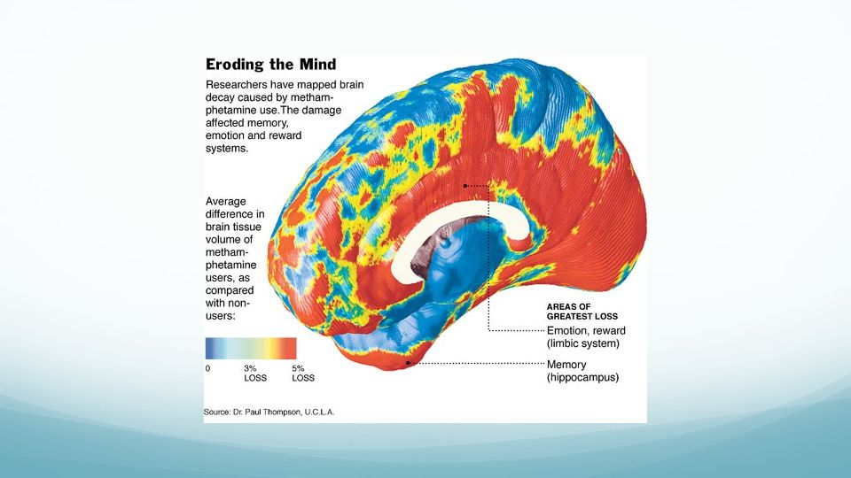
Article Study: 22 people who are in their thirties and have been using methamphetamine for 10 years 21 people as a control Used MRI machine to look at surface of the brains and the Limbic System Results: Part involved in drug craving, reward, mood and emotion lost 11% of its tissue Hippocampus lost 8% of its tissue Study in the new york times about the damage that meth can have on the brain very surprised with how much damage there is to the limbic system

36
Methamphetamine Addictive Stimulant
Stimulates the Limbic System causing the body to feel good and to want have that feeling again Gives users intense pleasure Releases dopamine Releases 12 to 13 times more than the normal amount of dopamine Destroys receptors so users cannot feel pleasure without the drugs cant feel same way without drugs most drugs: two to four times above normal

37
Brain Reward System Feelings of pleasure
Group of neurons in the Ventral Tegmental Area (VTA) These neurons connect to the Basal Ganglia Drugs activate VTA, dopamine is released into nucleus accumbens VTA in midbrain specific part of basal ganglia and what basal ganglia is
 These neurons connect to the Basal Ganglia. Drugs activate VTA, dopamine is released into nucleus accumbens. VTA in midbrain. specific part of basal ganglia and what basal ganglia is.")
38
Areas of Limbic System Addicts feel depressed, anxious and can’t concentrate because of the damage to the limbic system Hippocampus People did significantly worse on memory tests Symptoms similar to Alzheimer’s Amygdala Increase of Paranoia

39
References: Boeree, G. (n.d.). The limbic system: The emotional nervous system. Retrieved April 23, 2015, from Blakeslee, S. (2004, July 19). This Is Your Brain on Meth: A 'Forest Fire' of Damage. Retrieved April 23, 2015, from Carey, B. (2014, July 8). Probing Brain’s Depth, Trying to Aid Memory. Retrieved April 22, 2015, from Drugs and the Limbic System. (n.d.). Retrieved April 23, 2015, from Methamphetamine Brain Damage - Alcohol Rehab. (n.d.). Retrieved April 23, 2015, from Diagnosis | Autism Society. (1965, January 1). Retrieved April 23, 2015, from Edelson, S. (1995, January 1). Autism and the Limbic System. Retrieved April 23, 2015, from and the Limbic System Edelson.pdf Indian J Psychiatry Apr-Jun; 49(2): 132–139 Limbic System: Hippocampus (Section 4, Chapter 5) Neuroscience Online: An Electronic Textbook for the Neurosciences | Department of Neurobiology and Anatomy - The University of Texas Medical School at Houston. (n.d.). Retrieved from Limbic System: Amygdala (Section 4, Chapter 6) Neuroscience Online: An Electronic Textbook for the Neurosciences | Department of Neurobiology and Anatomy - The University of Texas Medical School at Houston. (n.d.). Retrieved from Links Between Methamphetamine Use, Paranoia, and Violence. (2013, February 11). Retrieved April 23, 2015, from
. The limbic system: The emotional nervous system. Retrieved April 23, 2015, from Blakeslee, S. (2004, July 19). This Is Your Brain on Meth: A Forest Fire of Damage. Retrieved April 23, 2015, from Carey, B. (2014, July 8). Probing Brain’s Depth, Trying to Aid Memory. Retrieved April 22, 2015, from _r=0. Drugs and the Limbic System. (n.d.). Retrieved April 23, 2015, from Methamphetamine Brain Damage - Alcohol Rehab. (n.d.). Retrieved April 23, 2015, from Diagnosis | Autism Society. (1965, January 1). Retrieved April 23, 2015, from Edelson, S. (1995, January 1). Autism and the Limbic System. Retrieved April 23, 2015, from and the Limbic System Edelson.pdf. Indian J Psychiatry Apr-Jun; 49(2): 132–139. Limbic System: Hippocampus (Section 4, Chapter 5) Neuroscience Online: An Electronic Textbook for the Neurosciences | Department of Neurobiology and Anatomy - The University of Texas Medical School at Houston. (n.d.). Retrieved from Limbic System: Amygdala (Section 4, Chapter 6) Neuroscience Online: An Electronic Textbook for the Neurosciences | Department of Neurobiology and Anatomy - The University of Texas Medical School at Houston. (n.d.). Retrieved from Links Between Methamphetamine Use, Paranoia, and Violence. (2013, February 11). Retrieved April 23, 2015, from")
40
References: Methamphetamine Brain Damage - Alcohol Rehab. (n.d.). Retrieved April 23, 2015, from Swenson (Ed.). (2006, January 1). Chapter 9 - Limbic System. Retrieved April 23, 2015, from
. Retrieved April 23, 2015, from Swenson (Ed.). (2006, January 1). Chapter 9 - Limbic System. Retrieved April 23, 2015, from")
Similar presentations


 CNS = Brain + spinal cord Surface anatomy includes.>")


