Download presentation
Presentation is loading. Please wait.

1
EXCRETORY AND REPRODUCTIVE
SYSTEM DEVELOPMENT

3
4. Degenerates by end of 4th week of development.
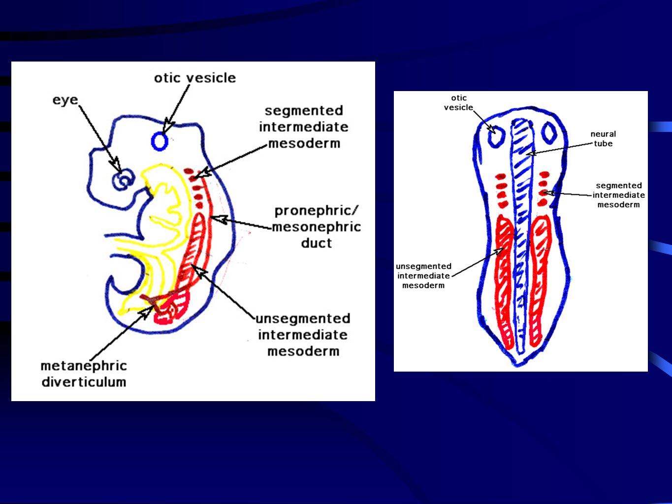
external glomerulus internal (glomus) 4. Degenerates by end of 4th week of development. 5. In species with functional pronephros, branch of aorta will form a glomerulus either adjacent to nephrostome in peritoneal coelom or adjacent to tubule Human embryo 1. Evident at about 20 days after fertilization pairs of rudimentary tubules in segmented mesoderm 3. Non-functional
 4. Degenerates by end of 4th week of development. 5. In species with functional pronephros, branch of aorta will form a glomerulus either adjacent to nephrostome in peritoneal coelom or adjacent to tubule. Human embryo. 1. Evident at about 20 days after fertilization pairs of rudimentary tubules in segmented mesoderm. 3. Non-functional.")
4
Human embryo: 1. Develops during second month pairs of functional mesonephric tubules in unsegmented intermediate mesoderm tubules adjacent to each somite 4. Extends from 10th - 26th somite 5. Induction - Mesonephric (pronephric) duct must be present. If removed, mesonephric tubules will not form. 6. Evocation - masked morphogen
 duct must be present. If removed, mesonephric tubules will not form. 6. Evocation - masked morphogen.")
5
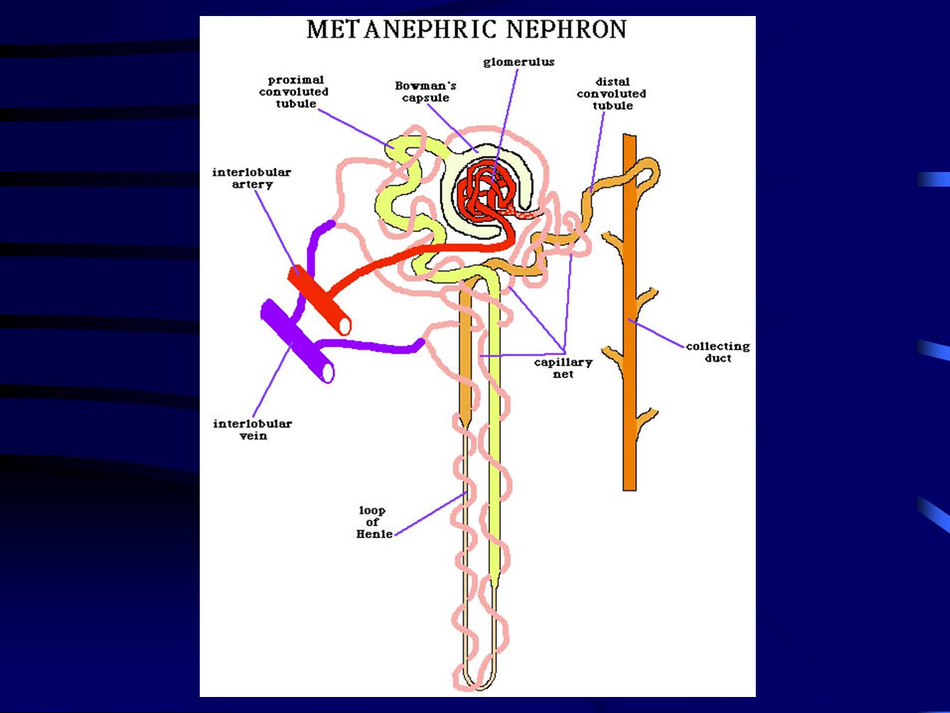
Metanephros Human embryo:
1. Development begins at end of 1st month with formation of ureteric bud 2. Development continues for next 2 months, but metanephric kidney is not functional until the 3rd month

6
View from ventral side of embryo. Allantois not shown
* * Urogenital sinus region End of first month

7
* View from ventral side of embryo. Allantois not shown
* Urogenital sinus region

8
* View from ventral side of embryo. Allantois not shown
* Urogenital sinus region Metanephric diverticulum will form 1. ureter 2. pelvis of kidney 3. major and minor calyxes 4. papillary collecting ducts Metanephrogenic blastema Cortex of kidney - metanephric nephrons

10
CLOACAL REGION - SEPARATION OF THE DIGESTIVE AND EXCRETORY SYSTEMS
Human Development ~4 weeks

11
CLOACAL REGION - SEPARATION OF THE DIGESTIVE AND EXCRETORY SYSTEMS
SEE LECTURE PACKET FOR FIGURES Formation of the urorectal septum involves the establishment and advance of the Rathke and Tourneux folds.

12
CLOACAL REGION - SEPARATION OF THE DIGESTIVE AND EXCRETORY SYSTEMS
SEE LECTURE PACKET FOR FIGURES As the Rathke and Tourneux folds meet and fuse, the establish the urorectal septum and separate the urinary and digestive tracts from each other.

13
SEE LECTURE PACKET FOR FIGURES
As the Rathke and Tourneux folds meet and fuse, the establish the urorectal septum and separate the urinary and digestive tracts from each other. • As the Rathke and Tourneux folds do their job, the metanephric diverticulum’s connection with the mesonephric duct is moved toward the forming bladder. • Eventually the metanephric diverticulum/ureter ends up connected to the bladder. • This movement of the metanephric diverticulum/ureter to a new connecting point is accomplished by differential growth of the surrounding tissues. SEE LECTURE PACKET FOR FIGURES

14
Rathke folds fail to form
These or similar sorts of defects occur in 1 in every 4000 births in the U.S.

15
Early in mammalian development, the primordial germ cells (PGCs) are found in a cluster in extraembryonic endoderm of the yolk sac. Prior to about 1990, they were considered to be endodermally derived. Subsequent research has identified these cells during the primitive streak stage in presumptive extraembryonic mesoderm of the epiblast. These cells can be identified by endogenous alkaline phosphatase activity that other cells of the early embryo lack. The alkaline phosphatase will react with a substrate that produces a colored reaction product. The PGCs are now considered to be mesodermally derived. They then migrate to the endoderm, and subsequently migrate to the developing gonads.

16
scrotum Corpus spongiosum

17
Human male reproductive tract
Tubuli recti

18
labia majora labia minora cervix

19
Human Development gonadal ridge genital ridge = ~ 4 weeks

20
subcardinal anatomosis (renal segment of inferior vena cava)
genital ridge left mesonephric duct umbilical (allantoic) arteries allantoic stalk left umbilical (allantoic) vein
 arteries. allantoic stalk. left umbilical (allantoic) vein.")
21
Human Development ~ 5 weeks
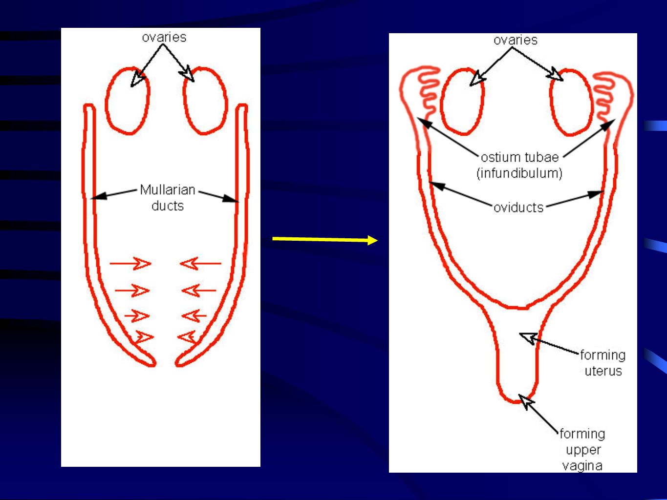
Primordial germ cells migrate from the yolk sac endoderm to the genital ridges during the 5th week of human development. mullarian duct Human Development ~ 5 weeks Gonads start to form during the 5th week, but at this point they are equivalent in males and females - i.e. not yet testis or ovary. Rete blastema forms from germinal epithelium of genital ridge in both males and females. Cells of rete blastema migrate closer to mesonephric ducts.

22
Human Development ~ 6-7 weeks
In human female embryos, proliferation of the primary sex cords stops by the end of the 6th week. Human Development ~ 6-7 weeks

23
Differentiation of the gonads (testis or ovary) will begin during the 7th week of development (tunica albuginea in males). The tissues of the genital ridge will bulge into the peritoneal cavity and eventually will hang from a mesentary - mesorchium in male and mesovarium in female. ~ 7 weeks

24
due to secretion of anti-Mullarian hormone by the sertoli cells in seminiferous tubules.

25
The ductwork of the male reproductive tract continues its development
The ductwork of the male reproductive tract continues its development. The germinal epithelium continues to degenerate

26
(epididymis & vas deferens)
")
27
(~6 - 7 weeks) Male and female reproductive tract - ductwork
Text Male and female reproductive tract - ductwork Fate of the mesonephric and Mullarian ducts. (~6 - 7 weeks)
")
28
SEE LECTURE PACKET FOR FIGURES
5-6 weeks

29
SEE LECTURE PACKET FOR FIGURES
In the male, the remnants of the Mullarian ducts are the vagina masculina and appendix testis The mesonephric tubules degenerate in female leaving some non-functional remnants called the epoophoron near the ovary. The mesonephric ducts degenerate in females; however, they sometimes leave a small non-functional duct called the canal of Gartner that extends from the epoophoron to the vagina.

30
ovary Female Female ~2.5 months

32
SEE LECTURE PACKET FOR FIGURES
Uterovaginal plate SEE LECTURE PACKET FOR FIGURES Uterovaginal plate Muller’s tubercle Vaginal plate Uterovaginal plate, vaginal plate, Muller’s Tubercle - other names for the sinovaginal bulb. Vestibule - the region surrounding the vaginal opening that is bordered by the labia minora. Bartholin’s glands - paired glands, one on either side of the vaginal opening, with ducts opening into the vestibule. Secrete a mucus that lubricates the vestibular walls. Developmentally homologous to Cowper’s (bulbourethral) glands in the male.
 glands in the male.")
33
MALE AND FEMALE EXTERNAL GENETLE DEVELOPMENT
SEE LECTURE PACKET FOR FIGURES

34
MALE AND FEMALE EXTERNAL GENETLE DEVELOPMENT
SEE LECTURE PACKET FOR FIGURES

35
Fusion Failures of the Urethral Folds
Hypospadias - abnormal opening of the end of the urethra on ventral side of penis. Occurs in about 1 in 125 to 250 new born males Cause of hypospadias is not always clear, but it can result from insufficient hormone production (i.e. testosterone, dihydrotestosterone) or lack of sufficient receptors for these hormones.
 or lack of sufficient receptors for these hormones.")
36
s

Similar presentations


 7 to 10 solid cell groups in the cervical region nephrotome.>")