Download presentation
Presentation is loading. Please wait.

1
DETERMINATION OF BLOOD GLUCOSE CONCENTRATION
SITI ANNISA DEVI TRUSDA BIOCHEMISTRY DEPARTMENT

2
INTRODUCTION Needed for supporting the D/ of some diseases, including DM. When to measure: fasting state or 2 hours after taking a meal. Sometimes at a present time detecting a present increased or decreased of blood glucose level (hyperglycemia or hypoglycemia).
.")
3
Two Methods of Measuring Blood Glucose Level
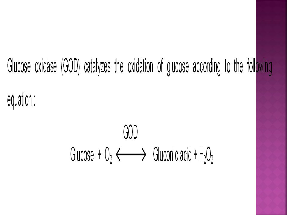
Reduction method, which is based on the ability of glucose to reduce Cu++ to Cu+ less sensitive, substances that could reduce Cu++ : fructose, galactose,vitamin C, creatinin, uric acid, glutathion, etc. Enzymatic method (more specific and precise result) : Glucose is oxidized by glucose oxidase gluconic acid + H2O2 red dye.
 : Glucose is oxidized by glucose oxidase gluconic acid + H2O2 red dye.")
5
.

6
Materials and equipments :
Color reagent glucose oxidase (GOD-PAP) : Glucose oxidase (GOD) > 15 KU/l Peroxidase (POD) > 1.5 KU/l 4-aminophenazone 0.25 mmol/l Phenol 0.75 mmol/l, mutarotase > 2.0 KU/l Phosphate buffer pH 7.5, 0.1 mol/l. Glucose standard solution 100 mg/dl (5.55 mmol/l). Serum or plasma Aquadest. Micropipette 10 l, 1.0 ml Reaction tubes Waterbath 37C Centrifuge Spectrophotometer (wavelength nm)
 : Glucose oxidase (GOD) > 15 KU/l. Peroxidase (POD) > 1.5 KU/l. 4-aminophenazone 0.25 mmol/l. Phenol 0.75 mmol/l, mutarotase > 2.0 KU/l. Phosphate buffer pH 7.5, 0.1 mol/l. Glucose standard solution 100 mg/dl (5.55 mmol/l). Serum or plasma. Aquadest. Micropipette 10 l, 1.0 ml Reaction tubes. Waterbath 37C Centrifuge. Spectrophotometer (wavelength nm)")
7
To make the GOD color reagent : dissolve the content of one bottle of GOD enzyme with its solvent available. This solution is stable for 30 days at 2 – 8 oC temperature. The absorbancy of the blank’s reagent must be about 0.0 – 0.4 AU if it is read at 505 nm wavelength compared with aquadest. For every series of measurement, only one standard and one blank are needed.

8
Procedure : 1. Centrifuge 3 ml EDTA blood, 2000 rpm for 10’. The plasma will be separated from the blood cells. Use plasma for sample 2. Pipette into each of the three reaction tubes according to the following table :

10
3. Mix the content of each tube well, then incubate them at 37oC for 10 minutes or let stand at room temperature for 25 minutes. Avoid direct sunlight 4. Using cuvette tube, read the sample’s and standard’s absorbancy against the blank at 505 nm.

12
Notes : By this method the blood glucose level can be measured linearly up to 600 mg/dl. If the blood glucose level is higher than 600 mg/dl, dilute the plasma three times by adding 2 volumes of aquadest, then repeat the procedure. Multiply the result three times. The result is not influenced by blood creatinin, fructose, galactose, uric acid, glutathion, ascorbic acid or bilirubin as long as these substances’s concentration are at normal range. Bilirubin concentration up to 10 mg/dl does not influence the result, but a high dose of oral ascorbic acid (vitamin C) could decrease the result. The color produced will be stable for 2 hours.
 could decrease the result. The color produced will be stable for 2 hours.")
13
Spectrophotometer

15
IDENTIFICATION OF GLUCOSE IN URINE
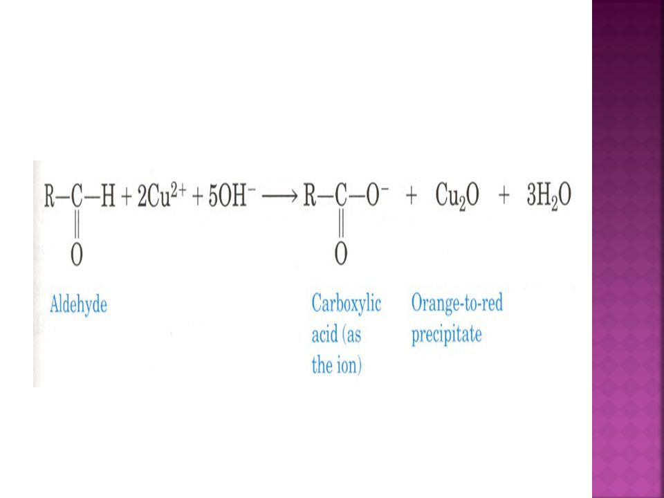
Urine Reduction Test (Benedict’s Test) : Principle : Glucose reduces the alkaline copper solution, Cu++ Cu+ and is precipitated as Cu2O, a red brick color substance. Depend on concentration of Cu2O produced, the shaked solution will produce different color that can be used to estimate the glucose concentration
 : Principle : Glucose reduces the alkaline copper solution, Cu++ Cu+ and is precipitated as Cu2O, a red brick color substance. Depend on concentration of Cu2O produced, the shaked solution will produce different color that can be used to estimate the glucose concentration.")
17
Materials and equipments :
Urine of a diabetic patient as sample Benedict’s qualitative solution (semi qualitative) : - 17 gr CuSO4 - 100 gr NaCO3 anhydride - 170 gr tri – Na- Citrate 2 H2O Dissolved in 1000 ml of aquadest Test tubes Waterbath 100 0C/ Bunsen burner
 : - 17 gr CuSO gr NaCO3 anhydride gr tri – Na- Citrate 2 H2O. Dissolved in 1000 ml of aquadest. Test tubes. Waterbath 100 0C/ Bunsen burner.")
18
Procedure : Mix 3 ml Benedict’s solution with 3 drops of urine in a test tube, heat it in 100oC waterbath or just boil it for a moment, note the color produced after shaking the tube. Repeat the same procedure with the urine diluted 2, 4 and 8 times.

20
Note : The positive result of Benedict’s test could be given by many reductors, such as : - Sugars : glucose, pentose, lactose, galactose. - Drugs : antipyrine, pyramidon, PAS, xanthonin. - High concentration of normally urine substances : indican, uric acid, creatinin. - Preservative agents :formalin, CHCl3.

21
To read the result, shake the tube and then note the solution’s color produced :
- Blue : (-) → means there is no glucose in the sample urine - Green : (+) → 0.5 – 1 gr % of glucose - Yellowish green : (++) → 1 – 1.5 gr % of glucose - Yellow : (+++) → 2 – 3.5 gr % glucose - Red brick color : (++++) → 4 gr % of glucose
 → means there is no glucose in the sample urine. - Green : (+) → 0.5 – 1 gr % of glucose. - Yellowish green : (++) → 1 – 1.5 gr % of glucose. - Yellow : (+++) → 2 – 3.5 gr % glucose. - Red brick color : (++++) → 4 gr % of glucose.")
23
Selamat Bekerja

Similar presentations


 is a very important fuel source to generate universal energy molecules (ATP). Blood glucose regulation I->")


![BCH302 [Practical] 1. There are three main methods of estimation the reducing sugar content in solution : 1. Reduction of cupric to cuprous salts. 2.](/24/7426540/big_thumb.jpg "BCH302 [Practical] 1. There are three main methods of estimation the reducing sugar content in solution : 1. Reduction of cupric to cuprous salts. 2.>")

 (AST) assay Biochemistry Clinical practice CLS 432 Dr. Samah Kotb Lecturer of Biochemistry 2015.>")

