Download presentation
Presentation is loading. Please wait.

1
VIROLOGY

2
INTRODUCTION Virology is the study of viruses. ☻Viruses are a heterogenous intracellular group of microorganisms that vary with respect to size, morphology, and chemical composition that contain either deoxyribonucleic acid (DNA) or ribonucleic acid (RNA). ☻ Viruses are unique life forms different from all other living organisms, either eukaryotes or prokaryotes, for three fundamental reasons: 1. The nature of environment in which they grow and multiply. they can function and multiply only inside another living organism, which may be either a prokaryotic or eukaryotic cell or any other living cells depending on the virus. Viruses are acellular and metabolically inert outside the host cell and are obligatory parasites.
 or ribonucleic acid (RNA). ☻ Viruses are unique life forms different from all other living organisms, either eukaryotes or prokaryotes, for three fundamental reasons: 1. The nature of environment in which they grow and multiply. they can function and multiply only inside another living organism, which may be either a prokaryotic or eukaryotic cell or any other living cells depending on the virus. Viruses are acellular and metabolically inert outside the host cell and are obligatory parasites.")
3
2. The nature of their genome.
whereas all other living forms can use only DNA (and not RNA) as their genetic material (genome) for information transmission from parent to progeny, viruses can use either DNA or RNA as their genome, that is, some viruses can use only RNA (and not DNA) as their genetic material. Therefore, these classes of RNA viruses have developed new sets of enzymes for replicating and transcribing RNA from an RNA template. 3. The mode of their multiplication. All eukaryotic and prokaryotic cells divide and multiply as a whole unit, that is, 1 ~ ~ 8 and so on. However, viruses do not multiply as a unit. In fact, they have developed a much more efficient way to multiply just as complex machines are made in a modern factory. Different viral components are made separately from independent templates, and then these components are assembled into the whole and infectious units, also called virus particles (virions).
 as their genetic material (genome) for information transmission from parent to progeny, viruses can use either DNA or RNA as their genome, that is, some viruses can use only RNA (and not DNA) as their genetic material. Therefore, these classes of RNA viruses have developed new sets of enzymes for replicating and transcribing RNA from an RNA template. 3. The mode of their multiplication. All eukaryotic and prokaryotic cells divide and multiply as a whole unit, that is, 1 ~ ~ 8 and so on. However, viruses do not multiply as a unit. In fact, they have developed a much more. efficient way to multiply just as complex machines are made in a modern factory. Different viral components are made separately from independent templates, and then these components are assembled into the whole and infectious units, also called virus particles (virions).")
4
CHEMICAL COMPOSITION: ☺The chemical composition of a virus depends on the nature of that virus, that is, the nature of the viral genome (RNA or DNA). ☺ The nucleic acid is the genome that contains the information necessary for viral function and multiplication. ☺ The composition of the protein shell called the viral "nucleocapsid" surrounding the genome. ☺ All viruses have nucleocapsids and therefore contain nucleic acids and proteins. ☺ The presence or absence of viral membrane depending on whether the virus is enveloped or naked. ☺ The enveloped is derived in part from the membrane of the host cell. Those viruses without envelope are described as naked viruses.

6
Size of viruses: ☼The size of virions ranges from 20 nm (parvovirus) to -300 nm (poxvirus) in diameter, as compared to the size of Escherichia coli, which is about 1000 nm in length, and therefore, too small to be seen with the light microscope. ☼ Viruses can, however, be studied with the electron microscope which can magnify up to X500000, but this microscope is very costly and special training facilities are required to use it. Dependence of viruses on host cells: Because viruses possess neither cellular structure nor organelles, they are unable to make their own proteins and essential enzymes. They are therefore completely dependent on their host cells for energy and multiplication. Outside of living cells, viruses are metabolically inactive. The information required for a virus to replicate is contained in its nucleic acid. This information is used by the host cell to produce new viruses.
 to -300 nm (poxvirus) in diameter, as compared to the size of Escherichia coli, which is about 1000 nm in length, and therefore, too small to be seen with the light microscope. ☼ Viruses can, however, be studied with the electron microscope which can magnify up to X500000, but this microscope is very costly and special training facilities are required to use it. Dependence of viruses on host cells: Because viruses possess neither cellular structure nor organelles, they are unable to make their own proteins and essential enzymes. They are therefore completely dependent on their host cells for energy and multiplication. Outside of living cells, viruses are metabolically inactive. The information required for a virus to replicate is contained in its nucleic acid. This information is used by the host cell to produce new viruses.")
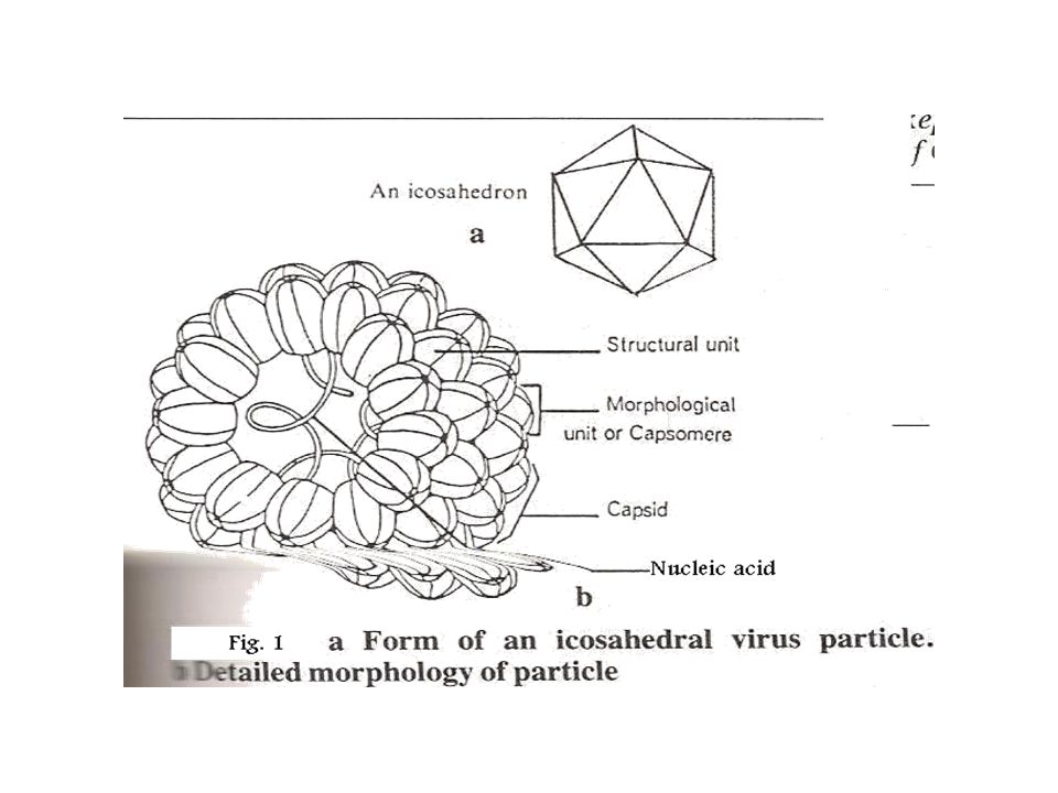
7
Shape and Structure of viruses:
♠Viral shape also varies. Some viruses are round (spherical), others filamentous, and still others pleomorphic. ♠ Usually, naked (non-enveloped) viruses have specific shapes and sizes, whereas some enveloped viruses (particularly enveloped viruses possessing helical nucleocapsids) are highly pleomorphic (e.g., orthomyxoviruses), with shapes varying from spherical to filamentous. ♠ All viruses consist of a mass (core) of single, or double stranded DNA or RNA surrounded by a protective protein coat called a capsid. ♠ The nucleic acid together with the capsid form the nucleocapsid. ♠ The capsid is antigenic and also contains the receptors which enable a virus to attach to the surface of its specific host cell. ♠ The capsid consists of a number of identical units called capsomers. ♠ The symmetry, or pattern, of viral capsides is used in the classification of viruses.
, others filamentous, and still others pleomorphic. ♠ Usually, naked (non-enveloped) viruses have specific shapes and sizes, whereas some enveloped viruses (particularly enveloped viruses possessing helical nucleocapsids) are highly pleomorphic (e.g., orthomyxoviruses), with shapes varying from spherical to filamentous. ♠ All viruses consist of a mass (core) of single, or double stranded DNA or RNA surrounded by a protective protein coat called a capsid. ♠ The nucleic acid together with the capsid form the nucleocapsid. ♠ The capsid is antigenic and also contains the receptors which enable a virus to attach to the surface of its specific host cell. ♠ The capsid consists of a number of identical units called capsomers. ♠ The symmetry, or pattern, of viral capsides is used in the classification of viruses.")
10
Capsid symmetry is described as being:
A. Helical Capsids ♦ Meaning the capsid is spiral in shape. ♦ It surrounds a spiral shaped core of nucleic acid ♦ Helical capsids are usually flexible and rodlike. ♦ The length of the helical capsid is usually determined by the length of the nucleic acids, that is, viruses having shorter nucleic acids will have a shorter helical nucleocapsid. ♦ Helical capsids can be naked, that is, without an envelope. ♦ However, such helical capsids when enclosed in an envelope, can appear spherical while others are elongated, filamentous, or pleomorphic (several different shapes), indicating that the helical capsid in these viruses is flexible. Some helical capsids can be further folded, forming. ♦ Helical capsids can package only single-stranded RNA, but not double-stranded DNA or RNA, possibly because of the rigidity of the double-stranded nucleic acids.
, indicating that the helical capsid in these viruses is flexible. Some helical capsids can be further folded, forming. ♦ Helical capsids can package only single-stranded RNA, but not double-stranded DNA or RNA, possibly because of the rigidity of the double-stranded nucleic acids.")
12
B. Icosahedral Capsids ♦ The term cubic is used to describe Icosahedral viruses. ♦ Viruses with icosahedral capsids possess a closed shell enclosing the nucleic acid inside. An icosahedron has 20 triangular faces, 30 edges. ♦ Unlike helical nucleocapsids that package only single-stranded nucleic acid, icosahedral capsids can be used to package either single- or double-stranded RNA and DNA molecules. ♦ An icosahedral virus can be either naked or enveloped; but, unlike the helical enveloped viruses, the enveloped icosahedral viruses are less pleomorphic in their shape because the icosahedron capsid structure is rather rigid, and in addition, with icosahedral capsids, the overall size is fixed for a particular virus. ♦ The virus particle's formation, stability, and size do not depend on the amount of nucleic acid in the capsid.

14
C. Complex, meaning the capsid symmetry is neither icosahedral nor helical. As indicated in the chart, only the poxviruses among the medically important viruses show complex symmetry. The overall shape of poxviruses is usually described as brick-shaped. Many of the helical viruses and a few of Icosahedral viruses are surrounded by an envelope.

16
FIVE BASIC STRUCTURAL FORMS OF VIRUSES IN NATURE
Naked icosahedral e.g. poliovirus, adenovirus, hepatitis A virus Naked helical e.g. tobacco mosaic virus. So far no human viruses with this structure are known Enveloped icosahedral e.g. herpes virus, yellow fever virus, rubella virus Enveloped helical e.g. rabies virus, influenza virus, parainfluenza virus, mumps virus, measles virus Complex e.g. poxvirus

17
CLASSIFICATION OF VIRUSES
The internationally agreed system of virus classification is based on the structure and composition of the virus particle (virion) (Figure 7). In some cases, the mode of replication is also important in classification. Viruses are classified into various families on this basis. INTERNATIONAL CLASSIFICATION OF VIRUSES Primary characteristics used in classification Viruses are classified according to the nature of their genome and their structure
 (Figure 7). In some cases, the mode of replication is also important in classification. Viruses are classified into various families on this basis. INTERNATIONAL CLASSIFICATION OF VIRUSES. Primary characteristics used in classification. Viruses are classified according to the nature of their genome and their structure.")
18
Secondary characteristics Replication strategy
VIRAL CLASSIFICATION Nucleic acid RNA or DNA single-stranded or double-stranded non-segmented or segmented linear or circular if genome is single stranded RNA, can it function as mRNA? whether genome is diploid (such as in retroviruses) Virion structure symmetry (icosahedral, helical, complex) enveloped or not enveloped number of capsomers Secondary characteristics Replication strategy Sometimes a group of viruses that seems to be a single group by the above criteria is found to contain a subgroup of viruses which have a fundamentally different replication strategy - in this case the group will be divided based on the mode of replication.
 Virion structure. symmetry (icosahedral, helical, complex) enveloped or not enveloped. number of capsomers. Secondary characteristics. Replication strategy. Sometimes a group of viruses that seems to be a single group by the above criteria is found to contain a subgroup of viruses which have a fundamentally different replication strategy - in this case the group will be divided based on the mode of replication.")
19
INFECTION OF CELLS BY VIRUSES:
♣When a virus infects a cell it usually replicates and causes the death of the host cell. The observable changes which lead to the death of the host cell are called Cytopathic effects (CPE). These may include the formation of inclusion bodies (sites of virus replication) or Syncytia (virus-infected cells massed together). In viruses cultured in the laboratory, CPE is observed by a shrinking or enlargement of infected cells, and the detachment of the dead cells from the glass surface on which they are growing. ♣ Occasionally, viruses infect cells and replicate without causing the death of their host cells. The new viruses are released through the cell membrane of the infected cells. Examples of such viruses include rubella virus and parainfluenza virus.
. These may include the formation of inclusion bodies (sites of virus replication) or Syncytia (virus-infected cells massed together). In viruses cultured in the laboratory, CPE is observed by a shrinking or enlargement of infected cells, and the detachment of the dead cells from the glass surface on which they are growing. ♣ Occasionally, viruses infect cells and replicate without causing the death of their host cells. The new viruses are released through the cell membrane of the infected cells. Examples of such viruses include rubella virus and parainfluenza virus.")
20
♣ Some viruses after infecting cells do not replicate, or they become active for a time and then become inactive (latent). In response to certain stimuli, latent viruses can be reactivated and become active replicating particles, for example herpesviruses. ♣ A small group of viruses are able to change, or transform their host cells from normal cells into tumor producing cells. Such viruses are changed to have neoplastic, or encogenic properties.

22
PRINCIPAL EVENTS INVOLVED IN REPLICATION
Adsorption The first step in infection of a cell is attachment to the cell surface. Attachment is via ionic interactions which are temperature-independent. The viral attachment protein recognizes specific receptors, which may be protein, carbohydrate or lipid, on the outside of the cell. Cells without the appropriate receptors are not susceptible to the virus. Penetration The virus enters the cell in a variety of ways according to the nature of the virus. Enveloped viruses A) Entry by fusing with the plasma membrane. Some enveloped viruses fuse directly with the plasma membrane. Thus, the internal components of the virion are immediately delivered to the cytoplasm of the cell (figure 1) Figure 1. Fusion of a virus with the plasma membrane after attachment to a cell surface receptor
 Entry by fusing with the plasma membrane. Some enveloped viruses fuse directly with the plasma membrane. Thus, the internal components of the virion are immediately delivered to the cytoplasm of the cell (figure 1) Figure 1. Fusion of a virus with the plasma membrane after attachment to a cell surface receptor.")
23
(B) Entry via endosomes at the cell surface (figure 2)
Some enveloped viruses require an acid pH for fusion to occur and are unable to fuse directly with the plasma membrane. These viruses are taken up by invagination of the membrane into endosomes. As the endosomes become acidified, the latent fusion activity of the virus proteins becomes activated by the fall in pH and the virion membrane fuses with the endosome membrane. This results in delivery of the internal components of the virus to the cytoplasm of the cell Non-enveloped viruses Non-enveloped viruses may cross the plasma membrane directly or may be taken up into endosomes. They then cross (or destroy) the endosomal membrane. Figure 2. Fusion of a virus with the membrane of an endosome
 the endosomal membrane. Figure 2. Fusion of a virus with the membrane of an endosome.")
24
3. Uncoating Nucleic acid has to be sufficiently uncoated that virus replication can begin at this stage. When the nucleic acid is uncoated, infectious virus particles cannot be recovered from the cell - this is the start of the ECLIPSE phase - which lasts until new infectious virions are made. 4. Synthesis of viral nucleic acid and protein Many strategies are used, some will be discussed in later chapters. 5. Assembly/maturation New virus particles are assembled. There may be a maturation step that follows the initial assembly process. 6. Release Virus may be released due to cell lysis, or, if enveloped, may bud from the cell. Budding viruses (figures 3 and 4) do not necessarily kill the cell. Thus, some budding viruses may be able to set up persistent infections. Not all released viral particles are infectious. The ratio of non-infectious to infectious particles varies with the virus and the growth conditions.
 do not necessarily kill the cell. Thus, some budding viruses may be able to set up persistent infections. Not all released viral particles are infectious. The ratio of non-infectious to infectious particles varies with the virus and the growth conditions.")
Similar presentations


 associated with proteins encoded by the nucleic acid. The virus.>")
 as their genome 3-They.>")
 replication of DNA viruses (2) the culture, growth and recognition of virus.>")


