Download presentation
Presentation is loading. Please wait.

1
The Integumentary System Chapter 6

2
Organs are two or more tissues which together perform a specialized function. Epithelial membranes are thin structures that usually contain both epithelial and connective tissue.

3
Three types of epithelial membranes Serous Membranes –Line cavities and cover organs –Simple squamous epi. over loose connective tissue –Parietal and visceral portions –Secrete a serous (watery) fluid for lubrication
 fluid for lubrication.")
4
Mucous membranes –Line cavities that open to the exterior –Layer of epithelium over connective tissue; epithelium varies with location –Tight junctions and goblet cells Cutaneous membrane is the skin –the major organ of the integumentary system

5
Integumentary system is the skin and the organs derived from it (hair, glands, nails) One of the largest organs –2 square meters; 10-11 lbs. –Largest sense organ in the body The study of the skin is Dermatology

6
Functions: 1. Regulation of body temperature –Cellular metabolism produces heat as a waste product. –High temperature Dilate surface blood vessels Sweating –Low temperature Surface vessels constrict shivering

8
2. Protection physical abrasion dehydration ultraviolet radiation 3. Sensation touch vibration pain temperature

9
4. Excretion 5. Immunity/ Resistance 6. Blood Reservoir 8-10 % in a resting adult 7. Synthesis of vitamin D uv light aids absorption of calcium

10
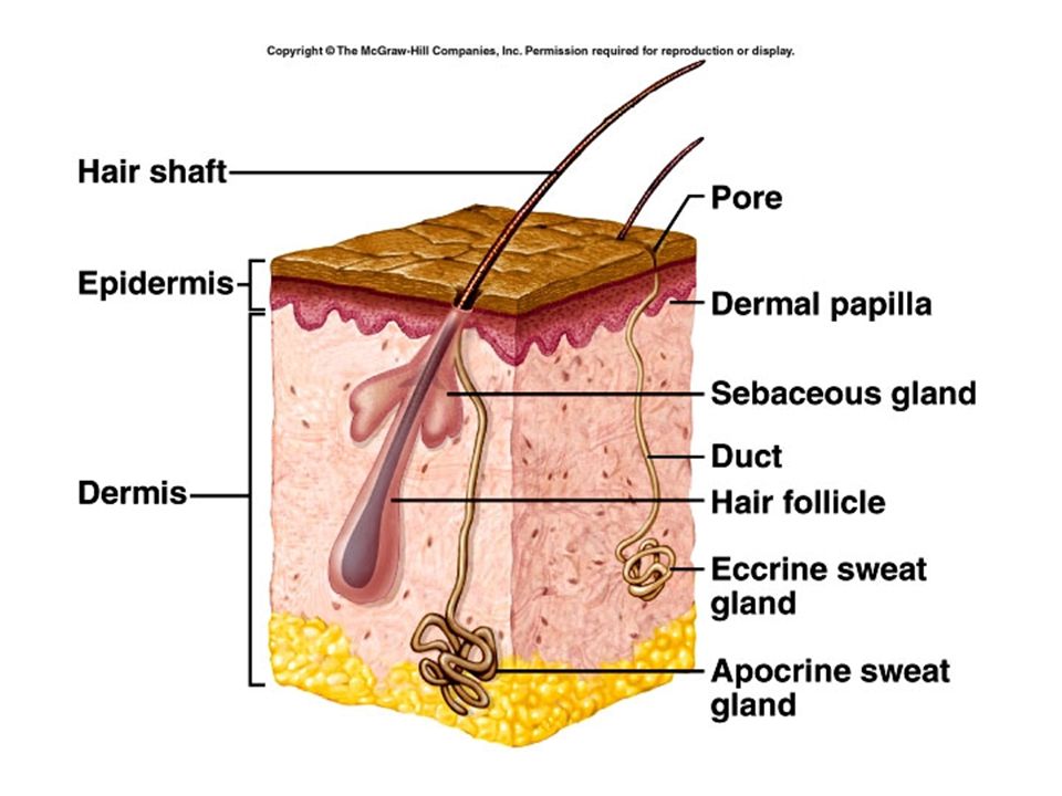
Anatomy Epidermis Skin Dermis Subcutaneous layer or hypodermis

16
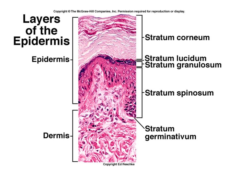
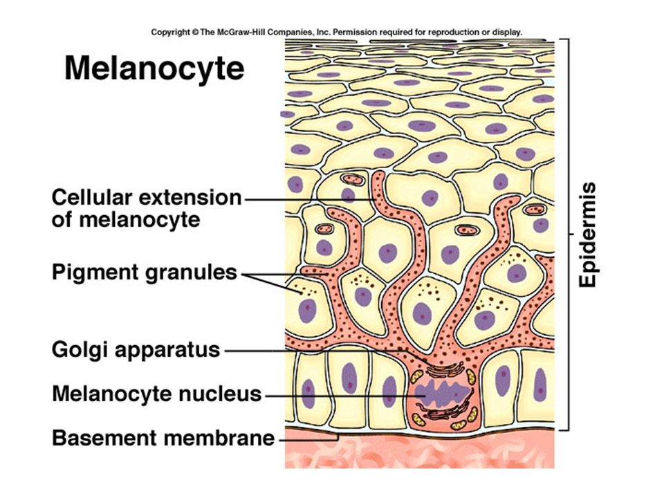
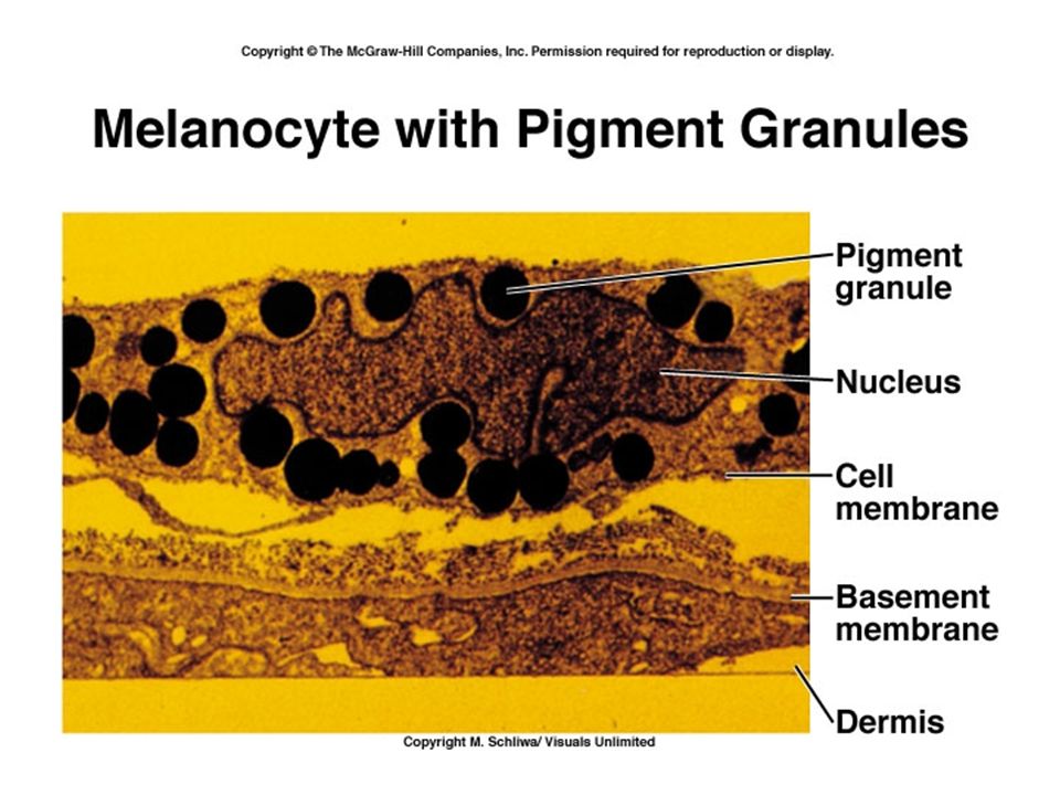
Epidermis Stratum basale (stratum germinativum) –Single layer of cuboidal to columnar cells –Stem cells that produce keratinocytes –Melanocytes - # the same for all races Melanin produced in a melanosome
 –Single layer of cuboidal to columnar cells –Stem cells that produce keratinocytes –Melanocytes - # the same for all races Melanin produced in a melanosome")
17
Stratum spinosum (thorn-like, prickly) –8-10 layers attached by desmosomes –See spines when cell is stained for microscopy –Keratinocytes take in melanin by cytocrine secretion
 –8-10 layers attached by desmosomes –See spines when cell is stained for microscopy –Keratinocytes take in melanin by cytocrine secretion")
20
Stratum granulosum –3-5 layers –Keratinization begins here –Keratohyalin found in granules –Cells beginning to die

21
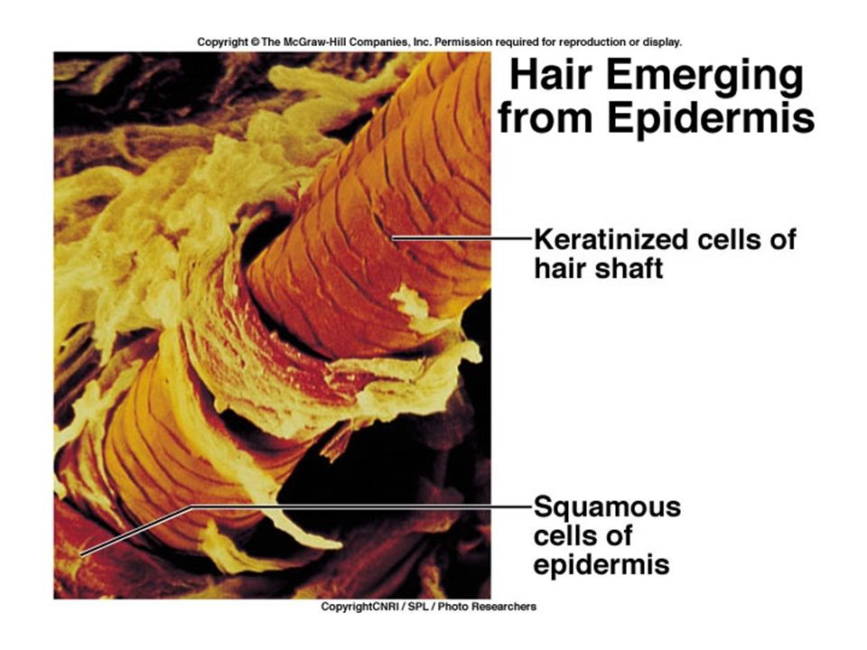
Stratum lucidum (lucid = clear) –More apparent in thick skin –3-5 layers of clear cells –Eleidin Stratum corneum (corneum means horny) –Dead, flat cells full of keratin –Keratin is waterproof –Cells are shed Basal cell to surface – about 2-4 weeks
 –More apparent in thick skin –3-5 layers of clear cells –Eleidin Stratum corneum (corneum means horny) –Dead, flat cells full of keratin –Keratin is waterproof –Cells are shed Basal cell to surface – about 2-4 weeks")
22
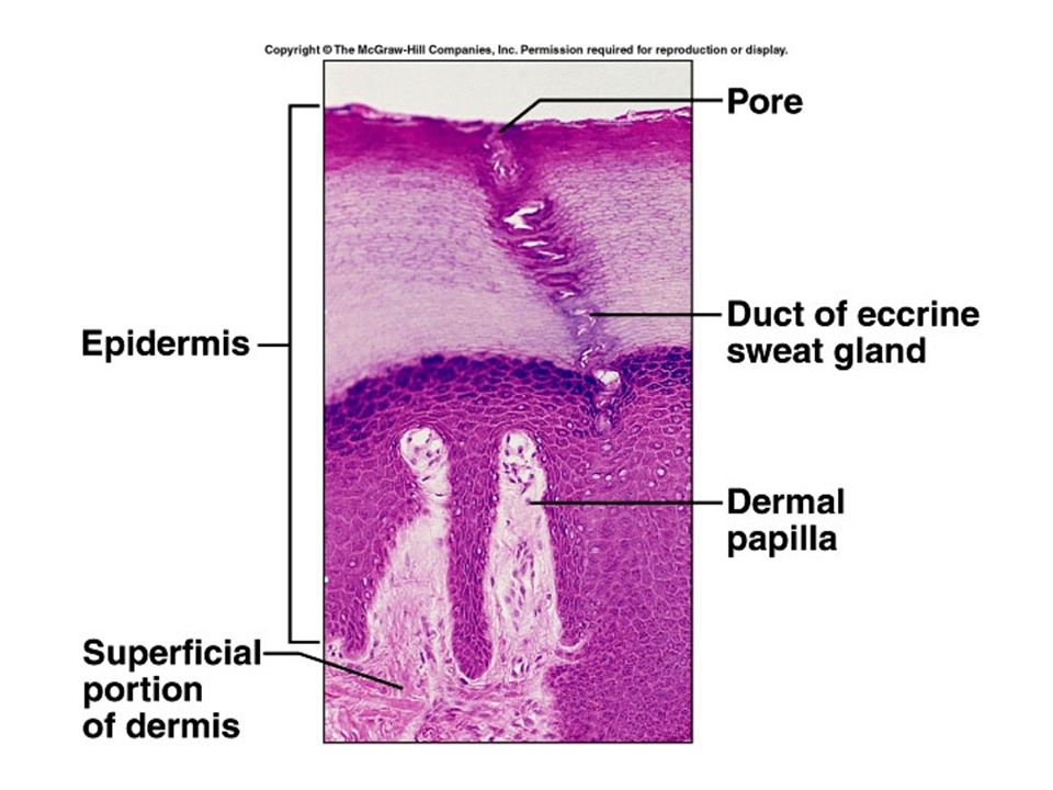
Dermis Connective tissue layer Collagen and elastic fibers, nerves, blood vessels, muscle fibers, adipose cells, hair follicles and glands. Papillary layer –1/5 of dermis – loose areolar connective tissue –Highly vascular –Dermal papillae - fingerprints

23
Reticular (net) layer –Dense irregular connective tissue –Sebaceous (oil) glands –Hair follicles –Ducts of sudoriferous (sweat) glands –Striae or stretch marks –Meissner’s corpuscles and Pacinian corpuscles
 layer –Dense irregular connective tissue –Sebaceous (oil) glands –Hair follicles –Ducts of sudoriferous (sweat) glands –Striae or stretch marks –Meissner’s corpuscles and Pacinian corpuscles")
24
Hypodermis Attaches the reticular layer to the underlying organs Loose connective tissue and adipose tissue Major blood vessels – rete cutaneum

25
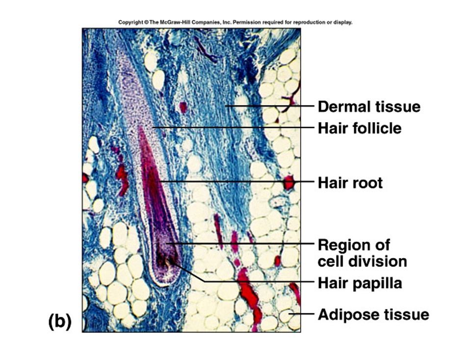
Accessory organs or epidermal derivatives Hairs –Epidermal growths that function in protection –Shaft, root, and folllicle –Sebaceous glands, arrector pili muscle, and hair root plexus (touch) –Hair growth and replacement have a cyclical pattern –‘male-pattern’ baldness
 –Hair growth and replacement have a cyclical pattern –‘male-pattern’ baldness")
29
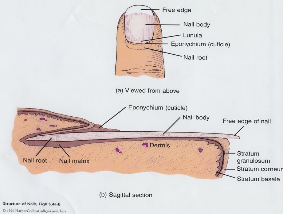
Nails Plates of highly packed, keratinized cells Protection, scratching, & manipulation Formed by cells in nail bed called the matrix ( in area of lunula) 1 mm / week Eponychium - cuticle
 1 mm / week Eponychium - cuticle")
32
Skin Glands Sebaceous (oil) glands –Usually connected to hair follicles –Holocrine glands –Fats, cholesterol, proteins, salts, and cell debris –Moistens hair and waterproofs skin
 glands –Usually connected to hair follicles –Holocrine glands –Fats, cholesterol, proteins, salts, and cell debris –Moistens hair and waterproofs skin")
33
Sweat (sudoriferous) glands –Eccrine sweat glands Merocrine glands Water, salt, wastes Function is to cool the body (also nervous) –Apocrine sweat glands Larger, merocrine glands Associated with hair follicles More viscous – fatty acids and proteins Odor occurs when broken down by bacteria
 glands –Eccrine sweat glands Merocrine glands Water, salt, wastes Function is to cool the body (also nervous) –Apocrine sweat glands Larger, merocrine glands Associated with hair follicles More viscous – fatty acids and proteins Odor occurs when broken down by bacteria")
34
Ceruminous glands –Modified sudoriferous glands –Secrete cerumen (ear wax) Mammary glands –Secrete milk
 Mammary glands –Secrete milk")
37
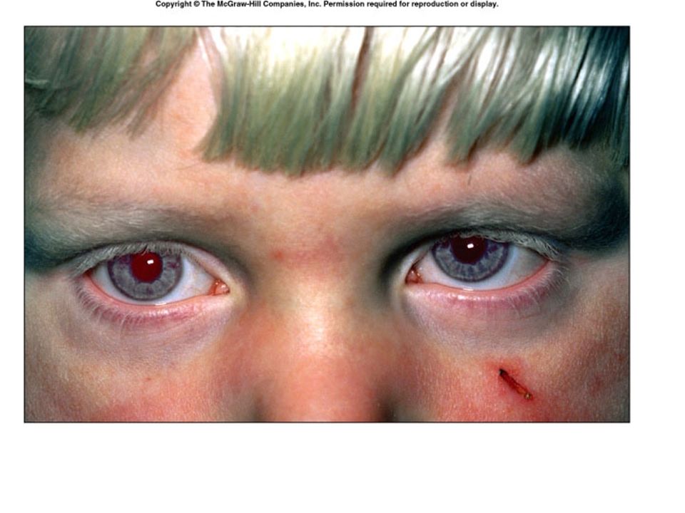
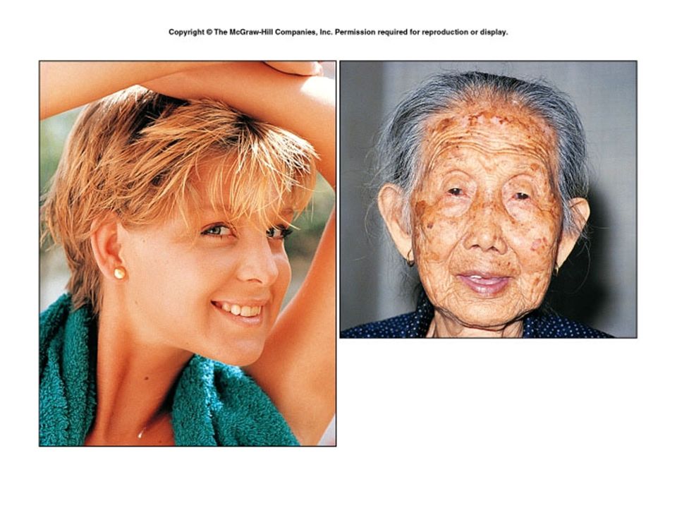
Skin color Genetic factors –Same number of melanocytes –Albinism Environmental factors –Uv light or x-rays

39
Physiological factors –Amount of blood –Amount of oxygen Cyanosis Carotene accumulation Jaundice – liver disorder

40
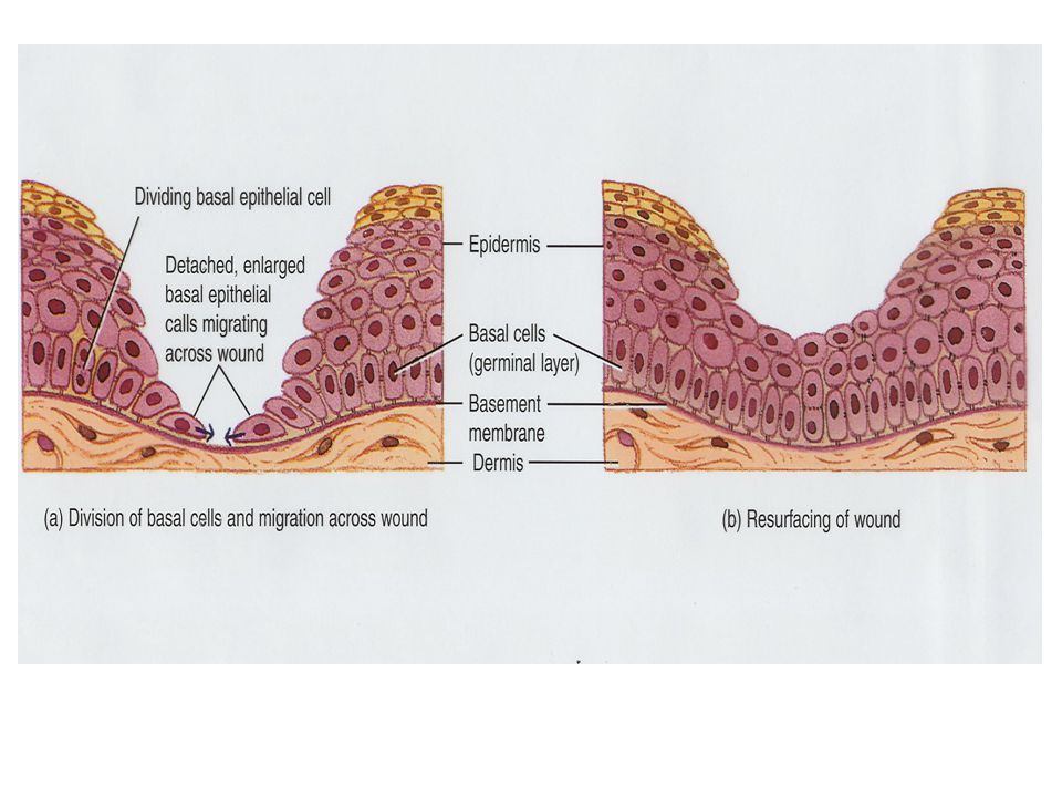
Wound healing Inflammation –Blood vessels dilate and become permeable Heat, redness, swelling and pain Shallow cuts –Epithelial cells migrate –Contact inhibition

42
Deeper wounds Inflammatory phase –Fibrin forms clot Migratory phase –Fibroblasts make granulation tissue Proliferative phase Maturation phase Scars – hypertrophic scar –keloid

47
Burns First degree or partial thickness burn –Only epidermis is damaged –Erythema, mild edema, surface layer shed –Healing – a few days to two weeks –No scarring

48
Second degree- deep partial-layer burn –Destroys epidermis –Blisters form –Healing depends on survival of accessory organs –No scars unless infected

49
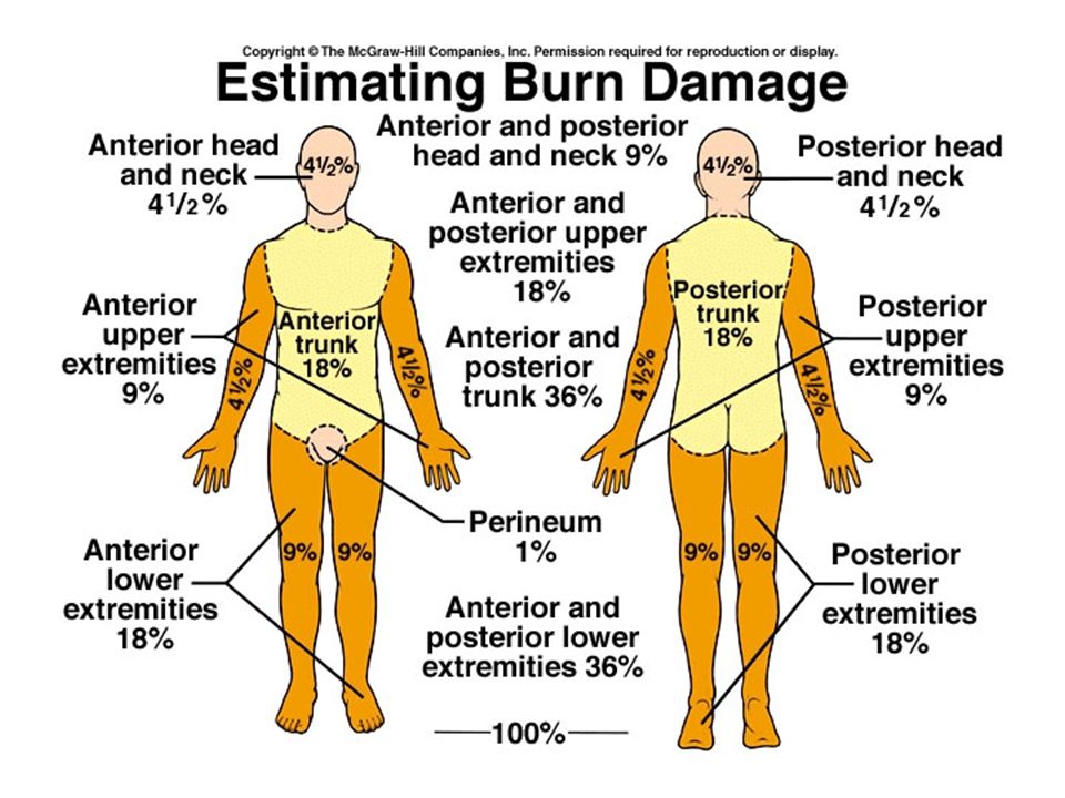
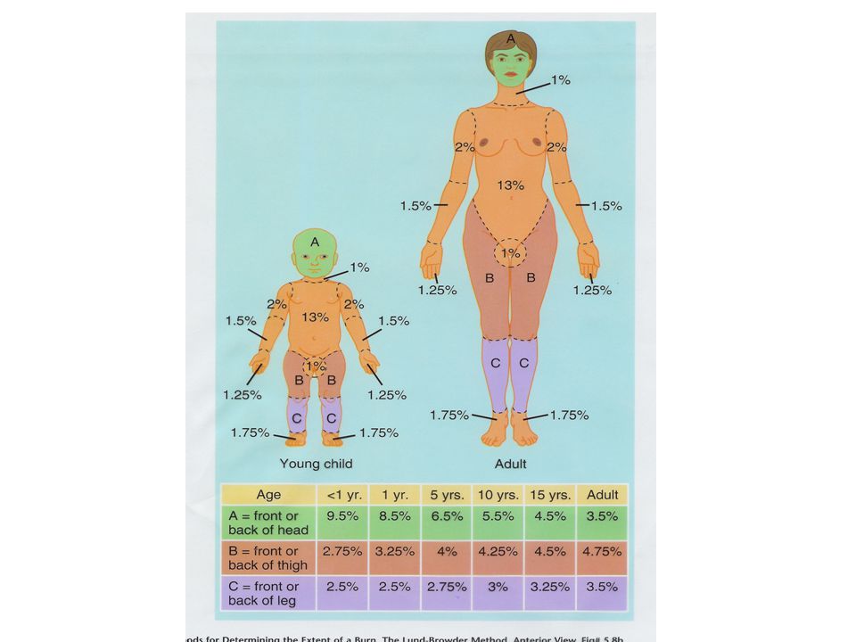
Third degree or full-thickness burn –Destroys epidermis, dermis and accessory organs of the skin –Healing occurs from margins inward –Skin grafting may be needed Autograft Homograft Rule of Nines

Similar presentations