Download presentation
Presentation is loading. Please wait.

1
Chapter 3 Macromolecules

2
Chemical Bonding and Water in Living Systems Strong and Weak Chemical Bonds

3
Covalent bonds (Figure 3
Covalent bonds (Figure 3.1) are strong bonds that bind elements in macromolecules.
 are strong bonds that bind elements in macromolecules.")
4
Covalent Bonding

5
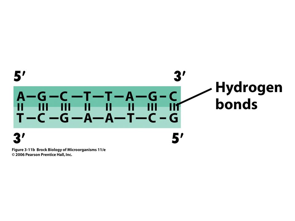
Weak bonds—such as hydrogen bonds (Figure 3
Weak bonds—such as hydrogen bonds (Figure 3.2), van der Waals forces, and hydrophobic interactions—also affect macromolecular structure, but through more subtle atomic interactions.
, van der Waals forces, and hydrophobic interactions—also affect macromolecular structure, but through more subtle atomic interactions.")
7
Hydrogen Bonding

9
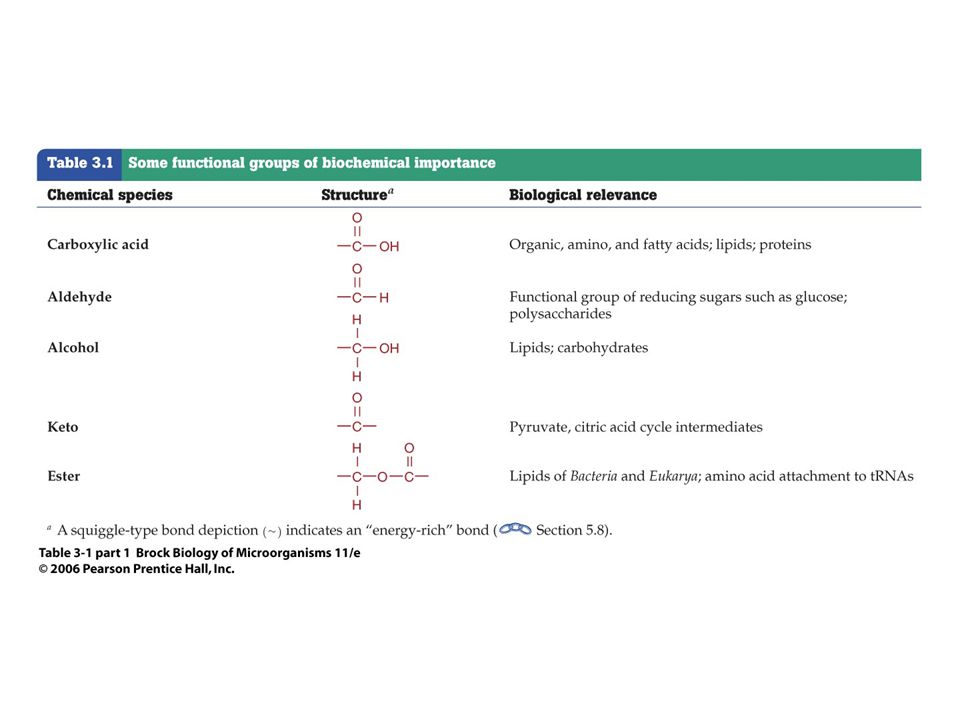
A variety of functional groups containing carbon atoms are common in biomolecules (Table 3.1) and in the folding of complex biomolecules.
 and in the folding of complex biomolecules.")
12
An Overview of Macromolecules and Water as the Solvent of Life

13
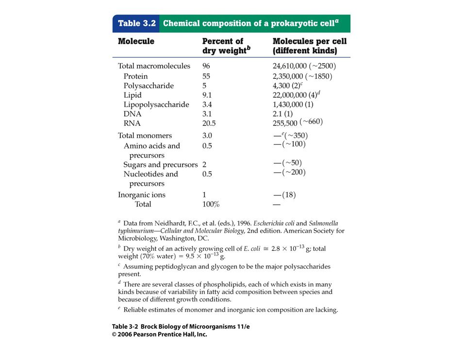
Understanding the relative composition of a bacterial cell (Table 3
Understanding the relative composition of a bacterial cell (Table 3.2) helps us to understand the metabolic needs of the organism.
 helps us to understand the metabolic needs of the organism.")
15
The bacterial cell is about 70% water, with over one-half of the dry portion being made up of protein and one-quarter being made up of nucleic acids.

16
Proteins (Figure 3. 3a) are polymers of monomers called amino acids
Proteins (Figure 3.3a) are polymers of monomers called amino acids. Nucleic acids (Figure 3.3b) are polymers of nucleotides and are found in the cell in two forms, ribonucleic acid (RNA) and deoxyribonucleic acid (DNA).
 are polymers of monomers called amino acids. Nucleic acids (Figure 3.3b) are polymers of nucleotides and are found in the cell in two forms, ribonucleic acid (RNA) and deoxyribonucleic acid (DNA).")
18
Lipids (Figure 3.3d) have both hydrophobic (nonpolar) and hydrophilic (polar) properties. They play crucial roles in membrane structure and as storage depots for excess carbon.

20
The cohesive and polar properties of water promote chemical interaction and help shape macromolecules into functional units.

21
PART II Noninformational Molecules Polysaccharides

22
Sugars combine into long polymers called polysaccharides.

23
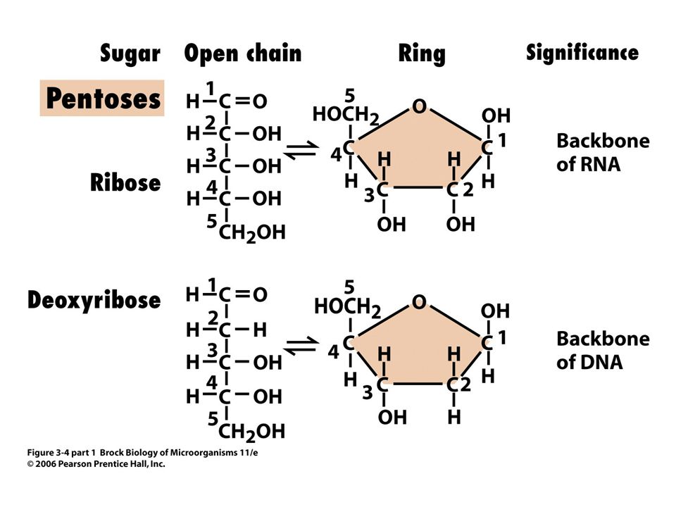
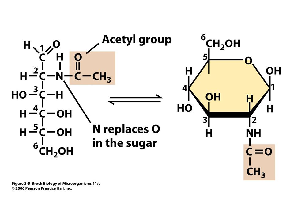
The relatively simple yet eloquent structure of the polysaccharides (Figure 3.4) and their derivatives (Figure 3.5) makes them the most abundant natural polymer on Earth and allows them to be used for metabolism, as a component of information transfer molecules (Figure 3.8), and for cellular structure.
 and their derivatives (Figure 3.5) makes them the most abundant natural polymer on Earth and allows them to be used for metabolism, as a component of information transfer molecules (Figure 3.8), and for cellular structure.")
28
Nucleotides

29
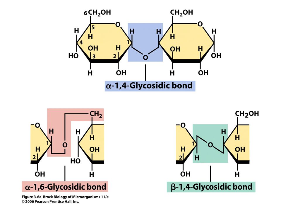
Glycosidic bonds (Figure 3
Glycosidic bonds (Figure 3.6) combine monomeric units (monosaccharides) into polymers (polysaccharides), all with a carbon-water (carbohydrate) chemical composition approaching (CH2O)n.
 combine monomeric units (monosaccharides) into polymers (polysaccharides), all with a carbon-water (carbohydrate) chemical composition approaching (CH2O)n.")
32
The two different orientations of the glycosidic bonds that link sugar residues impart different properties to the resultant molecules. Polysaccharides can also contain other molecules such as proteins or lipids, forming complex polysaccharides.

33
Lipids Lipids are amphipathic—they have both hydrophilic and hydrophobic components. This property makes them ideal structural components for cytoplasmic membranes.

34
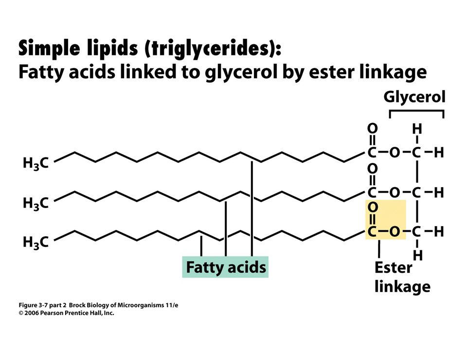
Simple lipids (triglycerides) are composed of a glycerol molecule with fatty acids (Figure 3.7) covalently linked in ester (Bacteria) or ether (Archaea) bonds.
 are composed of a glycerol molecule with fatty acids (Figure 3.7) covalently linked in ester (Bacteria) or ether (Archaea) bonds.")
39
Many lipids draw their polar characteristics from complex, non–fatty acid groups connected to carbon 1 or 3 of glycerol (Figure 3.7).
.")
40
Informational Molecules Nucleic Acids

41
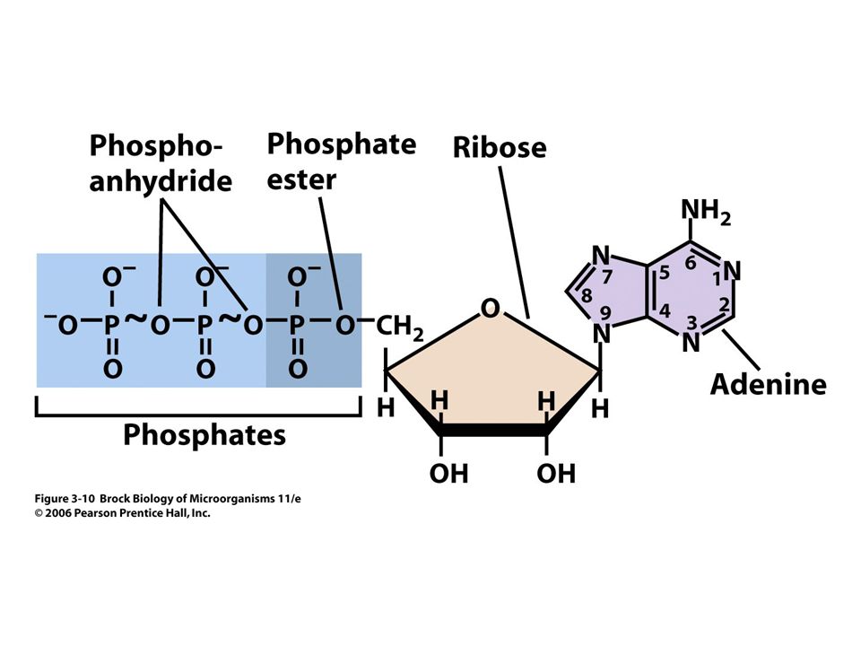
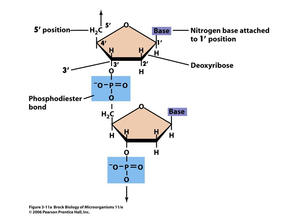
The nucleic acids deoxyribonucleic acid (DNA) and ribonucleic acid (RNA) are macromolecules composed of monomers called nucleotides. Therefore, DNA and RNA are polynucleotides. Without a phosphate, a base bonded to its sugar is referred to as a nucleoside.

42
All nucleotides have a phosphate group and a five-carbon sugar, with the sugar being ribose (–OH at carbon 2) in RNA or deoxyribose (–H at carbon 2) in DNA (Figure 3.10).
 in RNA or deoxyribose (–H at carbon 2) in DNA (Figure 3.10).")
44
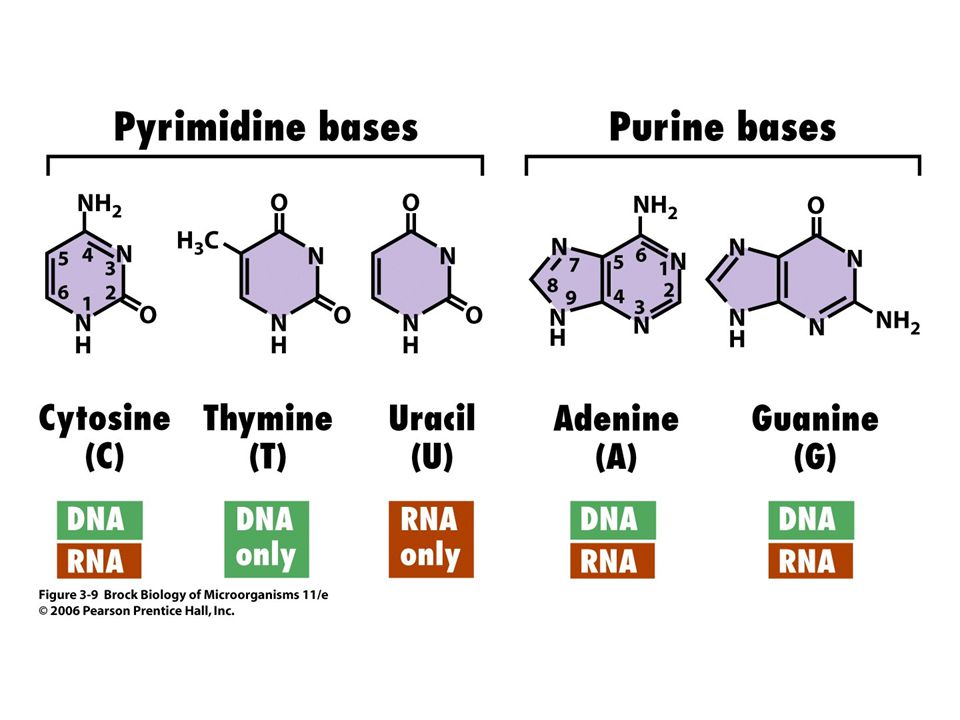
It is the primary structure, or order, of pyrimidine and purine bases (Figure 3.9) connected by the phosphodiester bond (Figure 3.11) that gives nucleic acids their information-storing capacity.
 connected by the phosphodiester bond (Figure 3.11) that gives nucleic acids their information-storing capacity.")
49
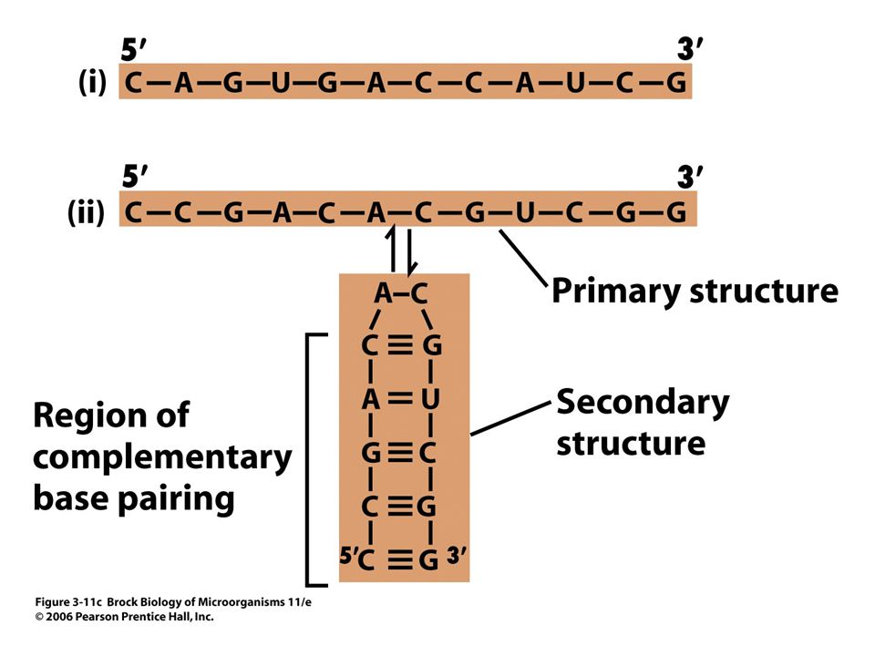
Both RNA and DNA are informational macromolecules
Both RNA and DNA are informational macromolecules. RNA can fold into various configurations to obtain secondary structure.

50
Amino Acids and the Peptide Bond
Although the -carbon of an amino acid can form four covalent bonds like other carbon atoms, the groups bonding to the -carbon are very specific.

51
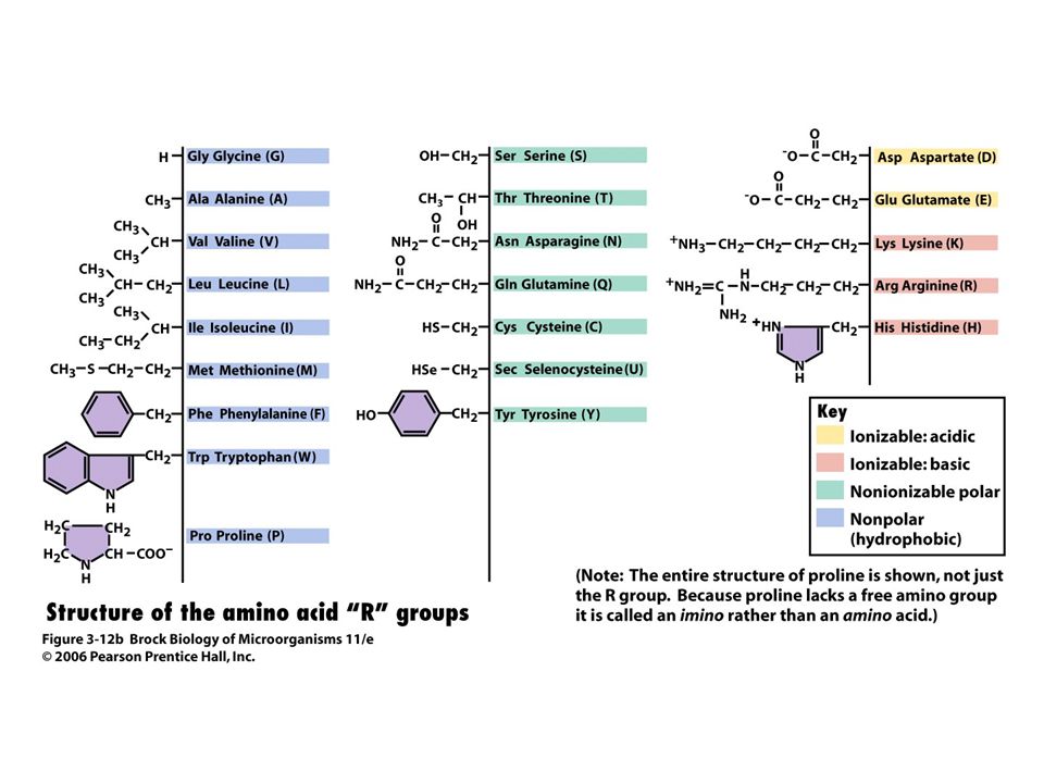
Hydrogen, an amino functional group (–NH2), and a carboxylic acid functional group (–COOH) are a part of each amino acid (Figure 3.12a).
, and a carboxylic acid functional group (–COOH) are a part of each amino acid (Figure 3.12a).")
53
The fourth bond can be one of 21 common side groups, which may be ionic, polar, or nonpolar (Figure 3.12b). It is the heterogeneity of these side groups that defines the properties of a peptide or protein.

55
Through a dehydration synthesis reaction, amino acids can bond covalently by forming a peptide bond between the amino and carboxylic acid groups.

56
Isomers are molecules that have the same molecular composition but have different structural form (Figure 3.15a).
.")
57
Isomers – Ball and Stick Model

58
Enantiomers contain the same molecular and structural formulas, except that one is a "mirror image" of the other; these are given the designations d and l (Figure 3.15b).
.")
59
Enantiomers

60
These different structural forms can greatly affect metabolism; for example, whereas sugars are typically d enantiomer, amino acids typically exist in the l form.

61
Proteins: Primary and Secondary Structure
The sequence of covalently linked amino acids in a polypeptide is the primary structure. When many amino acids are covalently linked via peptide bonds, they form a polypeptide.

62
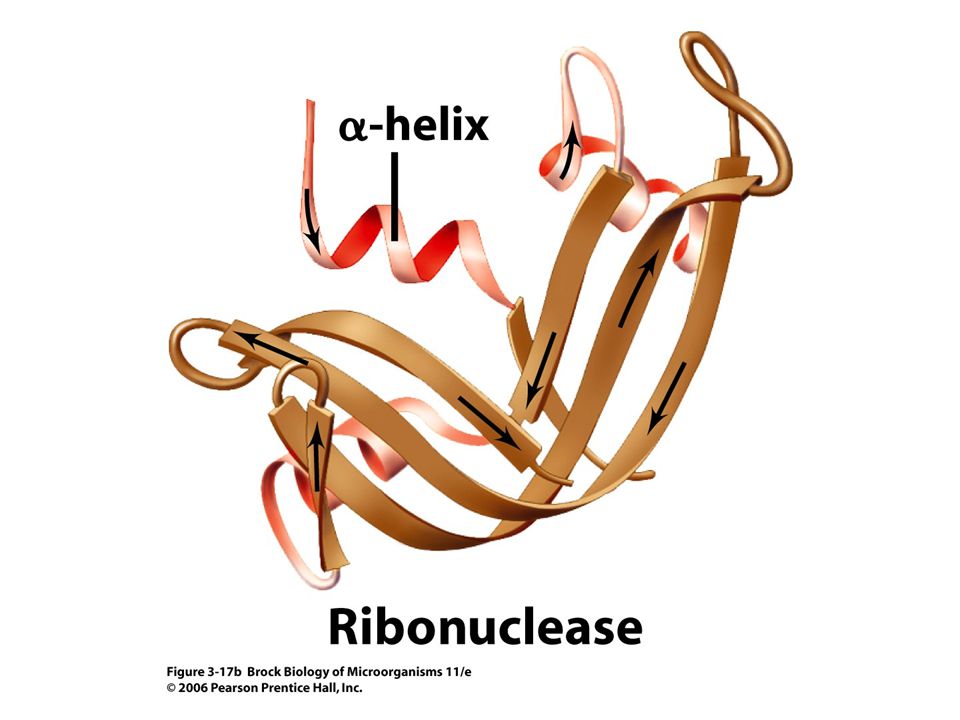
Secondary structure results from hydrogen bonding that produces an -helix ("corkscrew") or -sheet ("washboard") formation, or domain (Figure 3.16). Proteins may have an assortment of either or both domains.

63
Secondary structure of proteins- alpha-helix

64
Secondary structure of proteins- beta sheets

65
Proteins: Higher Order Structure and Denaturation
The polar, ionic, and nonpolar properties of amino acid side "R" chains cause regions of attraction and repulsion in the amino acid chain, thus creating the folding of the polypeptide (i.e., tertiary structure) (Figure 3.17).
 (Figure 3.17).")
66
Tertiary structure of polypeptides

69
Similarly, association of several polypeptides results in a unique, predictable final structure (quaternary structure) (Figure 3.18).
 (Figure 3.18).")
70
Quaternary structure of human hemoglobin

71
It is this final orientation and folding that dictate the usefulness of a protein as a catalyst (enzyme) or its structural integrity in the cell. Destruction of the folded structure by chemicals or environmental conditions is called denaturation (Figure 3.19).
..")
72
Denaturation and renaturation of ribonuclease

Similar presentations


>")


 The McGraw-Hill Companies, Inc. www.nicholls.edu/biol-qcf.>")


