Download presentation
Presentation is loading. Please wait.

1
Structure of Nerves (including roots and rami and plexuses)
Consist of Axon bundles/nerve fibers Schwann cells Connective tissue Blood vessels Endoneurium: surrounds individual neurons loose CT with capillaries (for neurons) Perineurium: Surrounds fascicles blood vessels Epineurium: surrounds the entire nerve Dense CT
 Perineurium: Surrounds fascicles. blood vessels Epineurium: surrounds the entire nerve. Dense CT.")
2
Spinal Cord and Spinal Nerves
Chapter 12 Spinal Cord and Spinal Nerves

3
Spinal Cord Functions carry/transmit sensory and motor impulses between spinal nerves and the brain in columns/white matter integration center for spinal reflexes in gray matter

4
Spinal Cord Extends from foramen magnum to ~L1-L2
Vertebral canal continues length of sacrum a portion of the vertebral canal is not occupied by the actual spinal cord creates the opportunity for spinal tap/lumbar puncture (see next side) Gives rise to 31 pairs of spinal nerves Exit through intervertebral and sacral foramina
 Gives rise to 31 pairs of spinal nerves. Exit through intervertebral and sacral foramina.")
5
Lumbar puncture

6
Spinal Cord Not uniform in diameter Conus medullaris: Cauda equina:
Cervical enlargement: supplies upper limbs Lumbar enlargement: supplies lower limbs Conus medullaris: Tapered/pointed inferior end of cord. Cauda equina: Roots and nerves extending down vertebral canal below L2 that exit intervertebral and sacral foramina

8
Spinal Nerves Thirty-one pairs of spinal nerves
First pair exit vertebral column between skull and atlas Exit vertebral canal through intervertebral and sacral foramina 8 pair cervical, 12 pair thoracic, 5 pair lumbar, 5 pair sacral, & coccygeal

10
Rami of spinal nerve (thoracic region): rami branch off the spinal nerve
: rami branch off the spinal nerve")
11
C1 2 3 Cervical 4 nerves 5 6 7 8 T1 Thoracic 9 10 11 12 L1 Lumbar
Sacral Coccygeal (a) Posterior view C1 T1 2 3 4 5 6 7 8 9 10 11 12 L1
 Posterior view. C1. T L1.")
12
Dermatomal Map Dermatomal map: skin area supplied with somatic sensory innervation by spinal nerves—general trends

13
Plexuses: intermingling nerves arising from multiple anterior rami

14
Branches of Spinal Nerves
Dorsal/posterior Ramus: innervate deep muscles of the trunk responsible for movements of the vertebral column and skin near dorsal midline Ventral/anterior Ramus: innervates structures anterior and lateral to spinal cord. Thoracic region: form intercostal nerves that innervate the intercostal muscles and the skin over the thorax Other regions they form plexuses (intermingling of nerves). Ventral rami C1-C4= cervical plexus Ventral rami C5-T1= brachial plexus Ventral rami of L1-L4= lumbar plexus Ventral rami of L4-S4= sacral plexus Ventral rami S4 & S5= coccygeal plexus 12-14 14
. Ventral rami C1-C4= cervical plexus. Ventral rami C5-T1= brachial plexus. Ventral rami of L1-L4= lumbar plexus. Ventral rami of L4-S4= sacral plexus. Ventral rami S4 & S5= coccygeal plexus")
16
Plexus

17
Cervical Plexus C1-C4 Innervates superficial neck structures, skin of neck, posterior portion of head Selected nerve of cervical plexus --Phrenic nerve Innervate diaphragm Sole motor innervation of diaphragm

18
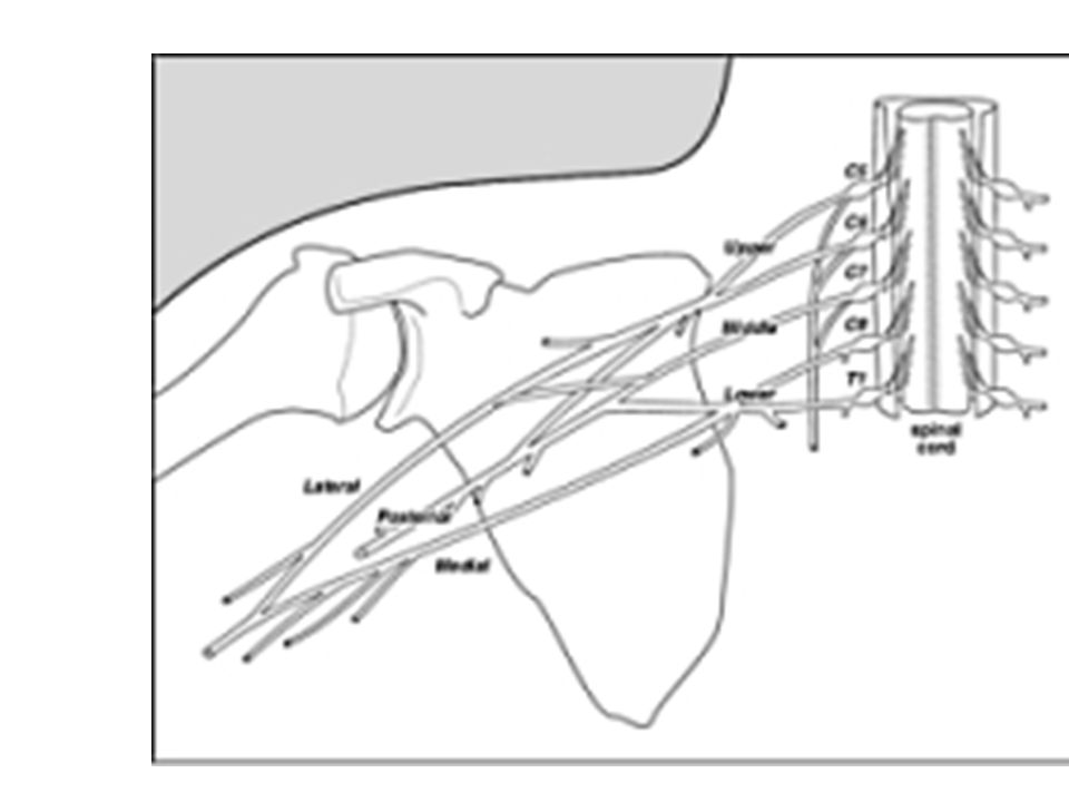
Brachial Plexus C5-T1 and some from C4 Nerves arising from: Axillary
Radial Musculocutaneous Ulnar Median

19
Lumbar Plexus Lumbar plexus: ventral rami of L1-L4 Selected Nerves:
Femoral Obturator

20
sacral Plexus Sacral plexus: ventral rami of L4-S4
sometimes considered together because of their close relationship major nerves exit and enter lower limb Selected Nerves: Sciatic Tibial Common fibular (peroneal)
")
21
Cross Section of Spinal Cord

22
Cross Section of Spinal Cord
Gray matter: mostly glial cell, cell bodies, dendrites Horns Posterior (dorsal) sensory neurons enter the cord Anterior (ventral) cell bodies of somatic motor neurons Lateral associated with ANS—cell bodies of visceral motor neurons distinct lateral horn not present in all regions of cord
 sensory neurons enter the cord. Anterior (ventral) cell bodies of somatic motor neurons. Lateral. associated with ANS—cell bodies of visceral motor neurons. distinct lateral horn not present in all regions of cord.")
23
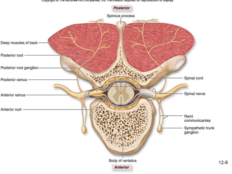
Commissures: connections between left and right halves (of CNS)
Gray & White Commisures Roots: nerves connecting to the cord Dorsal (posterior) root Sensory Nerve contains sensory neurons (unipolar) Dorsal Root Ganglion: cell bodies of sensory neurons
 root. Sensory Nerve. contains sensory neurons (unipolar) Dorsal Root Ganglion: cell bodies of sensory neurons.")
24
Ventral (anterior) Root motor nerve
axons of motor neurons (multipolar) Spinal Nerve: Two roots merge to form a spinal nerve then passes through intervertebral foramen mixed nerves axons of both motor and sensory neurons
 Spinal Nerve: Two roots merge to form a spinal nerve. then passes through intervertebral foramen. mixed nerves. axons of both motor and sensory neurons.")
25
Cross Section of Spinal Cord
White matter: myelinated axons forming nerve tracts columns (funiculi): divided into tracts (fasciculi; pathways) Carry information: to and from the brain (ascending and descending) to and from other regions of the spinal cord
: divided into tracts (fasciculi; pathways) Carry information: to and from the brain (ascending and descending) to and from other regions of the spinal cord.")
26
Figure 16.4 locations of various neurons within spinal cord (e.g., somatic motor, visceral motor [autonomic], somatic sensory) 26 26
![Figure 16.4 locations of various neurons within spinal cord (e.g., somatic motor, visceral motor [autonomic], somatic sensory)](http://slideplayer.com/slide/5264037/16/images/26/Figure+16.4+locations+of+various+neurons+within+spinal+cord+%28e.g.%2C+somatic+motor%2C+visceral+motor+%5Bautonomic%5D%2C+somatic+sensory%29.jpg)
27
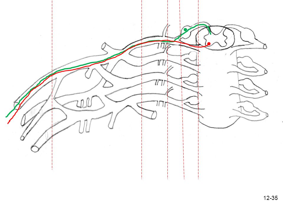
Pathways through roots and cord

28
Ascending and Descending Tracts/Pathways
= sensory = motor

29
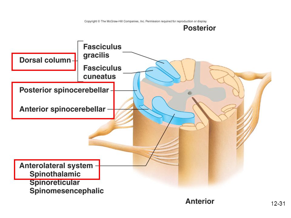
Specific Ascending Pathways within spinal cord white matter
Anteriolateral System — within anterior and lateral columns Spinothalamic Tracts: somatic sensory information from cutanous receptors to Thalamus pain, crude touch, temperature Dorsal Column System — in dorsal columns proprioception, fine touch, two point discrimination Spinocerebellar--periphery of lateral and dorsal columns proprioception to cerebellum

30
Spinocerebellar--periphery of lateral and dorsal columns
Anteriolateral System —Spinothalamic Tracts: somatic sensory information from cutanous receptors to Thalamus pain, pressure, crude touch, temperature Dorsal Column System proprioception, fine-touch, two point discrimination, pressure Spinocerebellar--periphery of lateral and dorsal columns proprioception to cerebellum

32
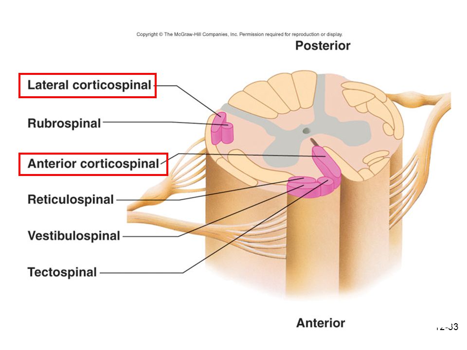
Descending Pathways through spinal cord white matter
Corticospinal (pyramidal)—within dorsal and anterior columns voluntary movements Indirect Pathways involuntary movement, upright posture, balance, walking, reflexive movements of head and neck in response to visual and auditory stimuli
—within dorsal and anterior columns. voluntary movements. Indirect Pathways. involuntary movement, upright posture, balance, walking, reflexive movements of head and neck in response to visual and auditory stimuli.")
34
ramus roots plexus “nerves” Spinal nerve
e.g., femoral, median, phrenic, sciatic

36
Spinal Meninges and Protection of cord

37
Cross Section of Spinal Cord

38
Figure 16.2 38 38

39
Meninges & Associated Spaces
Connective tissue membranes surrounding spinal cord and brain Epidural Space: Contains blood vessels, areolar CT and fat. Dura mater: continuous with epineurium of the spinal nerves Arachnoid mater: thin and wispy Subarachnoid space: Contains CSF and blood vessels within web-like strands of arachnoid tissue Pia mater: bound tightly to surface of brain and spinal cord. filum terminale: anchors spinal cord to coccyx—longitudinal support denticulate ligaments: attach the spinal cord to the dura mater laterally—lateral support

40
Protection of the Spinal Cord
Physical Protection Vertebrae rigid protection Epidural Space with adipose padding/cushioning Meninges CSF cushioning Filum Terminale longitudinal support Denticulate Ligaments lateral support Chemical Protection Blood Brain Barrier—blood CNS barrier

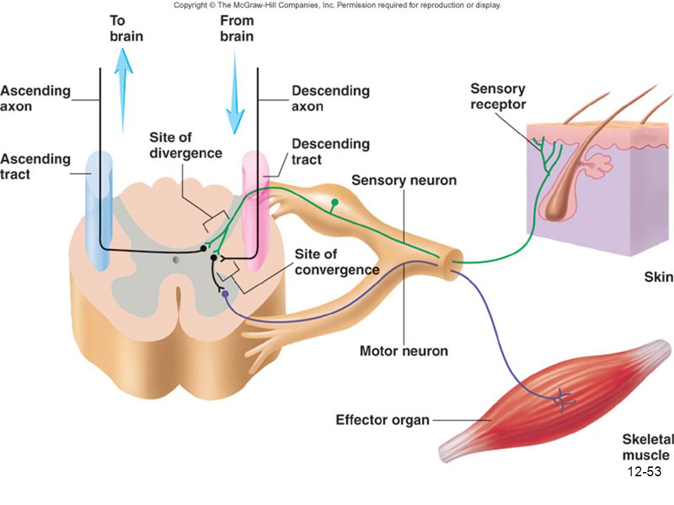
41
Somatic Motor and Sensory: --Single sensory neurons carry sensory impulses from the receptor/site of stimulus all the way into the spinal cord. --Single motor neurons carry motor impulses from the spinal cord all the way to the effector muscle

42
Pathways through roots and cord

43
Reflexes Automatic responses to specific stimuli (do not require conscious thought/processing) Higher brain centers can influence, suppress, or exaggerate reflex responses Types: Learned Innate (typically homeostatic) Spinal, integrated in spinal cord Cranial, integrated in brain
 Spinal, integrated in spinal cord. Cranial, integrated in brain.")
44
Spinal Reflexes represent some of the most basic nerve pathways and CNS integration Brain not necessary for spinal reflexes to occur Although brain can modify (suppress or enhance ) spinal reflexes. The nerve pathway is called a reflex arc
 spinal reflexes. The nerve pathway is called a reflex arc.")
45
Reflex Arc Components Electrical Impulse (Action potentials) produced in sensory receptors transmitted to Sensory neuron. To- Interneurons—in most cases. To- Motor neuron. To- Effector organ which responds with a reflex

46
Stretch/Extensor Reflexes
Monosynaptic: Two neurons One synapse Sensory neuron synapses directly with motor neuron Stretch Receptor (Muscle spindle): Sensory neuron synapse with motor neurons of the spinal cord Motor neuron innervates muscle that was stretch causing contraction
: Sensory neuron. synapse with motor neurons of the spinal cord. Motor neuron innervates muscle that was stretch causing contraction.")
47
Stretch/Extensor Reflex

48
Withdrawal/Flexor Reflex: Function is to remove a body limb or other part from a painful stimulus.
Polysynaptic: 2+ synapses 3+ neurons Interneuron(s) between sensory and motor neuron Variations: Reciprocal inhibition causes relaxation of antagonistic extensor muscle when flexor muscle contracts. Crossed extensor reflex: when a withdrawal reflex is initiated in one lower limb, the crossed extensor reflex causes extension of opposite lower limb.
 between sensory and motor neuron. Variations: Reciprocal inhibition. causes relaxation of antagonistic extensor muscle when flexor muscle contracts. Crossed extensor reflex: when a withdrawal reflex is initiated in one lower limb, the crossed extensor reflex causes extension of opposite lower limb.")
49
Withdrawal Reflex

50
Withdrawal Reflex with Reciprocal Inhibition

51
Withdrawal Reflex with Crossed Extensor Reflex

52
Relationship of Brain and Spinal Cord Reflexes
Sensory information goes to brain; e.g., pain. Descending tracts from brain carry info to reflexes. Neurotransmitters produce inhibitory or excitatory effects modifying the reflex.

54
PNS Disorders General disorders Anesthesia: loss of sensation
Hyperesthesia: increased sensitivity to pain, pressure, light Paresthesia: tingling, prickling, burning Neuralgia: nerve inflammation causing stabbing pain Sciatica: pain radiating down back of thigh and leg Infections Herpes: skin lesions Shingles or herpes zoster: adult disease of chickenpox, virus latent in peripheral ganglia Poliomyelitis: infantile paralysis Anesthetic leprosy: bacterial infection of peripheral nerves Diptheria: demylenation, motor/sensory decline, resp and heart failure Genetic and autoimmune disorders Myasthenia gravis: results in fatigue and muscular weakness due to inadequate ACh receptors

Similar presentations


 and Nerves. NERVOUS SYSTEM 1.Collect sensory input 2.Integrate sensory input 3.Motor output Functions of Nervous System.>")


