Download presentation
Presentation is loading. Please wait.

1
Lymphoma Dr Mohammed Alqahtani CSLT(CG), CLSp(CG), RT,MBA, Ph.D Genomic Medicine Unit Founder & Director Center of Excellence in Genomic Medicine Research Founder & Director
, CLSp(CG), RT,MBA, Ph.D Genomic Medicine Unit Founder & Director Center of Excellence in Genomic Medicine Research Founder & Director")
2
Overview Concepts, classification, biology Epidemiology Clinical presentation Diagnosis Staging Three important types of lymphoma

3
How Cancer Develops Normal cells are programmed to multiply, die when they’re old Signals to multiply and die are controlled by specific genes Mutations can occur in these genes If enough mutations occur in genes controlling growth or cell death a cell begins to multiply uncontrollably The cell has then become cancerous or “malignant ”

4
Features common to cancer cells Growth in the absence of “go” signals Growth despite “stop” signals Locally invasive growth and metastases to distant sites

5
Bone Marrow Present in the soft inner part of some bones such as the skull, shoulder, blade, ribs, pelvis, and backbones. (Occupies central cavity of bone) The bone marrow is made up of blood-forming stem cells, lymphoid tissue, fat cells, and supporting tissues that aid the growth of blood forming cells.
 The bone marrow is made up of blood-forming stem cells, lymphoid tissue, fat cells, and supporting tissues that aid the growth of blood forming cells..")
6
Bone Marrow Spongy tissue where development of all types of blood cells takes place All bones have active marrow at birth Adulthood - vertebrae, hip, shoulders, ribs, breast and skull contain marrow

7
Bone Marrow Aspiration/Biopsy

8
Hematopoietic Malignancies Lymphoma is a general term for hematopoietic solid malignancies of the lymphoid series. Leukemia is a general term for liquid malignancies of either the lymphoid or the myeloid series.

9
Conceptualizing lymphoma neoplasms of lymphoid origin, typically causing lymphadenopathy leukemia vs lymphoma lymphomas as clonal expansions of cells at certain developmental stages

10
What is Lymphoma Lymphomas are cancers that begin by the “malignant transformation” of a lymphocyte in the lymphatic system Many lymphomas are known to be due to specific genetic mutations Follicular lymphoma due to overexpression of BCL-2 (gene that blocks programmed cell death)
")
11
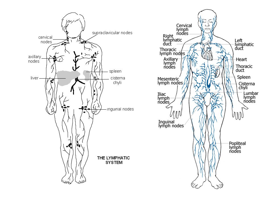
What is the Lymphatic System? Made up of organs, such as the tonsils, spleen, liver, bone marrow and a network of lymphatic vessels that connect glands, called lymph nodes Lymph nodes located throughout the body Lymph nodes filter foreign particles out of the lymphatic fluid Contain B and T lymphocytes

12
Lymphatic System Lymph nodes act as a filter to remove bacteria, viruses, and foreign particles Most people will have had “swollen glands” at some time as a response to infection

15
Blood Cell and Lymphocyte Development STEM CELLS Multipotential myeloid cells Differentiate & mature into 6 Types of blood cells red cells basophils neutrophils monocytes eosinophils platelets Multipotential lymphocytic cells Differentiate & mature into 3 Types of lymphocytes T lymphocytes B lymphocytes Natural Killer Cells

16
Lymphocytes Most lymphocytes are in lymph nodes, spleen, bone marrow and lymphatic vessels 20% of white blood cells in blood are lymphocytes T cells, B cells, natural killer cells B cells produce antibodies that help fight infectious agents T cells help B cells produce antibodies and they fight viruses

17
T-Cells and B-Cells Immature lymphocytes that travel to the thymus differentiate into T-Cells – “T” is for thymus Immature lymphocytes that travel to the spleen or lymph nodes differentiate into B cells –"B" stands for the bursa of Fabricius, which is an organ unique to birds, where B cells mature.

18
ALLMMCLLLymphomas Hematopoietic stem cell Neutrophils Eosinophils Basophils Monocytes Platelets Red cells Myeloid progenitor Myeloproliferative disorders AML Lymphoid progenitor T-lymphocytes Plasma cells B-lymphocytes naïve

19
B-cell development stem cell lymphoid progenitor progenitor-B pre-B immature B-cell memory B-cell plasma cell DLBCL, FL, HL ALL CLL MM germinalcenterB-cell mature naive B-cell

20
Clinically useful classification Diseases that have distinct clinical features natural history prognosis treatment Biologically rational classification Diseases that have distinct morphology immunophenotype genetic features clinical features Classification

21
Usually classified by how the cells look under a microscope and how quickly they grow and spread –Aggressive lymphomas (high-grade lymphomas) –Indolent Lymphomas (low-grade lymphomas)
 –Indolent Lymphomas (low-grade lymphomas)")
22
Lymphoma classification (2001 WHO) B-cell neoplasms –precursor –mature T-cell & NK-cell neoplasms –precursor –mature Hodgkin lymphoma Non- Hodgkin Lymphomas
 B-cell neoplasms –precursor –mature T-cell & NK-cell neoplasms –precursor –mature Hodgkin lymphoma Non- Hodgkin Lymphomas")
23
Three common lymphomas Follicular lymphoma Diffuse large B-cell lymphoma Hodgkin lymphoma

24
Relative frequencies of different lymphomas Hodgkin lymphoma NHL Diffuse large B-cell Follicular Other NHL Non-Hodgkin Lymphomas ~85% of NHL are B-lineage

25
Follicular lymphoma most common type of “indolent” lymphoma usually widespread at presentation often asymptomatic not curable (some exceptions) associated with BCL-2 gene rearrangement [t(14;18)] cell of origin: germinal center B-cell
![Follicular lymphoma most common type of indolent lymphoma usually widespread at presentation often asymptomatic not curable (some exceptions) associated with BCL-2 gene rearrangement [t(14;18)] cell of origin: germinal center B-cell](http://images.slideplayer.com/16/5245353/slides/slide_25.jpg "Follicular lymphoma most common type of indolent lymphoma usually widespread at presentation often asymptomatic not curable (some exceptions) associated with BCL-2 gene rearrangement [t(14;18)] cell of origin: germinal center B-cell")
26
defer treatment if asymptomatic (“watch-and-wait”) several chemotherapy options if symptomatic median survival: years despite “indolent” label, morbidity and mortality can be considerable transformation to aggressive lymphoma can occur
 several chemotherapy options if symptomatic median survival: years despite indolent label, morbidity and mortality can be considerable transformation to aggressive lymphoma can occur")
27
Diffuse large B-cell lymphoma most common type of “aggressive” lymphoma usually symptomatic extranodal involvement is common cell of origin: germinal center B-cell treatment should be offered curable in ~ 40%

28
B-Cell Lymphoma (80%) B-Cells help make antibodies, which are proteins that attach to and help destroy antigens Lymphomas are caused when a mutation arises during the B-cell life cycle Various different lymphomas can occur during several different stages of the cycle –Follicular lymphoma, which is a type of B-cell lymphoma is caused by a gene translocation which results in an over expressed gene called BCL-2, which blocks apoptosis.
 B-Cells help make antibodies, which are proteins that attach to and help destroy antigens Lymphomas are caused when a mutation arises during the B-cell life cycle Various different lymphomas can occur during several different stages of the cycle –Follicular lymphoma, which is a type of B-cell lymphoma is caused by a gene translocation which results in an over expressed gene called BCL-2, which blocks apoptosis.")
29
T-Cell Lymphoma (15%) The T-cells are born from stem cells, similar to that of B-cells, but mature in the thymus. They help the immune system work in a coordinated fashion. –These types of lymphomas are categorized by how the cell is affected Anaplastic Large cell Lymphoma, t-cell lymphoma caused by a gene translocation in chromosome 5

30
Mechanisms of lymphomagenesis Genetic alterations Infection Antigen stimulation Immunosuppression

31
Epidemiology of lymphomas males > females incidence –NHL increasing –Hodgkin lymphoma stable in NHL: 3 rd most frequently diagnosed cancer in males and 4 th in females in HL: 5 th most frequently diagnosed cancer in males and 10 th in females

37
Age distribution of new NHL

38
Risk factors for NHL immunosuppression or immunodeficiency connective tissue disease family history of lymphoma infectious agents ionizing radiation

39
Clinical manifestations Variable severity: asymptomatic to extremely ill time course: evolution over weeks, months, or years Systemic manifestations fever, night sweats, weight loss, anorexia, pruritis Local manifestations lymphadenopathy, splenomegaly most common any tissue potentially can be infiltrated

40
Other complications of lymphoma bone marrow failure (infiltration) CNS infiltration immune hemolysis or thrombocytopenia compression of structures (eg spinal cord, ureters) pleural/pericardial effusions, ascites
 CNS infiltration immune hemolysis or thrombocytopenia compression of structures (eg spinal cord, ureters) pleural/pericardial effusions, ascites")
41
Non-Hodgkin’s Lymphoma Staging Stage is the term used to describe the extent of tumor that has spread through the body ( I and II are localized where as III and IV are advanced. Each stage is then divided into categories A, B, and E –A: No systemic symptoms –B: Systemic Symptoms such as fever, night sweats and weight loss –E: Spreading of disease from lymph node to another organ

42
Stage IStage IIStage IIIStage IV Staging of lymphoma A: absence of B symptoms B: fever, night sweats, weight loss

43
Staging

44
Symptoms Painful Swelling of lymph nodes located in the neck, underarm and groin. Unexplained Fever Night Sweats Constant Fatigue Unexplained Weight loss Itchy Skin Cancer Sourcebook

45
Causes and Risk Factors The Exact causes are still unknown –Higher risk for individuals who: Exposed to chemicals such as pesticides or solvents Infected w/ Epstein-Barr Virus Family history of NHL (although no hereditary pattern has been established) Infected w/ Human Immunodeficiency Virus (HIV) Lymphoma.org
 Infected w/ Human Immunodeficiency Virus (HIV) Lymphoma.org")
46
Diagnosis Staging Studies Bone marrow aspiration and biopsy Radionuclide scans: GI x-rays Spinal fluid analysis CT scans Magnetic Resonance Imaging (MRI) Biopsy
 Biopsy")
47
Diagnosis requires an adequate biopsy Diagnosis should be biopsy-proven before treatment is initiated Need enough tissue to assess cells and architecture –open bx vs core needle bx vs FNA

48
Treatment Non-Hodgkin’s Lymphoma is usually treated by a team of physicians including hematologists, medical oncologists and a radiation oncologist. In some cases such as for Indolent lymphomas, the Doctor may wait to start treatment until the patient starts showing symptoms, known as “watchful waiting”

49
Treatment Options Chemotherapy Radiation Bone Marrow Transplantation Surgery Bortezomib (Velcade) Immunotherapy Using the bodies own immune system combined with material made in a lab.
 Immunotherapy Using the bodies own immune system combined with material made in a lab.")
50
Survival Rates Survival Rates vary widely by cell type and staging. –1 Year Survival Rate: 77% –5 Year Survival Rate: 56% –10 Year Survival Rate: 42% Cancer.org

51
Hodgkin lymphoma Thomas Hodgkin (1798-1866)
")
52
Classical Hodgkin Lymphoma

53
Hodgkin lymphoma cell of origin: germinal centre B-cell Reed-Sternberg cells (or RS variants) in the affected tissues most cells in affected lymph node are polyclonal reactive lymphoid cells, not neoplastic cells
 in the affected tissues most cells in affected lymph node are polyclonal reactive lymphoid cells, not neoplastic cells")
54
Reed-Sternberg cell

55
RS cell and variants popcorn celllacunar cellclassic RS cell (mixed cellularity)(nodular sclerosis) (lymphocyte predominance)
(nodular sclerosis) (lymphocyte predominance)")
56
A possible model of pathogenesis germinal centre B cell transforming event(s) loss of apoptosis RS cell inflammatory response EBV ? cytokines

57
Hodgkin lymphoma Histologic subtypes Classical Hodgkin lymphoma –nodular sclerosis (most common subtype) –mixed cellularity –lymphocyte-rich –lymphocyte depleted
 –mixed cellularity –lymphocyte-rich –lymphocyte depleted")
58
Epidemiology less frequent than non-Hodgkin lymphoma overall M>F peak incidence in 3rd decade

59
Age distribution of new Hodgkin lymphoma cases

60
Associated (etiological?) factors EBV infection smaller family size higher socio-economic status caucasian > non-caucasian possible genetic predisposition other: HIV? occupation? herbicides?

61
Clinical manifestations lymphadenopathy contiguous spread extranodal sites relatively uncommon except in advanced disease “B” symptoms

62
Treatment and Prognosis StageTreatmentFailure- free survival Overall 5 year survival I,IIABVD x 4 & radiation 70-80%80-90% III,IVABVD x 660-70%70-80%

63
Long term complications of treatment infertility –MOPP > ABVD; males > females –sperm banking should be discussed –premature menopause secondary malignancy –skin, AML, lung, MDS, NHL, thyroid, breast... cardiac disease

64
A practical way to think of lymphoma CategorySurvival of untreated patients CurabilityTo treat or not to treat Non- Hodgkin lymphoma IndolentYearsGenerally not curable Generally defer Rx if asymptomatic AggressiveMonthsCurable in some Treat Very aggressive WeeksCurable in some Treat Hodgkin lymphoma All typesVariable – months to years Curable in most Treat

65
Lab Diagnostic Studies Lymph node biopsy Bone marrow aspiration and biopsy Immunohistochemistry Flow cytometry Molecular Genetic studies FISH Cytogenetics

66
Cytogenetic Lab t(14,18) common (about 30%) – Bcl-2 –Follicular growth pattern t(8,14) ! common in Burkitt’s ! c-myc Multiple anomalies common Correlation between cytogenetic change and outcome is variable

67
In the next slide two examples of a lymphoma hybridised with a split-apart probe are shown. FISH analysis of paraffin embedded tissue sections

68
Truncated nucleus Truncated nuclei Myc split- apart probe: Probe 1+2 Large cell lymphoma Case 1 Large cell lymphoma Case 1

69
FISH analysis of paraffin embedded tissue Interpretation of results Case 1Case 2 Signals (even in truncated cells) are fused, excluding a translocation. Some nuclei contain split signals, indicating a translocation.

70
FISH analysis of paraffin embedded tissue Interpretation of results Case 1Case 2 Signals (even in truncated cells) are fused, excluding a translocation. Some nuclei contain split signals, indicating a translocation.

71
FISH analysis of paraffin embedded tissue sections There are now plentiful examples of how the FISH procedure is needed in routine lymphoma diagnosis. MALT lymphomas with the t(11;18)(q32;q21) translocation: For many laboratories FISH analysis is more convenient than a PCR procedure for detecting such cases. “Burkitt-like” lymphomas: Cases suggestive of Burkitt’s lymphoma but with atypical features should be analysed by the FISH technique for evidence of MYC translocation. What future applications of the FISH technique are likely to emerge in the future? One area lies in the detection of chromosomal amplifications and deletions of clinical significance. (CGH)
(q32;q21) translocation: For many laboratories FISH analysis is more convenient than a PCR procedure for detecting such cases. Burkitt-like lymphomas: Cases suggestive of Burkitt’s lymphoma but with atypical features should be analysed by the FISH technique for evidence of MYC translocation. What future applications of the FISH technique are likely to emerge in the future. One area lies in the detection of chromosomal amplifications and deletions of clinical significance. (CGH).")
72
Bea et al (2005) Blood 106:3183-3190 Tagawa et al Blood. (2005);106:1770-1777 For example specific patterns of chromosomal gains or losses have been noted in diffuse large B cell lymphoma.
;106: For example specific patterns of chromosomal gains or losses have been noted in diffuse large B cell lymphoma..")
73
Bea et al (2005) Blood 106:3183-3190 Tagawa et al Blood. (2005);106:1770-1777 For example specific patterns of chromosomal gains or losses have been noted in diffuse large B cell lymphoma.
;106: For example specific patterns of chromosomal gains or losses have been noted in diffuse large B cell lymphoma..")
74
Bea et al (2005) Blood 106:3183-3190 Tagawa et al Blood. (2005);106:1770-1777 For example specific patterns of chromosomal gains or losses have been noted in diffuse large B cell lymphoma.
;106: For example specific patterns of chromosomal gains or losses have been noted in diffuse large B cell lymphoma..")
75
Bea et al (2005) Blood 106:3183-3190 Tagawa et al Blood. (2005);106:1770-1777 For example specific patterns of chromosomal gains or losses have been noted in diffuse large B cell lymphoma.
;106: For example specific patterns of chromosomal gains or losses have been noted in diffuse large B cell lymphoma..")
77
Molecular Cytogenetic Lab Recurrent molecular abnormalities in lymphoma t(14;18) / Bcl2 - JH in follicular lymphoma t(11;14) / Bcl1 - JH in Mantle Zone lymphoma t(3;14) / Bcl6 - JH in Diffuse Large Cell lymphoma t(8;14) / cMyc - JH in Burkitt lymphoma t(2,5) / ALK-NPM in Anaplastic Large Cell Lymphoma
 / Bcl2 - JH in follicular lymphoma t(11;14) / Bcl1 - JH in Mantle Zone lymphoma t(3;14) / Bcl6 - JH in Diffuse Large Cell lymphoma t(8;14) / cMyc - JH in Burkitt lymphoma t(2,5) / ALK-NPM in Anaplastic Large Cell Lymphoma")
78
Histology Lab RS cell and variants popcorn celllacunar cellclassic RS cell (mixed cellularity)(nodular sclerosis) (lymphocyte predominance)
(nodular sclerosis) (lymphocyte predominance)")
Similar presentations


1 CHILDHOOD LEUKAEMIA. TA OGUNLESI (FWACP)2 LEUKAEMIA Heterogenous group of malignant disorders Characterised by uncontrolled clonal.>")


>")


>")


>")
