Download presentation
Presentation is loading. Please wait.

1
Dr Fawzia ALRoug, MBBS, Master, Ph.D
NEUROTRANSMITTERS Dr Fawzia ALRoug, MBBS, Master, Ph.D Assistant Professor, Department of Physiology, College of Medicine, King Khalid University Hospital, Riyadh, Saudi Arabia

2
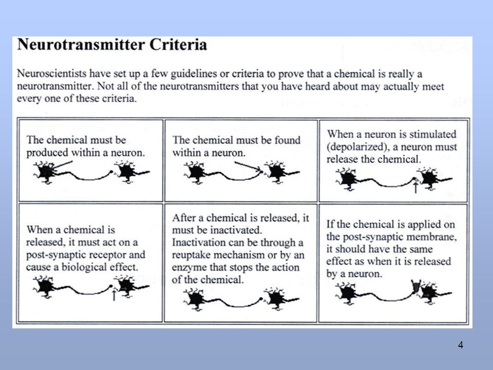
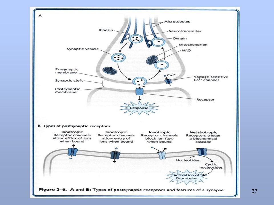
NEUROTRANSMITTERS DEFINITION: Are chemical transducers which are released by electrical impulse into the synaptic cleft from presynaptic membrane from synaptic vesicles. It then diffuse to the postsynaptic membrane and react and activate the receptors present leading to initiation of new electrical signals.

3
Discovery of neurotransmitters
Loewi, 1921 frog hearts in saline solution Stimulation of vagus nerve results in lower heart rate gave long vagal nerve stimulation Heart #2: Exposed to saline solution from heart #1 Slowed heart rate Conclusion: Neurotransmission is chemical nerve releases chemical that can influence other cells Fig 8.1, Zigmond “Fundamental Neuroscience”

7
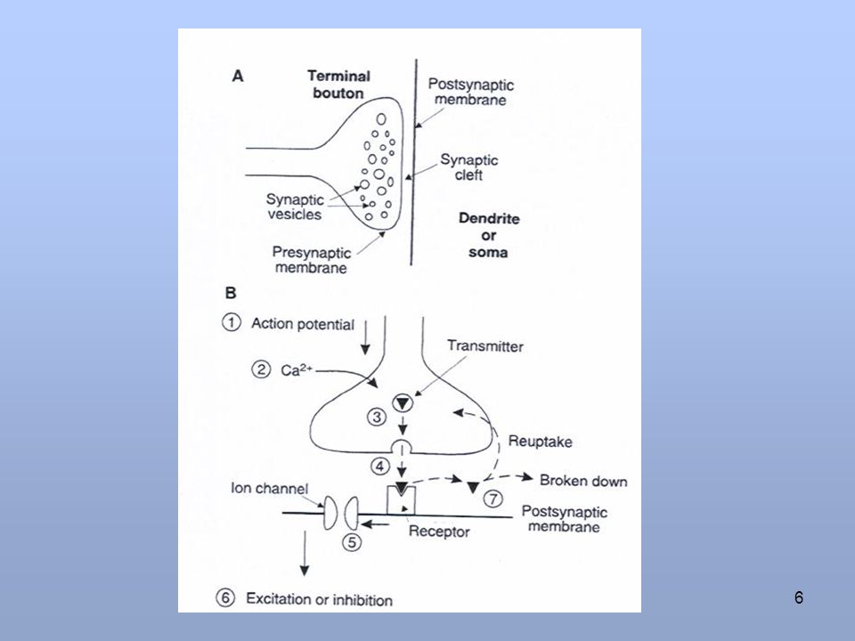
Fate of neurotransmitters
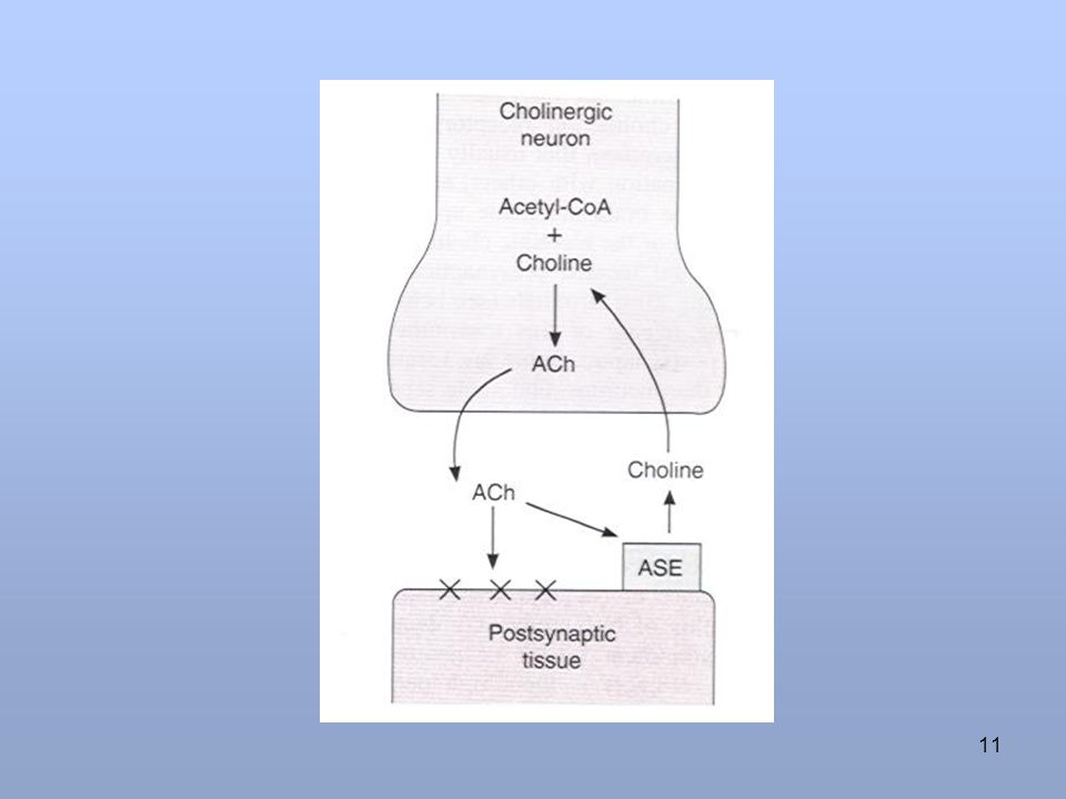
Are as , It is consumed ( broken down or used up) at postsynaptic membrane leading to action potential generation. Degraded by enzymes present in synaptic cleft. Reuptake mechanism( reutilization) this is the most common fate.
 at postsynaptic membrane leading to action potential generation. Degraded by enzymes present in synaptic cleft. Reuptake mechanism( reutilization) this is the most common fate.")
8
Types of responses on postsynaptic membrane
Excitatory postsynaptic potential (EPSPs) It is caused by depolarization. Inhibitory Postsynaptic potential (IPSPs) It is caused by hyperpolarization.
 It is caused by depolarization. Inhibitory Postsynaptic potential (IPSPs) It is caused by hyperpolarization.")
9
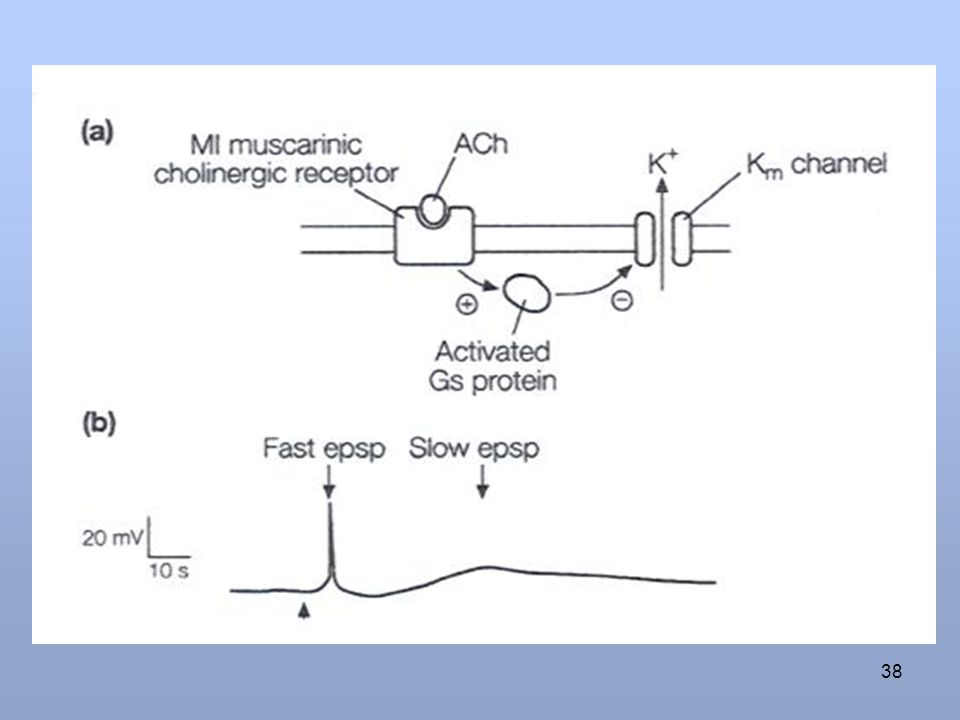
Fast & Slow Postsynaptic potentials
Fast EPSPs & IPSPs work through ligand gated ion channels.eg. Nicotinic receptors(at the level of neuromuscular junction) Slow EPSPs & IPSPs are produced by multi step process involving G protein eg. Muscarinic receptors ( at the level of autonomic gangila)
 Slow EPSPs & IPSPs are produced by multi step process involving G protein eg. Muscarinic receptors ( at the level of autonomic gangila)")
12
Acetyl Choline Receptors
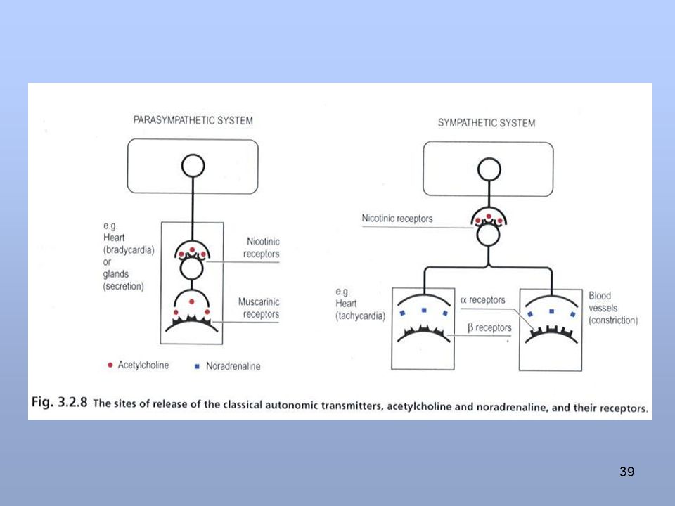
Nicotinic Muscarinic 1 Found at: Neuromuscular junction of skeletal muscle Postganglionic neurons of parasympathetic nervous system. Ventral tegmental area. Glands Neuromuscular junctions of cardiac and smooth muscle. Postganglionic neurons of sympathetic nervous system. 2 Agonist Nicotine Muscarine ( a toxin produced by certain mushroom) 3 Antagonist Curare ( paralyses skeletal muscle) Atropine
 3. Antagonist. Curare ( paralyses skeletal muscle) Atropine.")
14
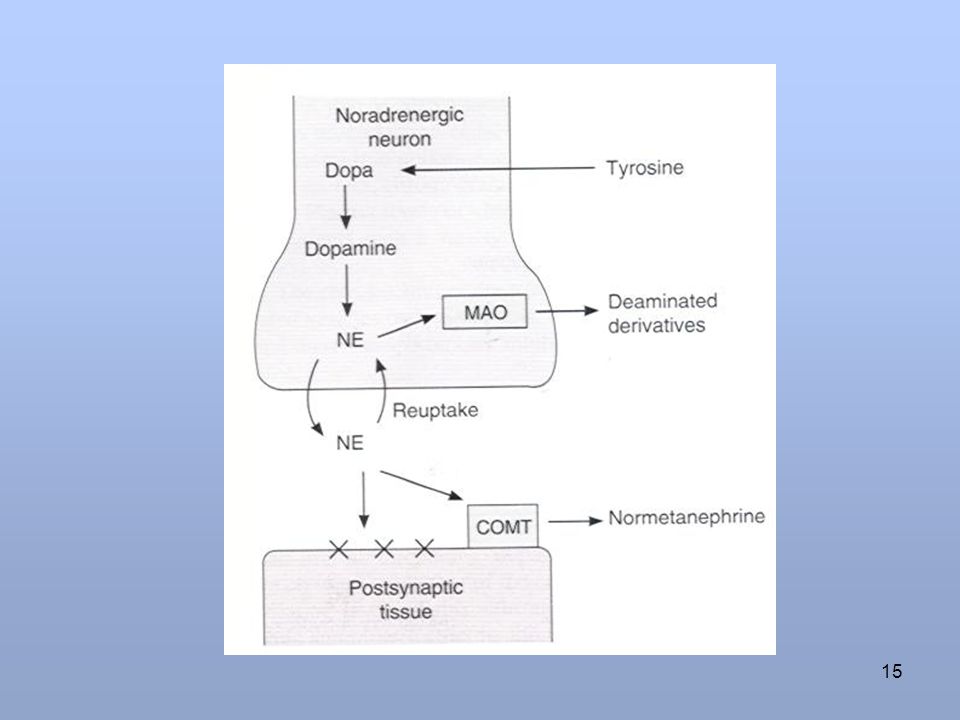
MAO=monoamine oxidase ,COMT=catechole-o-methyle-transferase

17
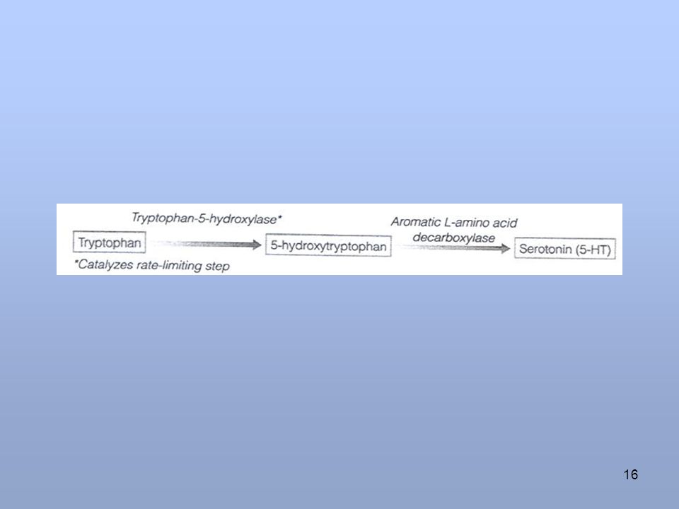
Formation of serotonin =5-HT
Hydroxy tryptamine HIAA=hydroxyindoleacetic acid

18
Histamine Histamine forming cells are in posterior hypothalamus also found in gastric mucosa and in mast cells. Formed by decarboxylation of amino acid histidine with the help of enzyme histaminase. Three known types of histamine receptors in found e.g. H1, H2, H3. H3 receptors are presynaptic. Its function in brain is not very certain. Its main function is that it is excitatory.

19
Glycine It is simplest of all aminoacids, consisting of amino group and a carboxyl group attached to a carbon atom C H3 N+ Coo- H+

20
Glycine…….. Its an inhibitory neurotransmitter.
It binds to a receptor which makes the post synaptic membrane more permeable to Cl- Ion and cause hyperpolarization (inhibition). The glycine receptor is primarily found in the ventral part of the spinal cord. Strychnine is glycine antagonist.
. The glycine receptor is primarily found in the ventral part of the spinal cord. Strychnine is glycine antagonist.")
21
Glutamic acid It is the most commonly found neurotransmitter in the brain. It is always excitatory. Glutamate is formed during Kreb’s cycle for α – ketoglutarate. Glutamate is carried into astrocytes where it is converted to glutamine and passed on to glutaminergic neurones. Glutamate is neurotoxic while glutamine is not. There are two types of receptors e.g. metabotropic and iontropic receptors.

22
NMDA =N methyl-D-aspartate receptors, when glutamate & glycine bind to receptor ion channels open,
Mg block channels

23
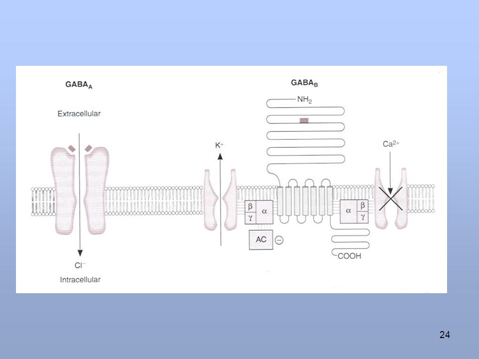
Gamma Aminobutyric acid (GABA)
It is one of the inhibitory neurotransmitter of CNS and is also found in retina. It is formed by decarboxylation of glutamate. The enzyme that catalyzes this reaction is glutamate decarboxylase(GAD) There are three types of GABA receptors e.g. GABAA B & C. GABA A & B receptors are widely distributed in CNS. GABAC are found in retina only. GABA B are metabotropic (G-protein) in function.
 There are three types of GABA receptors e.g. GABAA B & C. GABA A & B receptors are widely distributed in CNS. GABAC are found in retina only. GABA B are metabotropic (G-protein) in function.")
25
Postsynaptic receptor
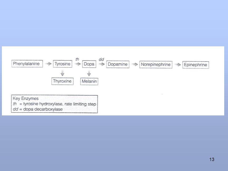
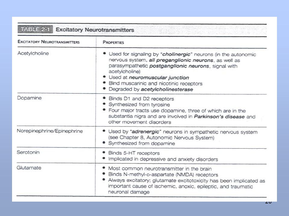
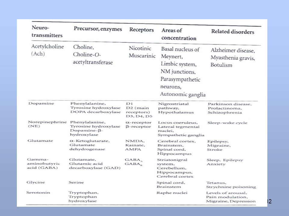
Neurotransmitter Postsynaptic effect Derived from Site of synthesis Postsynaptic receptor Fate Functions 1.Acetyl choline (Ach) Excitatory Acetyl co-A + Choline Cholinergic nerve endings Cholinergic pathways of brainstem Nicotinic Muscarinic Broken by acetyl cholinesterase Cognitive functions e.g. memory Peripheral action e.g. cardiovascular system 2. Catecholamines i. Epinephrine (adrenaline) Excitatory in some but inhibitory in other Tyrosine produced in liver from phenylalanine Adrenal medulla and some CNS cells Excites both alpha α & beta β receptors Catabolized to inactive product through COMT & MAO in liver Reuptake into adrenergic nerve endings Diffusion away from nerve endings to body fluid For details refer ANS. e.g. fight or flight, on heart, BP, gastrointestinal activity etc. Norepinehrine controls attention & arousal. ii.Norepinephrine Tyrosine, found in pons. Reticular formation, locus coerules, thalamus, mid-brain Begins inside axoplasm of adrenergic nerve ending is completed inside the secretary vesicles α1 α2 β1 β2 iii. Dopamine Tyrosine CNS, concentrated in basal ganglia and dopamine pathways e.g. nigrostriatal, mesocorticolimbic and tubero-hypophyseal pathway D1 to D5 receptor Same as above Decreased dopamine in parkinson’s disease. Increased dopamine concentration causes schizophrenia
 Excitatory. Acetyl co-A + Choline. Cholinergic nerve endings. Cholinergic pathways of brainstem. Nicotinic. Muscarinic. Broken by acetyl cholinesterase. Cognitive functions e.g. memory. Peripheral action e.g. cardiovascular system. 2. Catecholamines. i. Epinephrine. (adrenaline) Excitatory in some but inhibitory in other. Tyrosine produced in liver from phenylalanine. Adrenal medulla and some CNS cells. Excites both alpha α & beta β receptors. Catabolized to inactive product through COMT & MAO in liver. Reuptake into adrenergic nerve endings. Diffusion away from nerve endings to body fluid. For details refer ANS. e.g. fight or flight, on heart, BP, gastrointestinal activity etc. Norepinehrine controls attention & arousal. ii.Norepinephrine. Tyrosine, found in pons. Reticular formation, locus coerules, thalamus, mid-brain. Begins inside axoplasm of adrenergic nerve ending is completed inside the secretary vesicles. α1 α2. β1 β2. iii. Dopamine. Tyrosine. CNS, concentrated in basal ganglia and dopamine pathways e.g. nigrostriatal, mesocorticolimbic and tubero-hypophyseal pathway. D1 to D5 receptor. Same as above. Decreased dopamine in parkinson’s disease. Increased dopamine concentration causes schizophrenia.")
26
Postsynaptic receptor
Neurotransmitter Postsynaptic effect Derived from Site of synthesis Postsynaptic receptor Fate Functions 3. serotonin (5HT) Excitatory Tryptophan CNS, Gut (chromaffin cells) Platelets & retina 5-HT1 to 5-HT 7 5-HT 2 A receptor mediate platelet aggregation & smooth muscle contraction Inactivated by MAO to form 5-hydroxyindoleacetic acid(5-HIAA) in pineal body it is converted to melatonin Mood control, sleep, pain feeling, temperature, BP, & hormonal activity 4. Histamine Histidine Hypothalamus Three types H1, H2 ,H3 receptors found in peripheral tissues & the brain Enzyme diamine oxidase (histaminase) cause breakdown Arousal, pain threshold, blood pressure, blood flow control, gut secretion, allergic reaction (involved in sensation of itch) 5. Glutamate 75% of excitatory transmission in the brain By reductive amination of Kreb’s cycle intermediate α –ketoglutarate. Brain & spinal cord e.g. hippocampus Ionotropic and metabotropic receptors. Three types of ionotropic receptors e.g. NMDA, AMPA and kainate receptors. It is cleared from the brain ECF by Na + dependent uptake system in neurons and neuroglia. Long term potentiation involved in memory and learning by causing Ca++ influx.
 Excitatory. Tryptophan. CNS, Gut (chromaffin cells) Platelets & retina. 5-HT1 to 5-HT 7. 5-HT 2 A receptor mediate platelet aggregation & smooth muscle contraction. Inactivated by MAO to form 5-hydroxyindoleacetic acid(5-HIAA) in pineal body it is converted to melatonin. Mood control, sleep, pain feeling, temperature, BP, & hormonal activity. 4. Histamine. Histidine. Hypothalamus. Three types H1, H2 ,H3 receptors found in peripheral tissues & the brain. Enzyme diamine oxidase (histaminase) cause breakdown. Arousal, pain threshold, blood pressure, blood flow control, gut secretion, allergic reaction (involved in sensation of itch) 5. Glutamate. 75% of excitatory transmission in the brain. By reductive amination of Kreb’s cycle intermediate. α –ketoglutarate. Brain & spinal cord e.g. hippocampus. Ionotropic and metabotropic receptors. Three types of ionotropic receptors e.g. NMDA, AMPA and kainate receptors. It is cleared from the brain ECF by Na + dependent uptake system in neurons and neuroglia. Long term potentiation involved in memory and learning by causing Ca++ influx.")
27
Postsynaptic receptor
Neurotransmitter Postsynaptic effect Derived from Site of synthesis Postsynaptic receptor Fate Functions 6. Aspartate Excitatory Acidic amines Spinal cord Aspartate & Glycine form an excitatory / inhibitory pair in the ventral spinal cord 7. Gama amino butyric acid(GABA) Major inhibitory mediator Decarboxylation of glutamate by glutamate decarboxylase (GAD) by GABAergic neuron. CNS GABA – A increases the Cl - conductance, GABA – B is metabotropic works with G – protein GABA transaminase catalyzes. GABA – C found exclusively in the retina. Metabolized by transamination to succinate in the citric acid cycle. GABA – A causes hyperpolarization (inhibition) Anxiolytic drugs like benzodiazepine cause increase in Cl- entry into the cell & cause soothing effects. GABA – B cause increase conductance of K+ into the cell. 8. Glycine Inhibitory Is simple amino acid having amino group and a carboxyl group attached to a carbon atom Glycine receptor makes postsynaptic membrane more permeable to Cl- ion. Deactivated in the synapse by simple process of reabsorbtion by active transport back into the presynaptic membrane Glycine is inhibitory transmitted found in the ventral spinal cord. It is inhibitory transmitter to Renshaw cells.
 Major inhibitory mediator. Decarboxylation of glutamate by glutamate decarboxylase (GAD) by GABAergic neuron. CNS. GABA – A increases the Cl - conductance, GABA – B is metabotropic works with G – protein GABA transaminase catalyzes. GABA – C found exclusively in the retina. Metabolized by transamination to succinate in the citric acid cycle. GABA – A causes hyperpolarization (inhibition) Anxiolytic drugs like benzodiazepine cause increase in Cl- entry into the cell & cause soothing effects. GABA – B cause increase conductance of K+ into the cell. 8. Glycine. Inhibitory. Is simple amino acid having amino group and a carboxyl group attached to a carbon atom. Glycine receptor makes postsynaptic membrane more permeable to Cl- ion. Deactivated in the synapse by simple process of reabsorbtion by active transport back into the presynaptic membrane. Glycine is inhibitory transmitted found in the ventral spinal cord. It is inhibitory transmitter to Renshaw cells.")
29
RECEPTORS DYSFUNCTION
Presynaptic effect i) Botulinum toxin: Its an exotoxin that binds to the presynaptic membrane and prevents the release of Ach resulting in weakness and reduction of tone. It is used to control dystonia in which body shows overactive muscular activity.
 Botulinum toxin: Its an exotoxin that binds to the presynaptic membrane and prevents the release of Ach resulting in weakness and reduction of tone. It is used to control dystonia in which body shows overactive muscular activity.")
30
ii) Lumbert – Eaton syndrome
Antibodies directed against Ca++ channels located in presynaptic terminals and interfere with transmitter release causing weakness. iii)Neuromyotonia Patient complains of muscle spasm and stiffness resulting in continuous motor activity in the muscle. It is cased by antibody directed against the presynaptic voltage gated K+ channel so that the nerve terminal is always in a state of depolarization
Neuromyotonia. Patient complains of muscle spasm and stiffness resulting in continuous motor activity in the muscle. It is cased by antibody directed against the presynaptic voltage gated K+ channel so that the nerve terminal is always in a state of depolarization.")
31
2. Effects at Postsynaptic level:
Curare binds to the acetylcholine receptor (AchR) and prevents Ach from acting on it and so that it induces paralysis. Myasthenia gravis: is caused by an antibody against the Ach receptors and Ach receptors are reduced hence the Ach released has few Ach receptor available to work and patients complain of weakness that increases with exercise.
 and prevents Ach from acting on it and so that it induces paralysis. Myasthenia gravis: is caused by an antibody against the Ach receptors and Ach receptors are reduced hence the Ach released has few Ach receptor available to work and patients complain of weakness that increases with exercise.")
33
Synaptic strength Can be facilitated like long – term potentiation.
Can be depressed ( inhibited) by long-term depression.
 by long-term depression.")
34
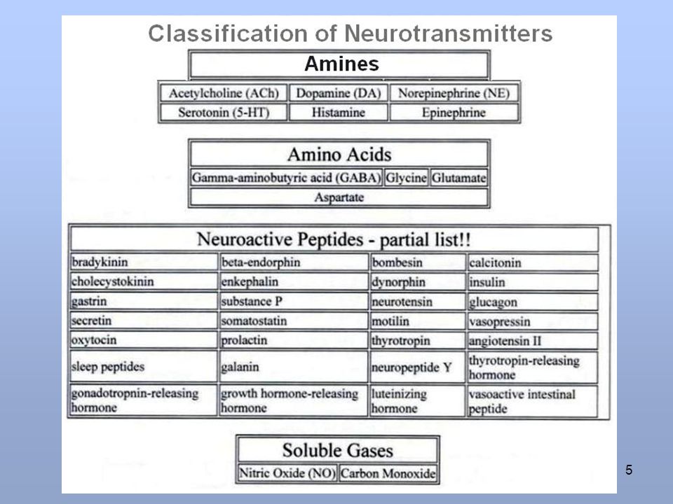
Classification of Neurotransmitters
Amines Acetyl choline (Ach) Monoamines Catecholamines Epinephrine Nor epinephrine Dopamine (Substantia nigra, sympathetic ganglia)
 Monoamines. Catecholamines. Epinephrine. Nor epinephrine. Dopamine (Substantia nigra, sympathetic ganglia)")
35
Serotonin ( hypothalamus, cerebellum, spinal cord, retina)
Histamine ( Hypothalamus) Amino acids: Excitatory eg. Glutamate ( cortex, brainstem) - Aspartate (visual cortex) Inhibitory eg. Gamma amino butaric acid GABA – cerebrum, cerebellum presynaptic inhibitory neurone in retina - Glycine – spinal cord.
 Amino acids: Excitatory eg. Glutamate ( cortex, brainstem) - Aspartate (visual cortex) Inhibitory eg. Gamma amino butaric acid GABA – cerebrum, cerebellum presynaptic inhibitory neurone in retina. - Glycine – spinal cord.")
36
Purine derivatives eg. Adinosine & ATP. Polypeptides ( a very long list of names) eg. Enkephaline, hormones ( VIP etc) ( refer to the list in Ganong 21st edition pg.97) Nonsynaptic transmitters eg. Gases, nitric oxide & cabon mono oxide.
 Nonsynaptic transmitters. eg. Gases, nitric oxide & cabon mono oxide.")
Similar presentations


 –Nicotinic –Muscarinic Adrenergic Receptors (receptors that respond.>")


 & Drugs n Receptor proteins l Control ion channels n NT.>")
