Download presentation
Presentation is loading. Please wait.

1
Active Vision Carol Colby Rebecca Berman Cathy Dunn Chris Genovese Laura Heiser Eli Merriam Kae Nakamura Department of Neuroscience Center for the Neural Basis of Cognition University of Pittsburgh Department of Statistics Carnegie Mellon University

2
Hermann von Helmholtz Treatise on Physiological Optics, 1866 Why does the world stay still when we move our eyes? “Effort of will”

4
1)Remapping in monkey area LIP and extrastriate visual cortex
Remapping in monkey area LIP and extrastriate visual cortex")
5
2) Remapping in split-brain monkeys Behavior Physiology
 Remapping in split-brain monkeys Behavior Physiology")
6
1)Remapping in monkey area LIP and extrastriate visual cortex 2) Remapping in split-brain monkeys Behavior Physiology 3) Remapping in human cortex Parietal cortex Striate and extrastriate visual cortex Remapping in a split brain human
Remapping in monkey area LIP and extrastriate visual cortex 2) Remapping in split-brain monkeys Behavior Physiology 3) Remapping in human cortex Parietal cortex Striate and extrastriate visual cortex Remapping in a split brain human")
7
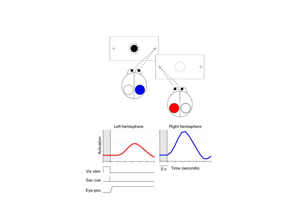
LIP memory guided saccade Stimulus OnSaccade

8
Stimulus appears outside of RF Saccade moves RF to stimulus location

9
Single step task

10
Spatial updating or remapping The brain combines visual and corollary discharge signals to create a representation of space that takes our eye movements into account

11
LIP Summary Area LIP neurons encode attended spatial locations.

12
LIP Summary Area LIP neurons encode attended spatial locations. The spatial representation of an attended location is remapped when the eyes move.

13
LIP Summary Area LIP neurons encode attended spatial locations. The spatial representation of an attended location is remapped when the eyes move. Remapping is initiated by a corollary discharge of the eye movement command.

14
LIP Summary Area LIP neurons encode attended spatial locations. The spatial representation of an attended location is remapped when the eyes move. Remapping is initiated by a corollary discharge of the eye movement command. Remapping produces a representation that is oculocentric: a location is represented in the coordinates of the movement needed to acquire the location.

15
LIP Summary Area LIP neurons encode attended spatial locations. The spatial representation of an attended location is remapped when the eyes move. Remapping is initiated by a corollary discharge of the eye movement command. Remapping produces a representation that is oculocentric: a location is represented in the coordinates of the movement needed to acquire the location. Remapping allows humans and monkeys to perform a spatial memory task accurately.

17
V1 LGN Retina V2 V3A LIP FEF SC Oculomotor System V3

18
Stimulus appears outside of RF Saccade moves RF to stimulus location

19
Stimulus alone controlSaccade alone control

20
Single step task

22
Extrastriate Summary Remapping occurs at early stages of the visual hierarchy.

23
Extrastriate Summary Remapping occurs at early stages of the visual hierarchy. Corollary discharge has an impact far back into the system.

24
Extrastriate Summary Remapping occurs at early stages of the visual hierarchy. Corollary discharge has an impact far back into the system. Remapping implies widespread connectivity in which many neurons have rapid access to information well beyond the classical receptive field.

25
Extrastriate Summary Remapping occurs at early stages of the visual hierarchy. Corollary discharge has an impact far back into the system. Remapping implies widespread connectivity in which many neurons have rapid access to information well beyond the classical receptive field. Vision is an active process of building representations.

26
1)Remapping in monkey area LIP and extrastriate visual cortex 2) Remapping in split-brain monkeys Behavior Physiology 3) Remapping in human cortex Parietal cortex Striate and extrastriate visual cortex Remapping in a split brain human
Remapping in monkey area LIP and extrastriate visual cortex 2) Remapping in split-brain monkeys Behavior Physiology 3) Remapping in human cortex Parietal cortex Striate and extrastriate visual cortex Remapping in a split brain human")
27
Stimulus appears outside of RF Saccade moves RF to stimulus location

28
What is the brain circuit that produces remapping?

29
The obvious pathway for visual signals: forebrain commissures

30
Are the forebrain commissures necessary for updating visual signals across the vertical meridian? Behavior in double step task Physiology in single step and double step task

31
Attain fixation FP T1 appears FP T1 T2 flashes briefly T1 T2 FP Saccade to T1 T1 Saccade to T2 T2

32
Attain fixation FP T1 appears FP T1 T2 flashes T1 T2 FP

33
WITHIN T1 T2 Transfer of visual signals

34
T2 WITHIN T1 T2 T2’

35
VISUAL-ACROSS T2 T1 T2 WITHIN T1 T2 T2’

36
VISUAL-ACROSS T2 T1 T2 T2’ WITHIN T1 T2 T2’

37
WITHIN T1 T2 Is performance impaired on visual-across sequences in split-brain monkeys? VISUAL-ACROSS T2 T1 T2 T2’T2T2’

38
Central AcrossWithin Central WithinAcross

39
Day 1: Initial impairment for visual-across WithinAcrossCentralWithinAcrossCentral Monkey C Monkey E correct incorrect

40
TRIALS 1-10 WithinCentralAcrossWithinCentralAcross 120-130 60-70

41
Horizontal eye position (degrees) Vertical eye position (degrees) Monkey C First day saccade endpoints Monkey E
 Vertical eye position (degrees) Monkey C First day saccade endpoints Monkey E")
42
Horizontal eye position (degrees) Vertical eye position (degrees) Monkey E Monkey C Last day saccade endpoints Monkey E
 Vertical eye position (degrees) Monkey E Monkey C Last day saccade endpoints Monkey E")
43
Are the forebrain commissures necessary for updating spatial information across the vertical meridian?

44
No. The FC are the primary route but not the only route.

45
Are the forebrain commissures necessary for updating spatial information across the vertical meridian? No. The FC are the primary route but not the only route. What are LIP neurons doing?

46
Stimulus appears outside of RF Saccade moves RF to stimulus location

47
SINGLE STEP STIMULUS ALONE SACCADE ALONE

48
Population activity in area LIP

49
SINGLE STEP DOUBLE STEP

50
Split Brain Monkey Summary The forebrain commissures normally transmit remapped visual signals across the vertical meridian but they are not required.

51
Split Brain Monkey Summary The forebrain commissures normally transmit remapped visual signals across the vertical meridian but they are not required. Single neurons in area LIP continue to encode remapped stimulus traces in split-brain animals.

54
1)Remapping in monkey area LIP and extrastriate visual cortex 2) Remapping in split-brain monkeys Behavior Physiology 3) Remapping in human cortex Parietal cortex Striate and extrastriate visual cortex Remapping in a split brain human
Remapping in monkey area LIP and extrastriate visual cortex 2) Remapping in split-brain monkeys Behavior Physiology 3) Remapping in human cortex Parietal cortex Striate and extrastriate visual cortex Remapping in a split brain human")
64
Functional Imaging Predictions 1) Robust activation in cortex ipsilateral to the stimulus.
 Robust activation in cortex ipsilateral to the stimulus.")
65
Functional Imaging Predictions 1) Robust activation in cortex ipsilateral to the stimulus. 2) Ipsilateral activation should be smaller than the contralateral visual response.
 Ipsilateral activation should be smaller than the contralateral visual response..")
66
Functional Imaging Predictions 1) Robust activation in cortex ipsilateral to the stimulus. 2) Ipsilateral activation should be smaller than the contralateral visual response. 3) It should not be attributable to the stimulus alone or to the saccade alone.
 Ipsilateral activation should be smaller than the contralateral visual response. 3) It should not be attributable to the stimulus alone or to the saccade alone..")
67
Functional Imaging Predictions 1) Robust activation in cortex ipsilateral to the stimulus. 2) Ipsilateral activation should be smaller than the contralateral visual response. 3) It should not be attributable to the stimulus alone or to the saccade alone. 4) Ipsilateral activation should occur around the time of the saccade.
 Ipsilateral activation should be smaller than the contralateral visual response. 3) It should not be attributable to the stimulus alone or to the saccade alone. 4) Ipsilateral activation should occur around the time of the saccade..")
71
Contralateral Visual Response

72
Ipsilateral Remapped Response

76
Visual and Remapped Responses

85
Human Parietal Imaging Summary Remapping in humans produces activity in parietal cortex ipsilateral to the visual stimulus.

86
Human Parietal Imaging Summary Remapping in humans produces activity in parietal cortex ipsilateral to the visual stimulus. Remapped activity is lower amplitude than visual activity.

87
Human Parietal Imaging Summary Remapping in humans produces activity in parietal cortex ipsilateral to the visual stimulus. Remapped activity is lower amplitude than visual activity. It cannot be attributed to the stimulus or the saccade alone.

88
Human Parietal Imaging Summary Remapping in humans produces activity in parietal cortex ipsilateral to the visual stimulus. Remapped activity is lower amplitude than visual activity. It cannot be attributed to the stimulus or the saccade alone. It occurs in conjunction with the eye movement.

89
1)Remapping in monkey area LIP and extrastriate visual cortex 2) Remapping in split-brain monkeys Behavior Physiology 3) Remapping in human cortex Parietal cortex Striate and extrastriate visual cortex Remapping in a split brain human
Remapping in monkey area LIP and extrastriate visual cortex 2) Remapping in split-brain monkeys Behavior Physiology 3) Remapping in human cortex Parietal cortex Striate and extrastriate visual cortex Remapping in a split brain human")
98
Contralateral Visual Response

99
Ipsilateral Remapped Response

100
Remapping in Multiple Visual Areas

101
1)Remapping in monkey area LIP and extrastriate visual cortex 2) Remapping in split-brain monkeys Behavior Physiology 3) Remapping in human cortex Parietal cortex Striate and extrastriate visual cortex Remapping in a split brain human
Remapping in monkey area LIP and extrastriate visual cortex 2) Remapping in split-brain monkeys Behavior Physiology 3) Remapping in human cortex Parietal cortex Striate and extrastriate visual cortex Remapping in a split brain human")
102
Intact Subjects Split Brain Subject

105
Strength of Parietal Responses in Split Brain and Intact Subjects

106
Human Imaging Summary Remapping in humans produces activity in the hemisphere ipsilateral to the stimulus.

107
Human Imaging Summary Remapping in humans produces activity in the hemisphere ipsilateral to the stimulus. Remapped activity is present in human parietal, extrastriate and striate cortex.

108
Human Imaging Summary Remapping in humans produces activity in the hemisphere ipsilateral to the stimulus. Remapped activity is present in human parietal, extrastriate and striate cortex. Remapped visual signals are more prevalent at higher levels of the visual system hierarchy.

109
Human Imaging Summary Remapping in humans produces activity in the hemisphere ipsilateral to the stimulus. Remapped activity is present in human parietal, extrastriate and striate cortex. Remapped visual signals are more prevalent at higher levels of the visual system hierarchy. Remapping occurs in parietal and visual cortex in a split brain human subject.

110
Conclusions Remapping of visual signals is widespread in monkey cortex.

111
Conclusions Remapping of visual signals is widespread in monkey cortex. Split-brain monkeys are able to remap visual signals across the vertical meridian.

112
Conclusions Remapping of visual signals is widespread in monkey cortex. Split-brain monkeys are able to remap visual signals across the vertical meridian. Remapped visual signals are present in area LIP in split-brain monkeys.

113
Conclusions Remapping of visual signals is widespread in monkey cortex. Split-brain monkeys are able to remap visual signals across the vertical meridian. Remapped visual signals are present in area LIP in split-brain monkeys. Remapped visual signals are robust in human parietal and visual cortex.

114
Conclusions Remapping of visual signals is widespread in monkey cortex. Split-brain monkeys are able to remap visual signals across the vertical meridian. Remapped visual signals are present in area LIP in split-brain monkeys. Remapped visual signals are robust in human parietal and visual cortex. In a split-brain human, remapped visual signals are found in parietal and visual cortex.

115
Conclusions Remapping of visual signals is widespread in monkey cortex. Split-brain monkeys are able to remap visual signals across the vertical meridian. Remapped visual signals are present in area LIP in split-brain monkeys. Remapped visual signals are robust in human parietal and visual cortex. In a split-brain human, remapped visual signals are found in parietal and visual cortex. Vision is an active process of building representations from sensory, cognitive and motor signals.

118
WithinAcross Central WithinAcross Central Learning? Or a monkey trick?

119
no monkey tricks..

120
Monkey EMMonkey CH Both monkeys really update the visual representation

123
Magnitude of Remapped Response

Similar presentations


![Read this article for Friday next week [1]Chelazzi L, Miller EK, Duncan J, Desimone R. A neural basis for visual search in inferior temporal cortex. Nature.](/5/1578404/big_thumb.jpg "Read this article for Friday next week [1]Chelazzi L, Miller EK, Duncan J, Desimone R. A neural basis for visual search in inferior temporal cortex. Nature.>")


 and monkey (cell recording) data together 1. Modality specific extrastriate cortex is modulated by attention (V4, IT, MT). 2. V1.>")

 2.Describe the organization of visual signals in extra-striate visual cortex and the specialization of.>")


 E. Salinas & T. Sejnowski(2001) E. Salinas & L.G. Abbott (1997, 1996) Pouget & T.>")


 Nature 363 Pg 345 - 347.>")