Download presentation
Presentation is loading. Please wait.

1
Cardiovascular Physiology
Dr. Abdulhalim Serafi, MB ChB,MSc,PhD,FESC Assistant Professor & Consultant Cardiologist Faculty of Medicine Umm Al-Qura University Makkah Al-Mukarramah Saudi Arabia

2
CARDIOVASCULAR PHYSIOLOGY LECTURE VII: CAPILLARY CIRCULATION
Part II CARDIOVASCULAR PHYSIOLOGY LECTURE VII: CAPILLARY CIRCULATION Outline: - Structure and functions of the blood capillaries - Capillary blood flow and capillary tone - Capillary blood pressure & capillary reactions to mechanical-stimuli. - The triple response. - Tissue fluid formation and drainage - Forces controlling fluid movement across the capillary wall. - Edema (definition, causes and types) Further Reading: Guyton: Textbook of Medical Physiology Ganong: Review of Medical Physiology
 Further Reading: Guyton: Textbook of Medical Physiology. Ganong: Review of Medical Physiology.")
3
CAPILLARY CIRCULATION
THE BLOOD CAPILLARIES are thin narrow vessels which form networks connecting the arterioles with the venules. Exchange of gases and materials between blood and tissue cells occurs only in the blood capillaries. Therefore, they are called exchange vessels. Structure of the blood capillaries: • The blood capillary is about 10 microns in diameter and about microns in length. • The human body contains about 10 billion capillaries with a total surface area estimated to be square meters.

4
• The wall of the capillary is formed of a single layer of
endothelial cells. These cells arranged close to each other and they are surrounded by a thin basement membrane on the outside. In the capillary wall and in between the endothelial cells, there are thin pores which determine the capillary permeability. The total thickness of the capillary wall is about 0.5-1 micron.

5
• The arteriolar ends of the blood capillaries are surrounded
by a small rings of smooth muscle fibers called precapillary sphincters. These sphincters: have sympathetic enervation and they regulate the blood flow to the capillaries. Like arterioles, the sphincters receive vasoconstrictor impulses from the VMC. - Capillary blood flow and capillary tone: • The flow of blood in the capillaries is very slow (about 0.5 mm/second) to give enough time for exchange of materials between blood and tissues. • The blood flow in the capillaries is intermittent, because they show. Alternating periods of closure and opening which occur 6-12 times/ minute. This is called alternation phenomenon or vasomotion.
 to give enough time for exchange of. materials between blood and tissues. • The blood flow in the capillaries is intermittent, because. they show. Alternating periods of closure and opening which. occur 6-12 times/ minute. This is called alternation. phenomenon or vasomotion.")
6
• At resting tissues, the majority of capillaries are closed
(about 90% closed and 10% open) During increased tissue activity, the number of the open capillaries is increased. If all capillaries open simultaneously, this leads to accumulation of blood in the dilated capillaries this leads to accumulation of blood in the dilated capillaries marked of VR marked of COP marked of ABP shock. This occurs after intravenous injection of histamine in experimental animals (histamine shock). In man, this may occur in extensive tissue injury e.g. in burns due to release of histamine like substances.
 During increased tissue activity, the number of the open. capillaries is increased. If all capillaries open simultaneously, this leads to. accumulation of blood in the dilated capillaries this leads. to accumulation of blood in the dilated capillaries marked of VR marked of COP marked of ABP shock. This occurs after intravenous injection of histamine. in experimental animals (histamine shock). In man, this. may occur in extensive tissue injury e.g. in burns due to. release of histamine like substances.")
7
CAPILLARY BLOOD PRESSURE
- The normal capillary blood pressure is about: mm Hg at the arterial ends of the capillaries. mm Hg at the venular ends of the capillaries. - It is affected by some factors e.g. Condition of the arterioles - Dilation of the arterioles cap. Blood flow cap. Blood pressure. - Constriction of the arterioles cap blood flow cap blood pressure. Venous pressure: - venous pressure capillary blood pressure

8
CAPILLARY REACTION TO MECHANICAL STIMULI:
1. White Line is produced by striking the skin gently with a blunt-pointed object. This occurs within 15 seconds. The white line response is due to contraction of the precapillary sphincters (produced directly by the mechanical stimulus) capillary blood flow i.e. capillary vasoconstriction. 2. Red Line is produced by scratching the skin firmly by a sharp-pointed object e.g. pin. It is the first reaction during a triple response.
 capillary blood flow i.e. capillary. vasoconstriction. 2. Red Line is produced by scratching the skin firmly by a. sharp-pointed object e.g. pin. It is the first reaction during. a triple response.")
9
THE TRIPLE RESPONSE: - Irritation or injury of the skin e.g. by scratching with sharp object leads to triple response which consists of 3 successive stages: 1. Red line due to capillary dilatation by histamine: The red line (local redness) appears immediately after the scratching. It is due to dilatation of the capillaries by histamine released from the injured skin cells. 2. Spreading flare due to arteriolar dilatation by local axon reflex: Spreading flare occurs within 30 seconds in the form of a diffuse irregular area of mottled redness surrounding the red line.
 appears immediately after the. scratching. It is due to dilatation of the capillaries by histamine. released from the injured skin cells. 2. Spreading flare due to arteriolar dilatation by local axon. reflex: Spreading flare occurs within 30 seconds in the form of a. diffuse irregular area of mottled redness surrounding the. red line.")
10
It is due to arteriolar vasodilatation which is produced by
local axon reflex I.e. impulses are conducted antidromically via a branch of the afferent neuron to the periphery where they cause a liberation of a vasodilator substance, most probably substance P, which produces arteriolar VD. 3. Local oedema (wheal) due to capillary dilatation by histamine: It is due to cap dilatation which causes cap permeability accumulation of excess tissue fluid.
 due to capillary dilatation by. histamine: It is due to cap dilatation which causes cap permeability. accumulation of excess tissue fluid.")
11
Dynamics of fluid exchange through the capillary wall (tissue fluid formation and drainage)
The cardiovascular system is a closed system I.e. the blood does not come into direct contact with the tissue cells. The nutrition of the tissues is maintained through a fluid filtered from the blood capillaries and surrounds the tissue cells and is called tissue fluid or interstitial fluid. Thus, the interstitial fluid acts as a medium for the passage of O2 and nutrients from the blood to the tissue cells and CO2 & waste products in the opposite direction. The interstitial fluid is filtered at the arterial end of the capillaries and is reabsorbed back to the blood at the venous end of the capillaries. Excess fluid is drained by the lymph vessels as lymph.

13
Forces controlling fluid movement through the
capillary membrane: There are 4 primary forces (called Starling forces) controlling fluid movement across the capillary membrane. 1. Capillary Blood Pressure: This acts as filtering force that drives fluid from the capillaries to the interstitial spaces. It is about: 30 mm Hg (25-40 mm Hg) at the arteriolar ends of the capillaries. 10 mm Hg (10-15 mm Hg) at the venular ends of the
 controlling fluid movement across the capillary membrane. 1. Capillary Blood Pressure: This acts as filtering force that drives fluid from the. capillaries to the interstitial spaces. It is about: 30 mm Hg (25-40 mm Hg) at the arteriolar ends of the. capillaries. 10 mm Hg (10-15 mm Hg) at the venular ends of the.")
14
2. Interstitial Fluid Pressure:
This is the pressure in the interstitial fluid outside the capillary wall. This pressure is negative (subatmospheric) in the most tissues, it is about-3 mm Hg. Thus it acts as suction force moving fluid from the capillary into the interstitial spaces. 3. Colloidal osmotic pressure of the interstitial fluid: It is caused by little amount of proteins present in the interstitial fluid (about 2 gm/dL). It is about 8 mm Hg and it acts as a force moving fluid from the capillaries into the interstitial spaces.
 in the most tissues, it is about-3 mm Hg. Thus it acts as. suction force moving fluid from the capillary into the. interstitial spaces. 3. Colloidal osmotic pressure of the interstitial fluid: It is caused by little amount of proteins present in the. interstitial fluid (about 2 gm/dL). It is about 8 mm Hg. and it acts as a force moving fluid from the capillaries. into the interstitial spaces.")
15
4. Colloid Osmotic Pressure of the plasma proteins:
The colloid or oncotic OP is produced by plasma proteins (7 gm%) mainly albumin & globulin. It acts as reabsorbing force i.e. it moves fluid from the interstitial spaces into the blood capillaries. It is about 28 mm Hg (25-30 mm Hg). The net force for fluid movement through the capillary wall: The net force is the difference between a. Out ward force (force moving fluid out) & b. Inward force (force moving fluid into the capillaries).
 mainly albumin & globulin. It acts as. reabsorbing force i.e. it moves fluid from the interstitial. spaces into the blood capillaries. It is about 28 mm Hg. (25-30 mm Hg). The net force for fluid movement through the capillary. wall: The net force is the difference between. a. Out ward force (force moving fluid out) & b. Inward force (force moving fluid into the capillaries).")
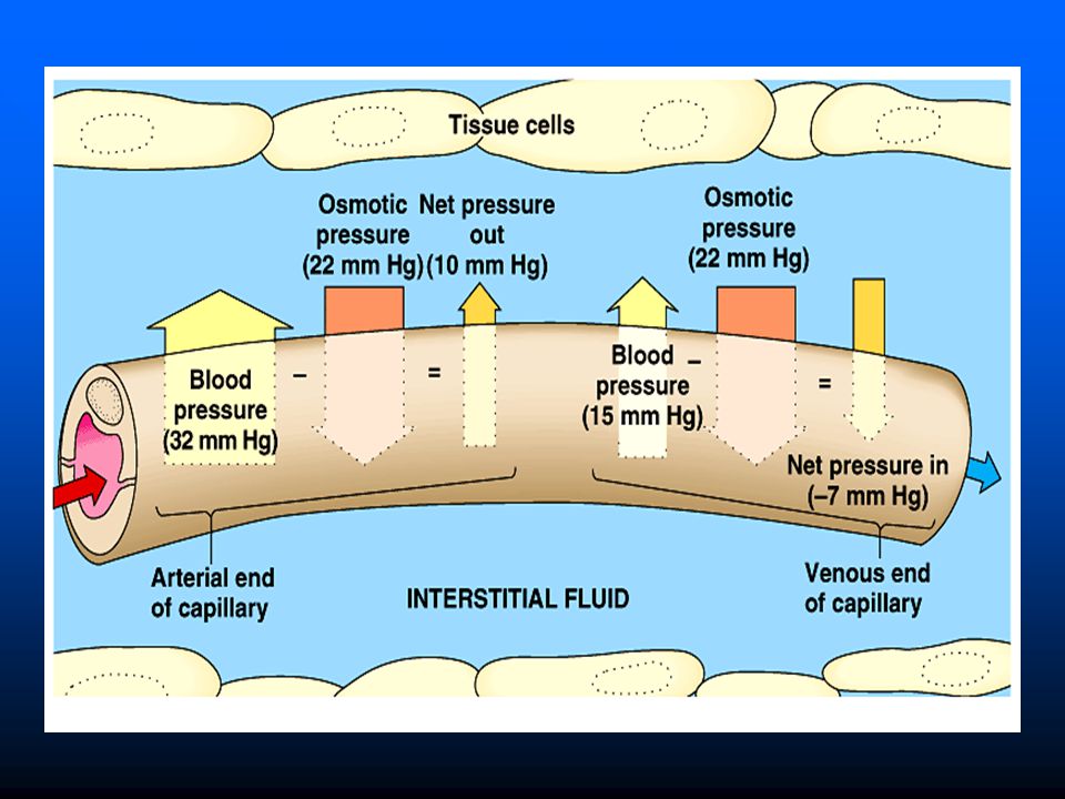
16
At the arteriolar end of the capillary:
Outward force > Inward force filtration The net force = (30+3+8) - (28). = = 13 mm Hg The direction of this force will cause fluid to move from the capillaries into the interstitial spaces i.e. filtration. At the venular end of the capillary: Inward force > outward force reabsorption The net force = (28) - (10+3+8) = = 7 mm Hg The direction of the net force will cause fluid to move from the interstitial spaces into the capillaries i.e. reabsorption.
 - (28). = = 13 mm Hg. The direction of this force will cause fluid to move from the capillaries into the interstitial spaces i.e. filtration. At the venular end of the capillary: Inward force > outward force reabsorption. The net force = (28) - (10+3+8) = = 7 mm Hg. The direction of the net force will cause fluid to move from the interstitial spaces into the capillaries i.e. reabsorption.")
17
EDEMA Edema means swelling of the tissues due to abnormal
accumulation of excess tissue fluid in the interstitial spaces. Causes and mechanisms of edema: Increase of capillary blood pressure (=increase of filtration force): Increase of capillary BP increases filtration of fluid from the capillaries to the interstitial spaces producing edema. Increase of capillary BP may be due to increase of venous pressure, Examples: a. Cardiac edema due to congestive heart failure (CHF); in CHF (= right-sided heart failure), there is increase of the venous pressure increase of capillary BP increase of filtration edema.
: Increase of capillary BP increases filtration of fluid from the capillaries to the interstitial spaces producing edema. Increase of capillary BP may be due to increase of venous pressure, Examples: a. Cardiac edema due to congestive heart failure (CHF); in CHF (= right-sided heart failure), there is increase of the venous pressure increase of capillary BP increase of filtration edema.")
18
b. Pregnancy edema during the last months of pregnancy;
b. Pregnancy edema during the last months of pregnancy; this is because the large uterus presses on the iliac veins increase of the venous pressure in the veins of the lower limbs increase of filtration edema in the lower limbs. Decrease of the colloidal osmotic pressure of the plasma proteins (=decrease of reabsorption force): Decrease of colloid osmotic pressure of plasma proteins usually occurs when the concentration of plasma proteins decreases to 5gm/dL or less, examples: a. Nutritional edema due to decrease of protein intake in diet or decrease of absorption of food proteins from the small intestine. b. Renal edema due to loss of proteins in urine as in a renal disease called nephrotic syndrome.
: Decrease of colloid osmotic pressure of plasma proteins usually occurs when the concentration of plasma proteins decreases to 5gm/dL or less, examples: a. Nutritional edema due to decrease of protein intake in diet or decrease of absorption of food proteins from the small intestine. b. Renal edema due to loss of proteins in urine as in a renal disease called nephrotic syndrome.")
19
Increase of capillary permeability (= increase of filtration):
- Increase of capillary permeability occurs if there is capillary dilatation due to release of vasodilator substances such as histamine & kinins, e.g. a. Allergic edema due to release of histamine. b. Inflammatory edema due to release of kinins and histamine. Obstruction of lymph vessels (=decrease of lymph drainage): Lymphatic obstruction decrease of lymphatic drainage from the affected part lack of drainage of excess tissue fluid which accumulates lymphatic edema, examples: a. Elephantiasis which is a marked lymphatic edema of the lower limbs due to obstruction of their lymph vessels from inside by filarial worms (parasites).
: Lymphatic obstruction decrease of lymphatic drainage from the affected part lack of drainage of excess tissue fluid which accumulates lymphatic edema, examples: a. Elephantiasis which is a marked lymphatic edema of the lower limbs due to obstruction of their lymph vessels from inside by filarial worms (parasites).")
20
b. Cancer edema due to obstruction of the lymph vessels
b. Cancer edema due to obstruction of the lymph vessels by malignant cells (tumour). Edema occurs in the part drained by the obstructed lymph vessels. Salt and water retention (=increase of plasma volume): Salt and water retention occurs in some conditions e.g. a. Excessive secretion of Aldosterone and glucocorticoids from the supra renal cortex in a disease called Cushing’s syndrome. b. Prolonged use of cortisone (cortisol) as a drug. c. During pregnancy due to high level of oestrogen and progesterone. Salt and water retention results from the effect of the previous hormones on the kidney increase of NA+ and H2O reabsorption from the renal tubules increase of plasma volume increase of filtration through the capillary wall edema.
. Edema occurs in the part drained by the obstructed lymph vessels. Salt and water retention (=increase of plasma volume): Salt and water retention occurs in some conditions e.g. a. Excessive secretion of Aldosterone and glucocorticoids from the supra renal cortex in a disease called Cushing’s syndrome. b. Prolonged use of cortisone (cortisol) as a drug. c. During pregnancy due to high level of oestrogen and progesterone. Salt and water retention results from the effect of the previous hormones on the kidney increase of NA+ and H2O reabsorption from the renal tubules increase of plasma volume increase of filtration through the capillary wall edema.")
21
TYPES OF EDEMA: According to its nature, edema may be
Soft “pitting” edema i.e it pits on pressure. Most types of edema are pitting e.g. cardiac edema, nutritional edema… etc. Hard “non-pitting” edema which occurs in some conditions e.g. in hypothyroidism (myxedema) due to presence of excess mucoproteins and fluids in the interstitial spaces. According to its location or distribution, edema may be Local edema e.g. Edema of the triple response. Inflammatory edema Edema in one limb (due to deep vein thrombosis in one lower limb). Generalized edema e.g. Cardiac edema. Nutritional edema Renal edema
 due to presence of excess mucoproteins and fluids in the interstitial spaces. According to its location or distribution, edema may be. Local edema e.g. Edema of the triple response. Inflammatory edema. Edema in one limb (due to deep vein thrombosis in one lower limb). Generalized edema e.g. Cardiac edema. Nutritional edema. Renal edema.")
Similar presentations


RBC 2)WBC 3)Platelet.>")


