Download presentation
Presentation is loading. Please wait.

1
Techniques in Protein Biochemistry Chapter 3

2
Problem: isolation & analysis of protein or aa found in cell Assumption: can somehow analyze for wanted protein –Common – Colorimetric indicator (chemical rxn color form’n; can be monitored spectrophotometrically) –BUT others available Functional indicator (biological endpoint) –This ex – colorimetric (breakdown of fats purple color) Activity assay Use at each step of separation
 –BUT others available Functional indicator (biological endpoint) –This ex – colorimetric (breakdown of fats purple color) Activity assay Use at each step of separation")
3
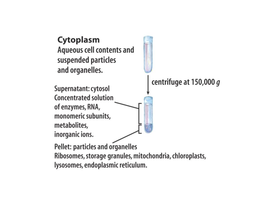
Isolation of Wanted Protein from Brain Cells Brain cells contain wanted protein Open cells –Homogenization, sonication, grinding –Maintain cold, pH, osmolality Centrifugation often used Known speeds/ conditions for diff organelles

6
Test each fraction for activity –Save most active fractions Sep’n wanted protein from other mol’s, types of mol’s –Dialysis against physiological buffer –Sep’n lipids into hydrophobic sol’n –Others

7
Separation from Other Proteins Chromatography –Gel filtration –Ion exchange –Affinity Electrophoresis –Zone –Isoelectric focusing

8
Chromatography Solid or aqueous support –Wanted protein has some affinity Aqueous or gaseous mobile phase –Wanted prot has affinity diff from that of support –Also moves molecules through/past support

9
If wanted prot affinity for support > affinity for mobile phase, prot “adheres” to support phase If wanted prot affinity for mobile phase > affinity for support, prot moves with mobile phase through/ away from support

11
Gel Filtration Chromatography (= Size Exclusion) Sep’n by MW Solid support = porous beads (ex: sephadex, sepharose) –Held in column –Beads have microscopic pores/pits/spaces Mobile phase = buffer of physio pH, ionic strength
 Sep’n by MW Solid support = porous beads (ex: sephadex, sepharose) –Held in column –Beads have microscopic pores/pits/spaces Mobile phase = buffer of physio pH, ionic strength")
12
Sample = sol’n of wanted prot + other (unwanted) prot’s; most have different MWs Apply sample to column Begin slow mobile phase flow –Smaller prot’s enter spaces in beads –Larger prot’s flow w/ buffer around beads (so emerge 1 st from column) Collect fractions; test each fraction by activity assay
 prot’s; most have different MWs Apply sample to column Begin slow mobile phase flow –Smaller prot’s enter spaces in beads –Larger prot’s flow w/ buffer around beads (so emerge 1 st from column) Collect fractions; test each fraction by activity assay")
14
Ion Exchange Chromatography Sep’n by diff’s in overall charges of prot’s Solid support = resin (charged microscopic beads) suspended in buffer Mobile phase = buffer of partic pH, ionic strength Sample = sol’n of wanted prot + other (unwanted) prot’s –Most prot’s have diff overall + or – charges –Charges of most prot’s vary in strength
 suspended in buffer Mobile phase = buffer of partic pH, ionic strength Sample = sol’n of wanted prot + other (unwanted) prot’s –Most prot’s have diff overall + or – charges –Charges of most prot’s vary in strength")
15
Apply sample to column, slow mobile phase flow –Prot’s of charge opposite that of resin and of sim strength of charge of resin: Good affinity for resin Bind electrostatically –Prot’s of same charge or different strength of charge of resin No good affinity for resin Flow through column quickly Eluted first Result: prot similar to resin is held in column

17
To elute prot held to resin in column –Buffer of higher ionic strength or stronger pH Changes ionic environment Ions in new (elution) buffer “exchange” for prot (more attractive to resin, so take the place of prot on resin in column) Collect fractions from mobile phase + elution buffer Test all fractions by activity assay
 buffer exchange for prot (more attractive to resin, so take the place of prot on resin in column) Collect fractions from mobile phase + elution buffer Test all fractions by activity assay")
18
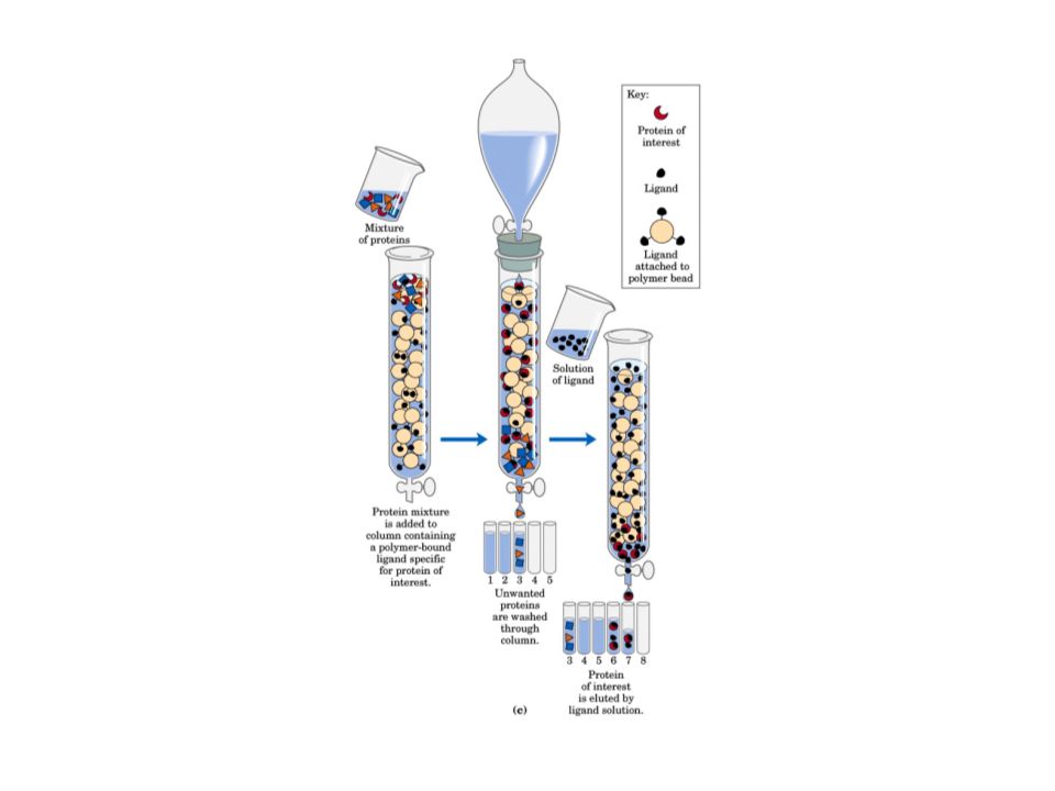
Affinity Chromatography Basis: specificity of wanted prot for some molecule(s) to which it alone will bind –Ex: Ab binds only spec Ag BUT binding must be reversible Solid support = specific binding mol (=ligand) covalently bound to beads, etc. Mobile phase = buffer of proper pH, ionic strength to maintain activity of wanted prot

19
Pack column; apply sample Begin slow mobile phase flow Prot of interest ONLY will bind to ligand –Types of binding (reversible): ionic, H- bonds, hydrophobic interactions To elute, may use sol’n of ligand (competes w/ solid phase ligand) OR buffers of diff strength, pH (disrupt protein/ligand interactions)
: ionic, H- bonds, hydrophobic interactions To elute, may use sol’n of ligand (competes w/ solid phase ligand) OR buffers of diff strength, pH (disrupt protein/ligand interactions)")
21
Electrophoresis Sep’n AND identification Based on overall charge of prot movement under influence of electric field Zone –Semisolid or gelatinous medium (plate or slab) –Spot prot mixture (wanted + unwanted prot’s in sol’n) onto gel –Apply electric field
 –Spot prot mixture (wanted + unwanted prot’s in sol’n) onto gel –Apply electric field")
23
Prot’s migrate toward anode (+ charged) Distance traveled depends on prot charge, size –Most impt = size –Gel support acts as molecular sieve; smaller mol’s go faster toward anode, so migrate further Also, prot’s more strongly charged move closer to anode Use chem to stain aa’s bands representing prot’s of decreasing MW
 Distance traveled depends on prot charge, size –Most impt = size –Gel support acts as molecular sieve; smaller mol’s go faster toward anode, so migrate further Also, prot’s more strongly charged move closer to anode Use chem to stain aa’s bands representing prot’s of decreasing MW")
25
Run stds simultaneously – Mixture of prot’s of known MW; spot on one or several lanes –Electrophorese under same cond’s as unknown protein mixture –Stain “ladder” of bands (lowest to highest MW proteins traveling some distance under these conditions)
")
27
Determine distance traveled for each band from origin Plot x = distance migrated for each std of known MW; y = log MW of stds Get std curve –Use to find distance traveled by unknown prot(s) on curve; deter MW Can also cut gel, dissolve to free prot’s
 on curve; deter MW Can also cut gel, dissolve to free prot’s")
28
Moving Boundary = IsoElectric Focusing (IEF) Sep’n due to charge; based on isoelectric pt of each prot Gel made of ampholytes (regions of different pHs) Spot sample Apply electric field
 Sep’n due to charge; based on isoelectric pt of each prot Gel made of ampholytes (regions of different pHs) Spot sample Apply electric field")
29
Each prot in mixture will migrate toward + or – electrode, based on overall charge Each prot stops moving when reaches pH region of gel = its isoelectric pH Stain aa’s of prot’s w/ chemical

30
Run stds simultaneously –Mixture of prot’s of known pI’s; spot in one or several lanes –Electrophorese under same cond’s as unknown protein mixture –Stain “ladder” of bands (lowest to highest MW proteins traveling some distance under these conditions) –Determine distance traveled for each band from origin Plot x = distance migrated for each std of known pI y = pH Yields std curve; find distance traveled by unknown prot(s) on curve pI
 –Determine distance traveled for each band from origin Plot x = distance migrated for each std of known pI y = pH Yields std curve; find distance traveled by unknown prot(s) on curve pI")
31
Characterize Wanted Prot by aa Sequence Once wanted prot isolated from all other cell mol’s Old method: Break all peptide bonds solution of aa’s Analyze aa’s by chromatography –Thin Layer Chromatography (TLC) – coated plate or paper support; various mobile phases sep aa’s from each other –High Pressure Liquid Chromatography (HPLC) – force sample through small column packed w/ support; mobile phases forced through column by high pressure pumps; sep aa’s from each other
 – coated plate or paper support; various mobile phases sep aa’s from each other –High Pressure Liquid Chromatography (HPLC) – force sample through small column packed w/ support; mobile phases forced through column by high pressure pumps; sep aa’s from each other")
32
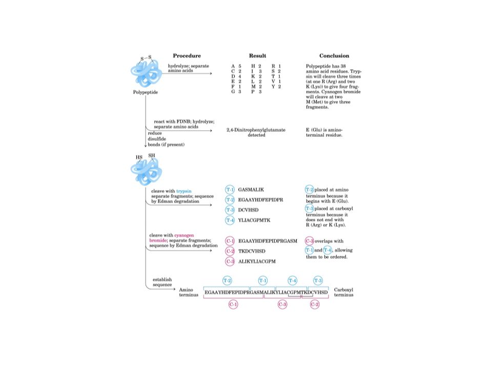
Now have identified all aa’s in protein With original protein, use chem. rxn to label amino terminal aa –Use various enz’s to cleave prot at partic aa’s along peptide chain peptide fragments –Analyze fragments for overlap; use knowledge of all aa’s in protein sequence

34
New method: Automated Chem rxn to label amino terminal aa Cleave amino terminal aa –Analyze for identity of last aa –Rest of prot now has diff amino terminal aa (second to last in original prot) –Chem rxn to label second to last aa of amino terminal –Cleave this terminal aa – Analyze for identity of second-to-last aa –Etc. etc. etc.

36
Common now: identify gene for prot of interest –Isolate mRNAs (found w/in cell rich in wanted protein) w/ message for that prot –Use mRNAs to identify gene mRNA will have complimentary sequence to gene in DNA, so will pair in that region of DNA only –Analyze gene or mRNA for nucleotide sequence –Use genetic code to determine aa sequence of wanted protein from gene which codes for it
 w/ message for that prot –Use mRNAs to identify gene mRNA will have complimentary sequence to gene in DNA, so will pair in that region of DNA only –Analyze gene or mRNA for nucleotide sequence –Use genetic code to determine aa sequence of wanted protein from gene which codes for it")
Similar presentations