Download presentation
Presentation is loading. Please wait.

1
Tour of the Cell

2
Robert Hooke (1635-1703)
")
3
Robert Hooke -- 1665: examined thinly sliced cork and coined term “cell”

4
van Leeuwenhoek --1674: first to observe living cells

5
van Leeuwenhoek

6
Schleiden and Schwann + others 1. all organisms consist of one or more cells 2. the cell is the basic unit of structure for all organisms 3. all cells arise only from preexisting cells The cell theory

7
Light microscopy

8
Cilia Electron microscopy

9
Cilia Light microscopy Electron microscopy

10
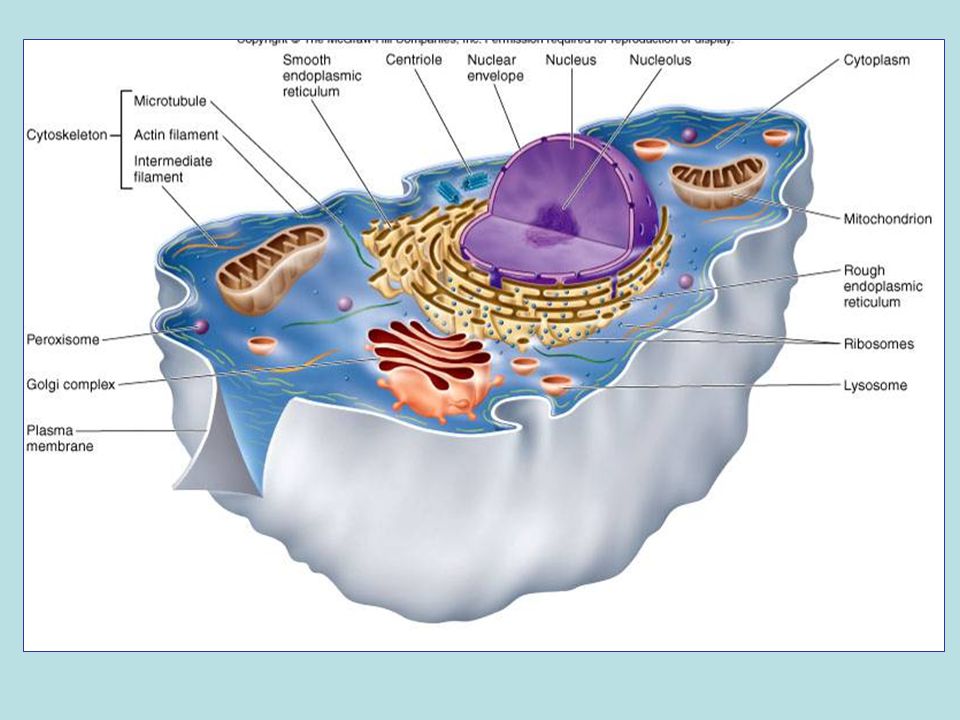
Chromosomes Light microscopy

11
Electron microscopy

12
Fluorescence microscopy Fluorescent micrograph of dividing cells Green: DNA Red: Microtubules

13
Units of measurement in cell biology micrometer (micron, )- 1/1,000,000 of a meter, or 1/1000 of a millimeter. useful for measuring the sizes of cells and organelles. “typical” animal cell: 20-40 m “typical” nucleus: 5-10 m bacteria, mitochondria, chloroplasts - few m in diameter

14
nanometer - 1/1000 of a m or 1/1,000,000 of a mm. used for molecules and subcellular structures too small to be seen with light microscopy. ribosomes - diameter of 25-30 nm. cell membranes: ~10nm cytoskeletal structures such as microtubules, microfilaments are generallly measured in nm. DNA helix ~2nm

15
Measurements 1 centimeter (cm) = 10 –2 meter (m) = 0.4 inch 1 millimeter (mm) = 10 –3 m 1 micrometer (µm) = 10 –3 mm = 10 –6 m 1 nanometer (nm) = 10 –3 µm = 10 –9 m 10 m 1 m Human height Length of some nerve and muscle cells Chicken egg 0.1 m 1 cm Frog egg 1 mm 100 µm Most plant and animal cells 10 µm Nucleus 1 µm Most bacteria Mitochondrion Smallest bacteria Viruses 100 nm 10 nm Ribosomes Proteins Lipids 1 nm Small molecules Atoms 0.1 nm Unaided eye Light microscope Electron microscope Sizes
 = 10 –2 meter (m) = 0.4 inch 1 millimeter (mm) = 10 –3 m 1 micrometer (µm) = 10 –3 mm = 10 –6 m 1 nanometer (nm) = 10 –3 µm = 10 –9 m 10 m 1 m Human height Length of some nerve and muscle cells Chicken egg 0.1 m 1 cm Frog egg 1 mm 100 µm Most plant and animal cells 10 µm Nucleus 1 µm Most bacteria Mitochondrion Smallest bacteria Viruses 100 nm 10 nm Ribosomes Proteins Lipids 1 nm Small molecules Atoms 0.1 nm Unaided eye Light microscope Electron microscope Sizes")
16
Why are cells so small?

17
1 1 5 Surface area increases while Total volume remains constant Surface to volume ratio

18
A typical rod-shaped bacterium A thin section through the bacterium Bacillus coagulans (TEM) 0.5 µm Pili Nucleoid Ribosomes Plasma membrane Cell wall Capsule Flagella Bacterial chromosome Prokaryotic Cells
 0.5 µm Pili Nucleoid Ribosomes Plasma membrane Cell wall Capsule Flagella Bacterial chromosome Prokaryotic Cells")
19
There are two types of cells…..

20
Prokaryotic Cell Example: bacteria cell

21
Eukaryotic Cells Animal Plant

22
Cell parts and functions….

23
Plasma Membrane (cell membrane)
")
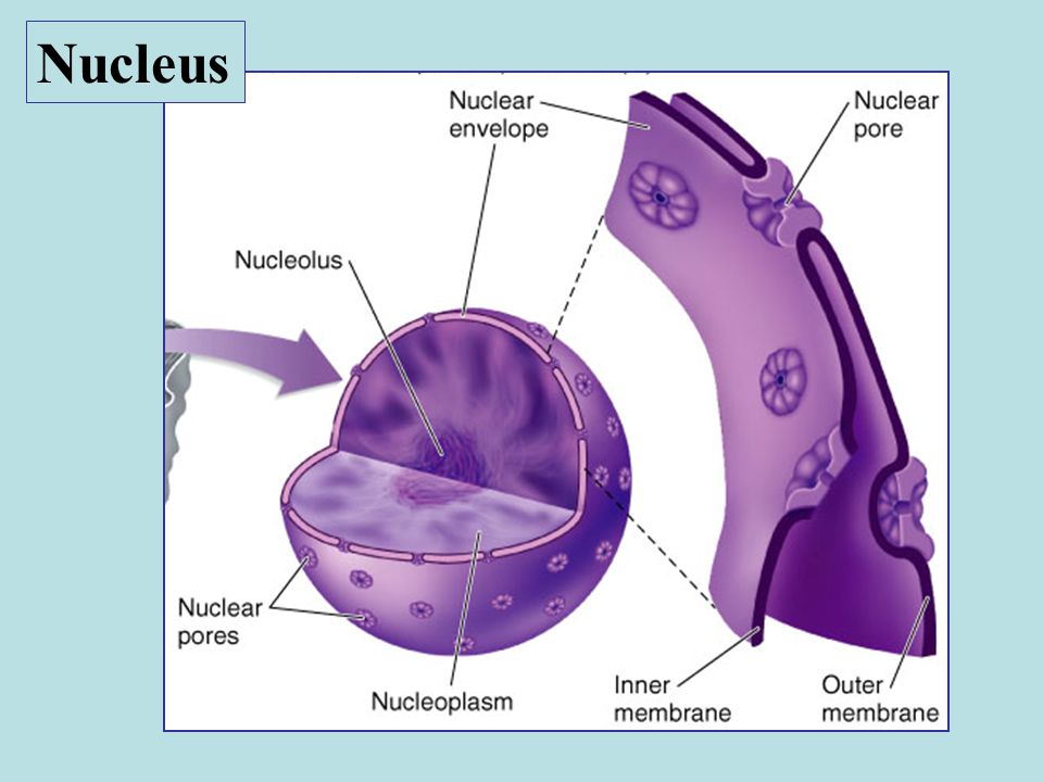
25
Close-up of nuclear envelope Nucleus Nucleolus Chromatin Nuclear envelope: Inner membrane Outer membrane Nuclear pore Pore complex Ribosome Pore complexes (TEM)Nuclear lamina (TEM) 1 µm Rough ER Nucleus 1 µm 0.25 µm Surface of nuclear envelope Nucleus
Nuclear lamina (TEM) 1 µm Rough ER Nucleus 1 µm 0.25 µm Surface of nuclear envelope Nucleus")
28
Ribosomes 0.5 µm ER Cytosol Endoplasmic reticulum (ER) Free ribosomes Bound ribosomes Large subunit Small subunit Diagram of a ribosome TEM showing ER and ribosomes Ribosomes
 Free ribosomes Bound ribosomes Large subunit Small subunit Diagram of a ribosome TEM showing ER and ribosomes Ribosomes")
30
Smooth ER Rough ER ER lumen Cisternae Transport vesicle Smooth ER Rough ER Transitional ER 200 nm Nuclear envelope Endoplasmic reticulum

31
Endoplasmic reticulum smooth ER and rough ER

33
Golgi apparatus Glycosylation, sorting and distribution of proteins and lipids

34
Golgi apparatus Camillo Golgi (1843 – 1926)
")
36
1 µm Lysosome Nucleus Phagocytosis: lysosome digesting food Plasma membrane Food vacuole Digestive enzymes Digestion Lysosome Vesicles

38
5 µm Central vacuole Cytosol Tonoplast Central vacuole Nucleus Cell wall Chloroplast Vacuoles

39
Contractile Vacuole

40
Nuclear envelope Nucleus Rough ER Smooth ER

41
Nuclear envelope Nucleus Rough ER Smooth ER Transport vesicle cis Golgi trans Golgi

42
Nuclear envelope Nucleus Rough ER Smooth ER Transport vesicle cis Golgi trans Golgi Plasma membrane

43
Organelles

44
Mitochondrion Chloroplasts

45
Mitochondrion Intermembrane space Outer membrane Inner membrane Cristae Matrix 100 nm Mitochondrial DNA Free ribosomes in the mitochondrial matrix Mitochondria

46
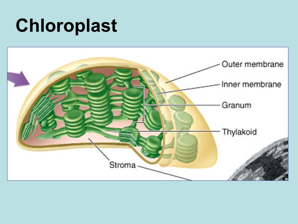
Chloroplast DNA Ribosomes Stroma Inner and outer membranes Granum Thylakoid 1 µm Chloroplast

48
Cytoskeleton microfilament

49
8-10 nm 25 nm 7 nm Cytoskeleton

50
Microtubules

51
Microfilaments

52
Microfilaments (actin filaments) Microvillus Plasma membrane Intermediate filaments 0.25 µm Lining of intestine Microfilaments
 Microvillus Plasma membrane Intermediate filaments 0.25 µm Lining of intestine Microfilaments")
53
Intermediate filaments

54
Extracellular components

55
Central vacuole of cell Plasma membrane Secondary cell wall Primary cell wall Middle lamella 1 µm Central vacuole of cell Central vacuole Cytosol Plasma membrane Plant cell walls Plasmodesmata Plant Cell Walls

56
Plant Cells cellulose

57
Animal Cell exterior Extra cellular matrix

58
Extracellular components: intercellular junctions

59
Interior of cell Interior of cell 0.5 µm PlasmodesmataPlasma membranes Cell walls Plasmodesmata: found in plants

60
Tight junctions prevent fluid from moving across a layer of cells Tight junction 0.5 µm 1 µm 0.1 µm Gap junction Extracellular matrix Space between cells Plasma membranes of adjacent cells Intermediate filaments Tight junction Desmosome Gap junctions Animals tight junctions, desmosomes and gap junctions

61
Tight junctions

62
Desmosome

63
Gap junctions

Similar presentations


 date = Wednesday November 12 Test Date= Friday November 14.>")


 I. Cell Background II. Organelles>")


>")
