Download presentation
Presentation is loading. Please wait.

1
Vision

2
Vision 1: Filling-in, Color, Motion, Form Visual Paths Filling-In –Perceptual Completion –Conceptual Completion Color Motion Form –Agnosia –Prosopagnosia

4
Filling-In Usually, function of a brain area is deduced from deficits correlated with damage to that area In the case of filling-in, brain function is deduced from intact abilities (perceptions) in the absence of sensory input
 in the absence of sensory input")
5
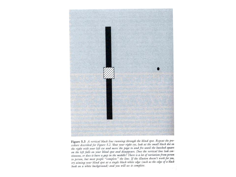
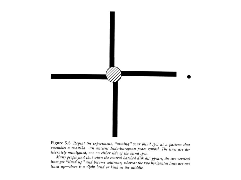
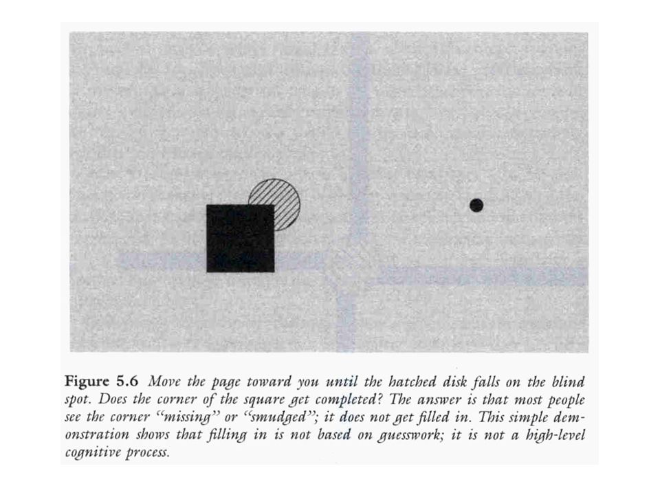
Filling-In: Perceptual Completion

10
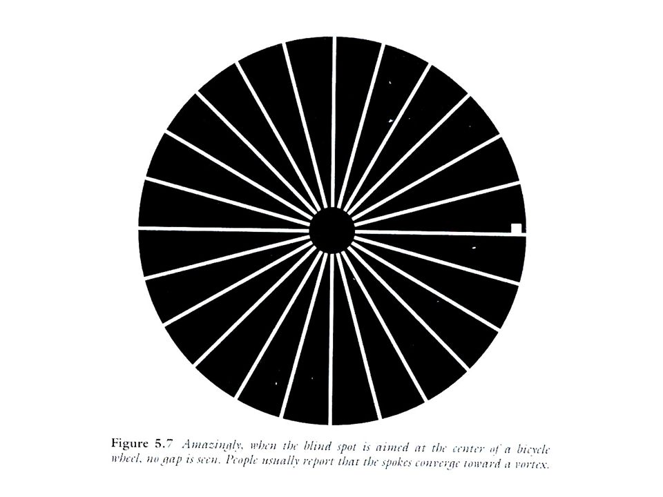
Perceptual Completion in Normals 1.Probably due to excitatory horizontal connections in V1

11
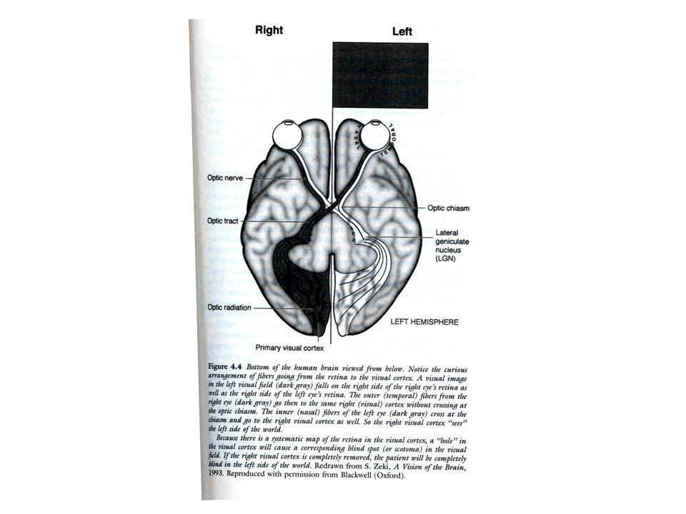
Filling-In Resulting From V1 Damage From scotomas (“hole” in V1) Hemianopias –***DIGITAL VIDEO: Hemianopia
 Hemianopias –***DIGITAL VIDEO: Hemianopia")
12
Perceptual Filling-In Ramachandran patient: Filled in texture/surfaces but not objects Filled in numbers but looked like hieroglyphics/couldn’t identify Filling in occurs at different speeds for different perceptual attributes Couldn’t fill in faces

13
Lessons From Filling In: Perceptual Completion 1.Brain uses statistical regularities to fill in. 2.This act of interpolation saves an enormous amount of computation. 3.Perhaps due to lateral horizontal connections in cortical areas higher than V1?

14
Conceptual Completion Additional parietal damage

15
Charles Bonnet Syndrome

16
Patients Know Hallucinations Aren’t Real Because: 1.Others correct them 2.Fade after a few seconds 3.Highly improbably 4.Something odd about the images (too vivid, cartoonish, etc.)
")
19
Lessons From Filling-In: Conceptual Completion Parietal Damage? Back-Projections?

21
Disorders of Color Processing

22
Central Achromatopsia Deficit in color perception caused by an acquired cerebral lesion

23
Tested With Color plate test –(e.g., Ishihara Color Plate Test) Color arrangement test –(e.g., Farnsworth-Munsell 100-Hue Test) Pass color chips across the visual field
 Color arrangement test –(e.g., Farnsworth-Munsell 100-Hue Test) Pass color chips across the visual field")
24
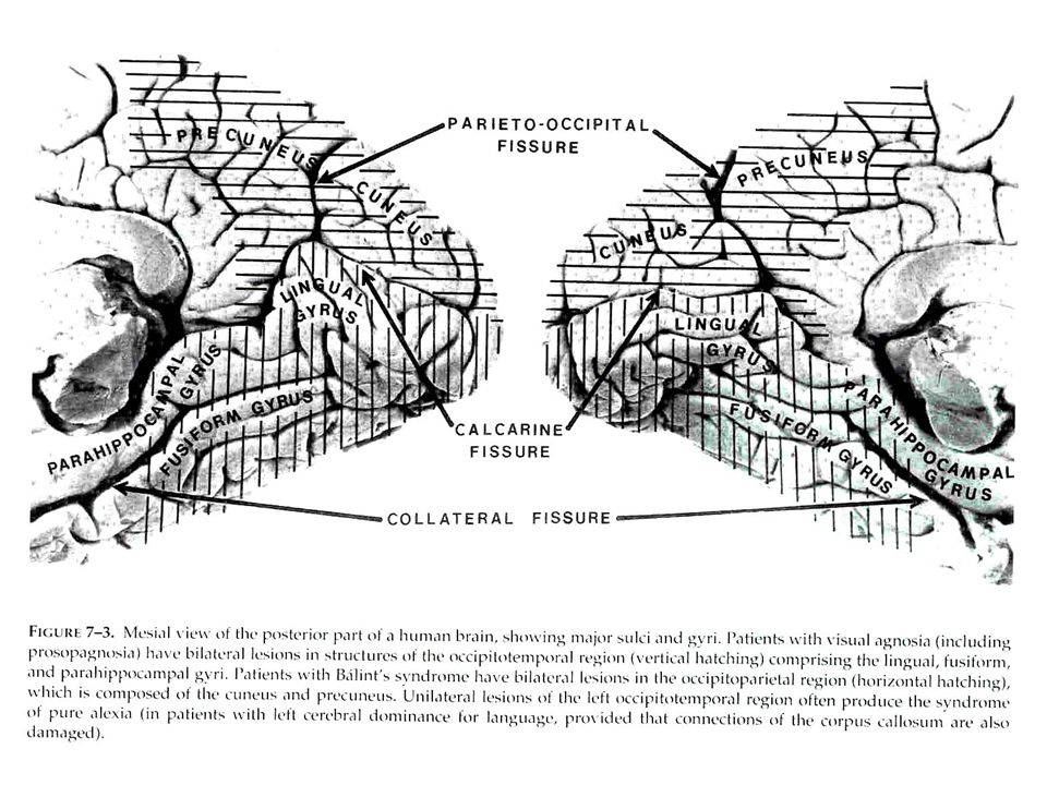
Central Achromatopsia – Disorder of Color Perception Nature of the impairment uncertain –Reduced hue discrimination –Deficient color constancy Co-occurs often with alexia or visual agnosia V4 damage most likely site –Lingual gyrus, fusiform gyrus, or white matter between the regions

25
V4 Damage

26
Disorder of Color Imagery Seems that defective color perception invariably results in defective color imagery Imagining an object’s color (e.g., a yellow banana) requires an least some of the neural representations required to perceive color Patient cannot “remember” the color of items that need to be imagined
 requires an least some of the neural representations required to perceive color Patient cannot remember the color of items that need to be imagined")
27
Color Agnosia – Disorder Of Color Recognition Perform fine on color matching tasks Exhibit errors in matching colors to objects May still have semantic knowledge about colors Not yet well-distinguished from color perception disorder –Behavioral manifestations –Site of damage

28
Cerebral Akinetopsia: Motion Blindness

29
Deficit of motion processing caused by acquired cerebral lesions Because motion cues serve many purposes, a range of deficits can result –E.g., Difficulty using motion to find objects (structure for motion or kinetic depth) Pursuit eye movements impaired
 Pursuit eye movements impaired")
30
L.M. Case Description Could see slowly moving targets Faster ones materialized at successive positions with no movement in between Did not perceive apparent motion Reduced perception of motion after-effects Saw changes in position not depth for objects moved towards her ***Motion after-effect illusion for demo

31
L.M. Case Description Good static visual acuity perception Perception of tactile and acoustic motion Accurate localization of visual targets by saccadic eye movements No visual field defect for form No neglect of visual targets flashed simultaneously in both hemi field Relative preservation of face and object recognition, reading, and color vision

32
V5 Damage

33
Motion Blindness: Neuroanatomical Locus Damage –Parietal-temporo-occipital, near angular gyrus –Parieto-occipital –As part of a more pervasive disturbance (Balint's syndrome or Alzheimer's disease) L M and others: superior temporoparietal –Includes the cortical areas of 19 and 37, which are adjacent (may resemble monkeys area MT / V5) The homologies between motion processing areas in monkey and human may not be as close as they once appeared. Severe deficits of motion perception can also occur with lesions in parietal insula and midline cerebellum

34
WHAT, WHERE, & HOW SYSTEMS

35
What, Where, & How Systems

36
What

37
Visual Agnosia

38
Visual Object Agnosia Apperceptive Associative

39
Apperceptive Agnosia Intact vision: –Acuity, brightness discrimination, color vision, & other elementary visual capabilities –Sometimes preserved shape from motion Deficits: –Abnormal shape perception (pictures, letters, simple shapes) –Grouping process deficit (that operates over an array of local features representing contour, color, depth, etc.)
 –Grouping process deficit (that operates over an array of local features representing contour, color, depth, etc.)")
40
Apperceptive Agnosia VIDEO: Apperceptive Agnosia, impaired triangle recognition, subject 1 VIDEO: Apperceptive Agnosia, impaired object recognition, subject 1 VIDEO: Object Agnosia 2: Impaired Visual but not tactile identification (naming), subject 2 VIDEO: Object Agnosia 3: Intact visual movement identification, subject 2 VIDEO: Object Agnosia 1: Impaired Visual identification (subject given name & array of objects), can’t see objects
, subject 2 VIDEO: Object Agnosia 3: Intact visual movement identification, subject 2 VIDEO: Object Agnosia 1: Impaired Visual identification (subject given name & array of objects), can’t see objects")
41
Associative Agnosia Cannot recognize objects by sight alone Intact general knowledge of objects Can recognize objects by touch or definition Visual perception better than in apperceptive agnosia Not a naming deficit (cannot indicate recognition by nonverbal means)
")
42
Theories of Associative Agnosia 1.Disconnection between visual representations and language areas 2.Disconnection between visual representations and memory areas 3.Stored visual memories have been damaged 4.A perceptual and memory problem, and the two are inseparable

43
Intertwined Perception & Memory Some visual problems Copying drawings on line by line On matching tasks, they rely on slow, sequential featured-by-feature checking In the PDP system, the memory of the stimulus would consist of a pattern of connections strengths among a number of neuron like units. The " perceptual" representation resulting from presentation of the stimulus will depend upon the pattern of connection strengths among the units directly or indirectly activated by the stimulus. Thus, if a memory is altered by damaging the network, perception will be altered as well. Thus, Associative Agnosia may not be the results of an impairment to perception or to memory; rather, the two are in principle inseparable, and the impairment is better described as a loss of high level visual perceptual representations that were shaped by, and embody the memory of, visual experience.

44
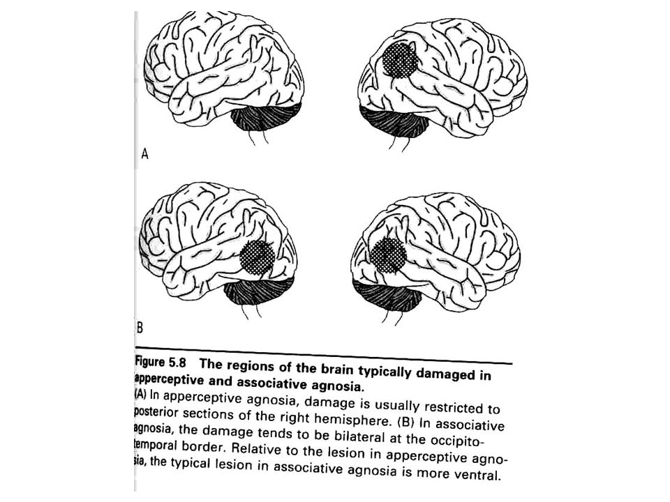
Apperceptive: Localization of Damage Diffuse brain damage, often from carbon monoxide poisoning

45
Apperceptive Associative

46
Associative Agnosia

50
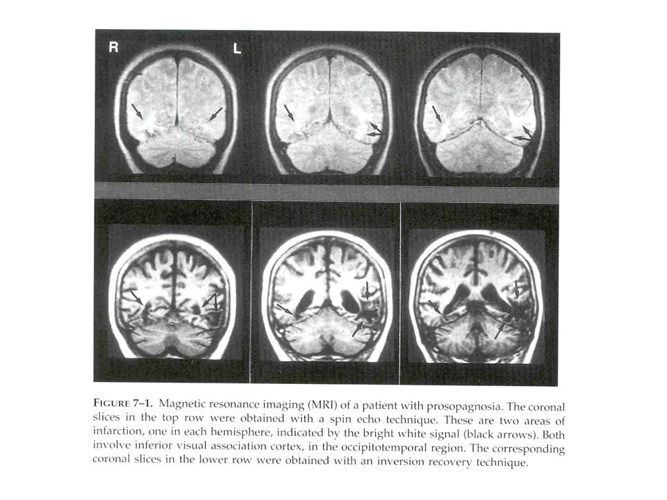
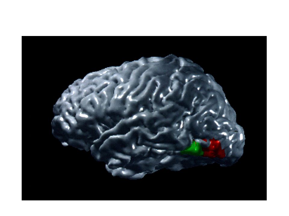
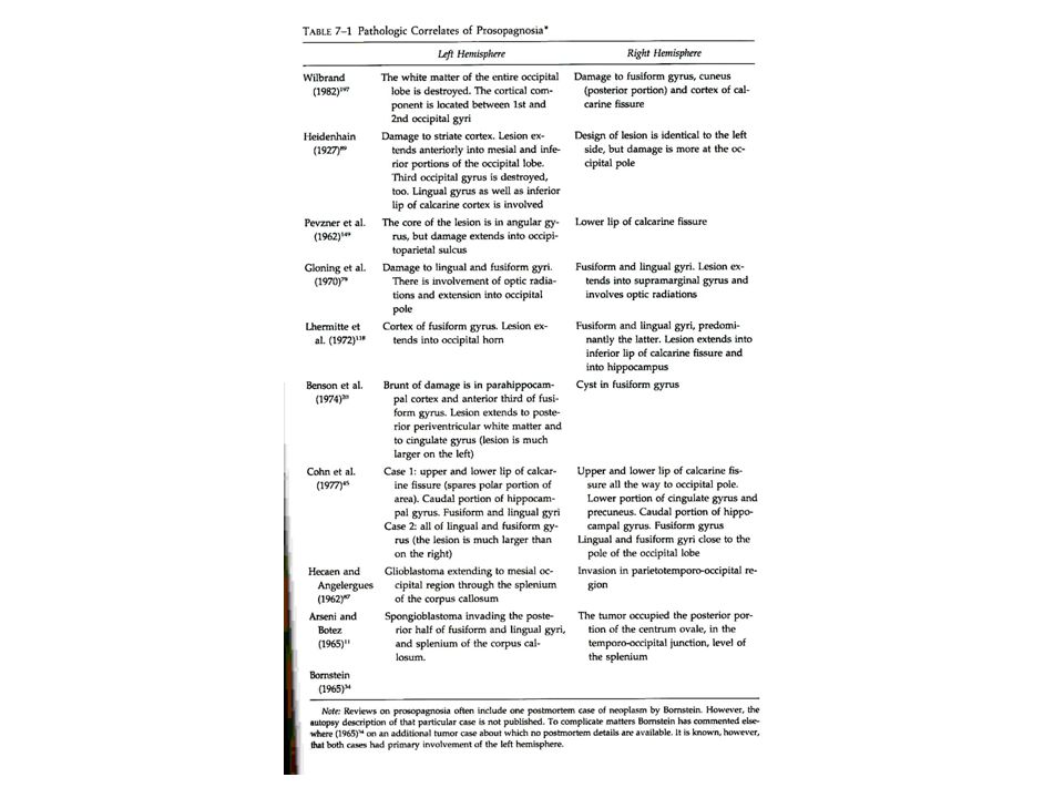
Prosopagnosia

51
Compensate by relying on nonfacial cues (voice, gait, clothing..) With a few exceptions, they can discriminate a face’s gender, ethnicity, approximate age, and emotion conveyed. Patients who do not have problems recognizing faces may have difficulty recognizing the emotion.

52
Matching Faces Task

53
Test of Famous Faces

54
Skin Conductance Response (SCR)
")
55
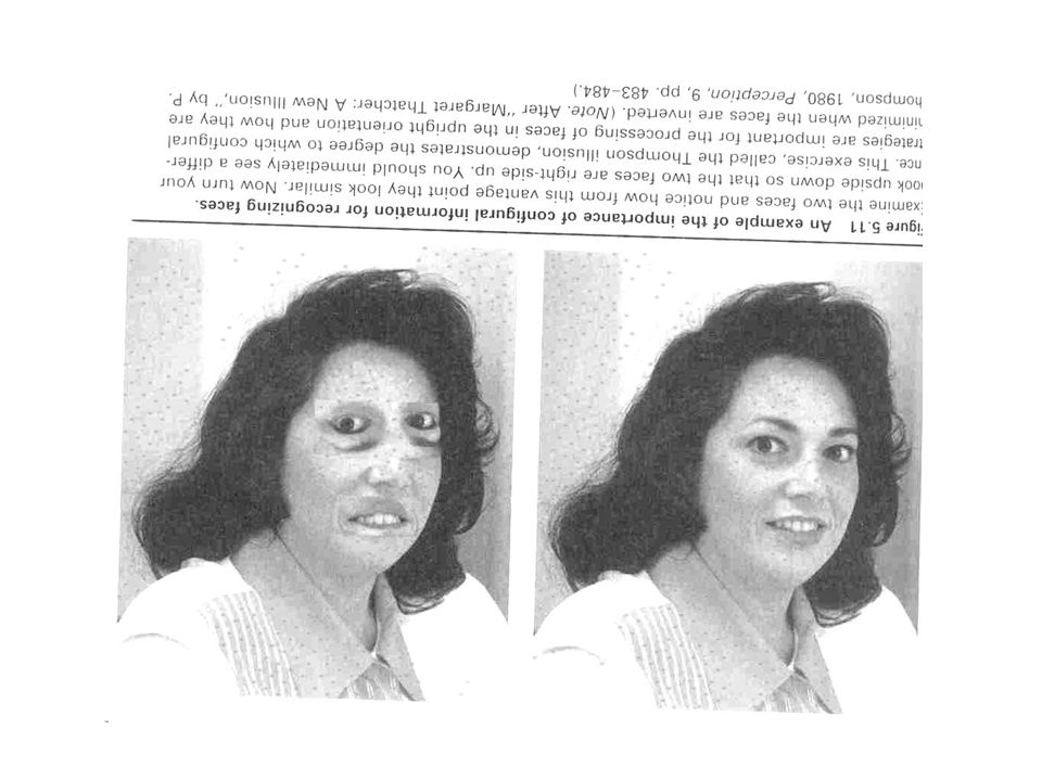
Farah Ch. 7: Are Faces Really Unique?

60
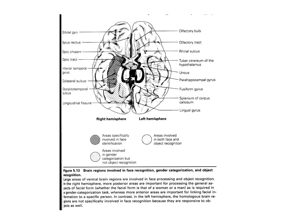
Types Of Agnosia Face Object Printed Word Face, or face and object -- right or bilateral Word, or word and object – left Maximum overlap in left inferior medial region (including parahippocampal, fusiform, and lingual gyri)
")
62
Capgras Patients have both left and right hemisphere damage Possible Damage Sites: – Disconnection between IT & amygdala (limbic system, emotion)
")
Similar presentations


![Sensory systems in the brain The visual system. Organization of sensory systems PS 103 Peripheral sensory receptors [ Spinal cord ] Sensory thalamus Primary.](/11/3241431/big_thumb.jpg "Sensory systems in the brain The visual system. Organization of sensory systems PS 103 Peripheral sensory receptors [ Spinal cord ] Sensory thalamus Primary.>")


 of the inferior temporal cortex lead to disorders of memory for people and things.>")

 23, 571 - 579 Hint #1: there are at least 3 ways of getting this article Hint #2: none.>")


