Download presentation
Presentation is loading. Please wait.

1
Chapter 7 Structure of Nucleic Acids
"Scherzo in D & E" (detail) by David E. Rodale ( )
 by David E. Rodale ( )")
2
7.1 · The Primary Structure of Nucleic Acids

3
Structure and Nomenclature of Nucleic Acids
Shorthand notation of a nucleic acid 5’ P T A G C OH 5’ 3’ Two major classes of nucleic acids: DNA and RNA 3’

4
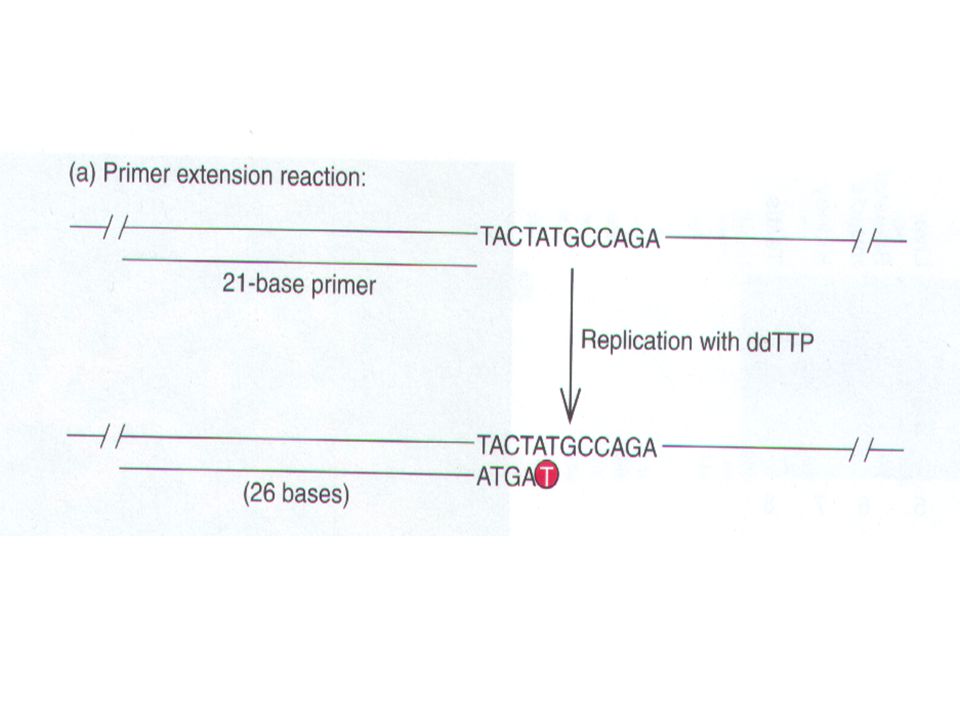
Sequencing Nucleic Acids
1 chain termination or dideoxy method base-specific chemical cleavage

5
DNA sequencing The Sanger DNA sequencing method uses dideoxy nucleotidcs to terminate DNA synthesis, yielding a series of DNA fragments whose sizes can be measured by electrophoresis. The last base in each of these fragments is known, because we know which dideoxy nucleotide was used to terminate each reaction. Therefore, ordering these fragments by size--each fragment one (known) base longer than the next--tells us the base sequence of the DNA.
 base longer than the next--tells us the base sequence of the DNA.")
11
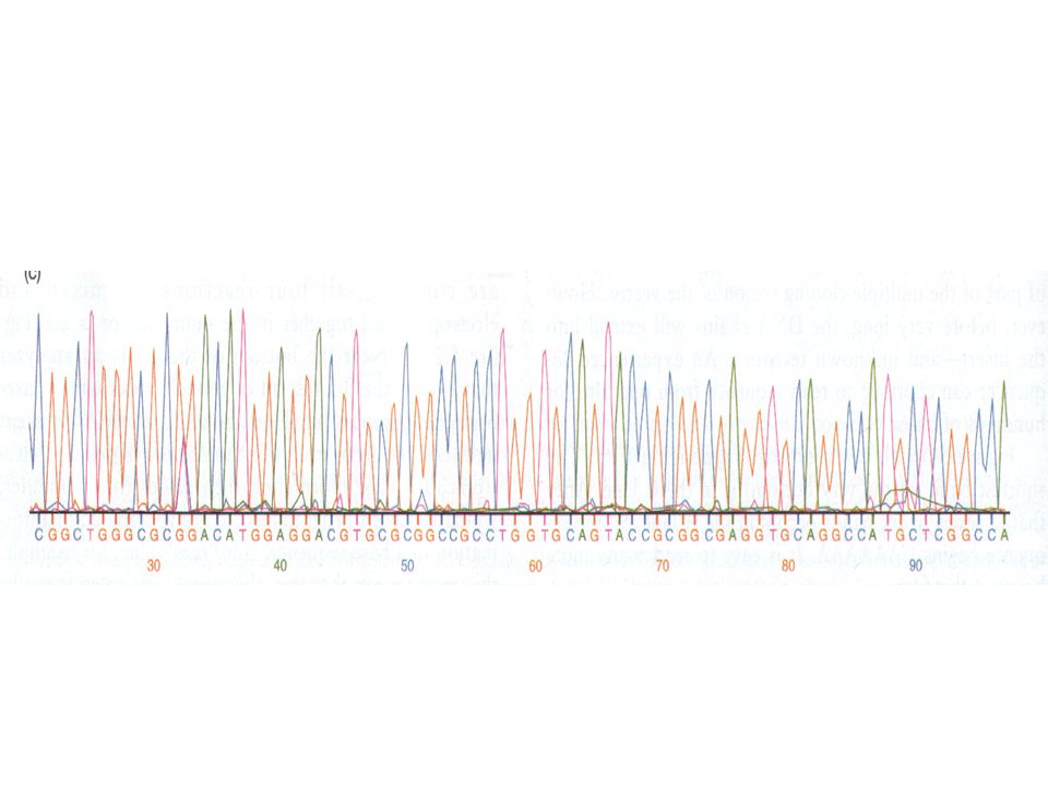
DNA sequencing can be automated

13
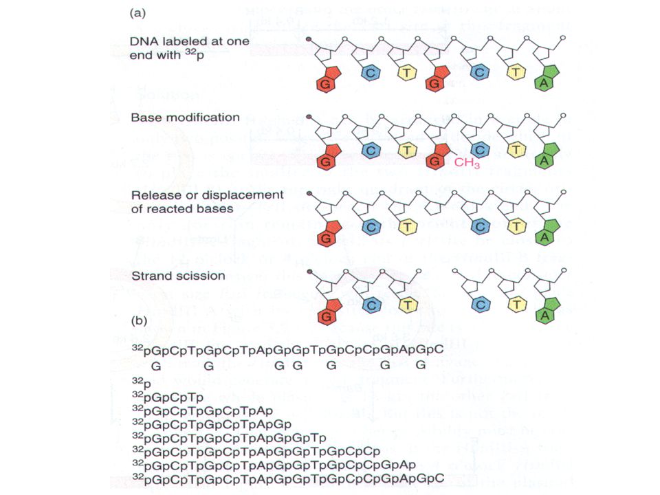
Maxam-Gilbert Sequencing
The Maxam-Gilbert sequencing method follows a very different strategy. Instead of synthesizing DNA in vitro and stopping the synthesis reactions with chain terminators, this method starts with full length, end-labeled DNA and cleaves it with base-specific reagents

15
TO G We use dimethyl sulfate (DMS) to methylate guanines. we do the methylation under mild conditions that lead to an average of only one methylated guanine per DNA strand. Next, we use a reagent (piperidine,哌啶) that does two things: It causes loss of the mcthylated base, then it breaks the DNA backbone at the site of the lost base (the apurinic site).
 to methylate guanines. we do the methylation under mild conditions that lead to an average of only one methylated guanine per DNA strand. Next, we use a reagent (piperidine,哌啶) that does two things: It causes loss of the mcthylated base, then it breaks the DNA backbone at the site of the lost base (the apurinic site).")
17
TO A+G we can weaken the glycosidic bonds to both adenine and guanine with acid; then piperidine will cause depurination and strand breakage after both A's and G's.

18
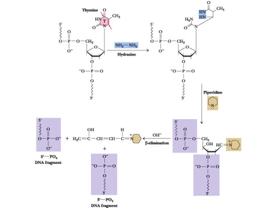
TO C+T Similarly, hydrazine(肼)opens both thymine and cytosine rings, and piperidine can then remove these bases and break the DNA strand at the resulting apyrlmidinic sites. TO C In the presence of 1 M NaC1, hydrazine is specific for cytosine only, so we can run this reaction next to the C + T reaction and obtain the T's by comparison.
opens both thymine and cytosine rings, and piperidine can then remove these bases and break the DNA strand at the resulting apyrlmidinic sites. TO C. In the presence of 1 M NaC1, hydrazine is specific for cytosine only, so we can run this reaction next to the C + T reaction and obtain the T s by comparison.")
20
Maxam-Gilbert sequencing is used only in special cases these days, but the methods of modifying and then breaking DNA used by Maxam and Gilbert are still useful for other purposes

21
7.2 · The ABZs of DNA Secondary Structure
Structure Overview of Nucleic Acids Unlike three dimensional structures of proteins, DNA molecules assume simple double helical structures independent on their sequences. There are three kinds of double helices that have been observed in DNA: type A, type B, and type Z, which differ in their geometries. The double helical structure is essential to the coding functional of DNA. Watson (biologist) and Crick (physicist) first discovered double helix structure in 1953 by X-ray crystallography. RNA, on the other, can have as diverse structures as proteins, although they can also hand form double helix of type A. The ability of being both informational and diverse in structure suggests that RNA was the prebiotic molecule that could function in both replication and catalysis (The RNA World Hypothesis). In fact, some virus encode their genetic materials by RNA (retrovirus)
 and Crick (physicist) first discovered double helix structure in 1953 by X-ray crystallography. RNA, on the other, can have as diverse structures as proteins, although they can also hand form double helix of type A. The ability of being both informational and diverse in structure suggests that RNA was the prebiotic molecule that could function in both replication and catalysis (The RNA World Hypothesis). In fact, some virus encode their genetic materials by RNA (retrovirus)")
22
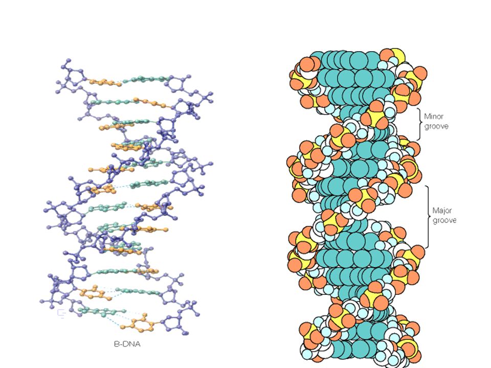
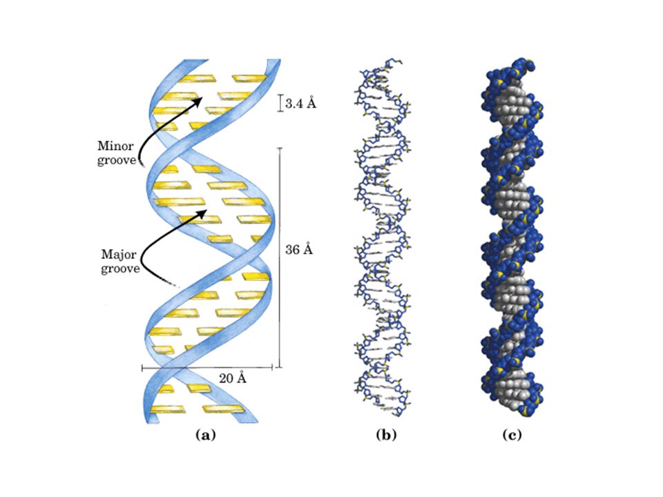
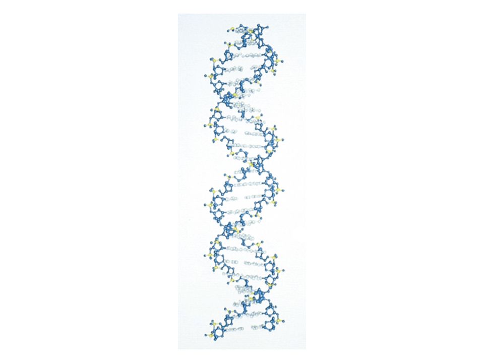
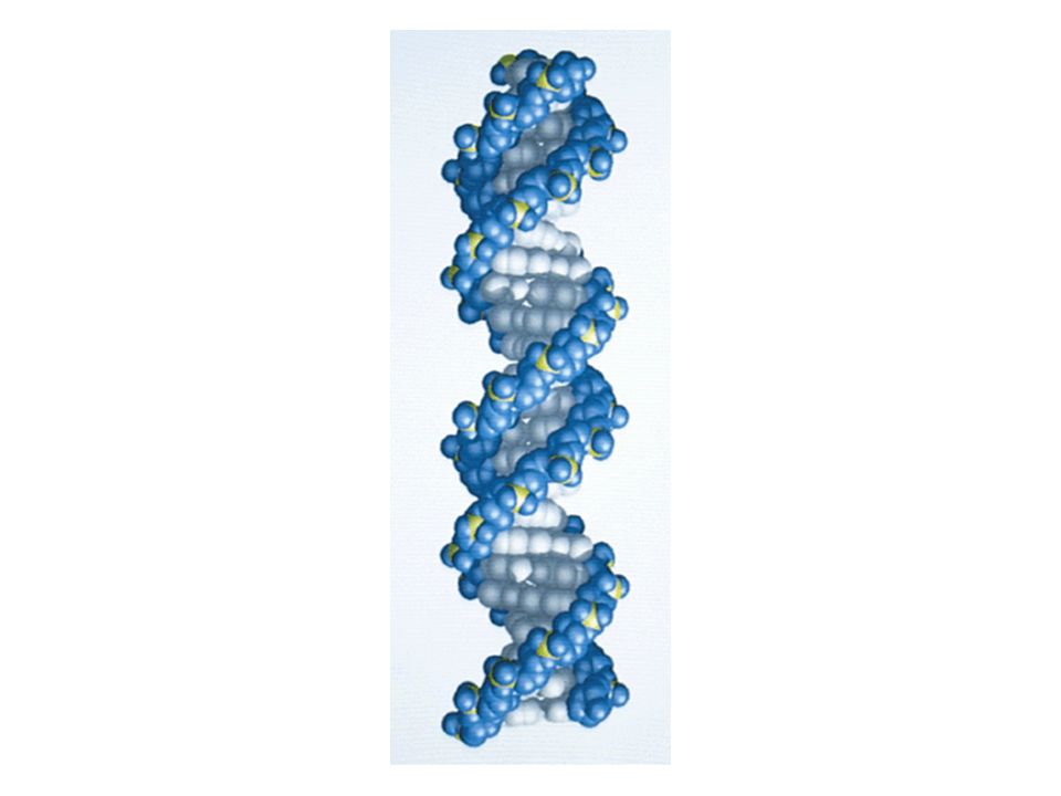
Structure of DNA DNA is a double helix

25
Structural Equivalence of Watson–Crick Base Pairs ;
the A:T pair and G:C pair have virtually identical dimensions.

29
The DNA Double Helix Is a Stable Structure
Forces That Stabilize Nucleic Acid Double Helix Hydrogen bonding in base-pairing Hydrophobic interactions in base stacking Electrostatical Interaction with cations in solution such as Mg2+. 5’ 3’ Same strand stacking cross-strand stacking

30
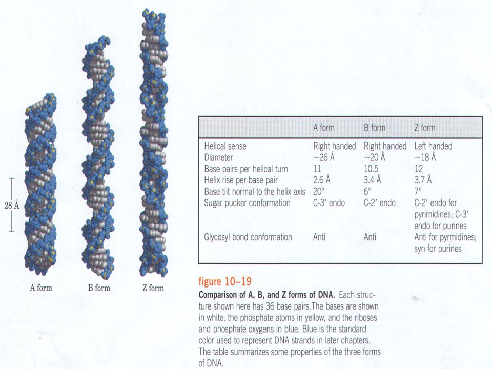
Types of DNA Double Helix
Type A: major conformation of RNA, minor conformation of DNA; Type B: major conformation of DNA; Type Z: minor conformation of DNA 5’ 3’ 5’ 5’ 3’ 3’ Z A B 3’ 5’ Narrow tight Wide Less tight Left-handed Least tight

31
Types of DNA Double Helix
A-DNA A-RNA Minor Groove Major Groove

33
Comparison of the Structural Properties of A-, B-, and Z-DNA

34
Edges of Base-paired Nucleotides
Edges of basepairs have specific relationships to grooves; Major groove edges are sequence specific thus provide sequence recognition sites; Many DNA-binding proteins bind to major grooves of DNA in gene transcription and regulation;

35
Intercalating Agents Distort the Double Helix
Several hydrophobic molecules containing flat aromatic and fused heterocyclic rings can insert between the stacked base pairs of DNA. These molecules are called intercalating agents. Intercalating agents are potential Cancer-inducing reagents.

36
7.3 Denaturation and Renaturation of DNA
When duplex DNA molecules are subjected to conditions of pH, temperature or ionic strength that disrupt hydrogen bonds, the strands are no longer held together. The double helix is denatured. If the temperature is the denaturing agent, the double helix is said to melt; The phenomenon that the relative absorbance of the DNA solution at 260 nm increases as the bases unstack is called hyperchromic shift; If one fellows the absorbance as a function of temperature, the midpoint temperature of the absorbance curve is termed melting temperature, Tm.

37
Denaturation and Renaturation of DNA
DNA of different sequences have different Tm. Tm is higher for DNA that contain more GC pairs; Tm is also directly proportional to the ionic strength of the solution. (salts can shield repulsions of negatively charged phosphate groups) Tm
 Tm.")
38
Structural Changes in DNA Melting

39
pH Extremes or Strong H-Bonding Solutes Also Denature DNA Duplexes At pH values greater than 10, extensive deprotonation of the bases occurs, destroying their hydrogen bonding potential and denaturing the DNA duplex. Similarly, extensive protonation of the bases below pH 2.3 disrupts base pairing. Alkali is the preferred denaturant because, unlike acid, it does not hydrolyze the glycosidic linkages in the sugar–phosphate backbone. Small solutes that readily form H bonds are also DNA denaturants at temperatures below Tm if present in sufficiently high concentrations to compete effectively with the H-bonding between the base pairs. Examples include formamide and urea.

40
Renaturation (or Annealing)
Renaturation refers to the process of DNA strands associate into a double helix; Renaturation can be analyzed quantitatively: C is the amount of single stranded DNA remaining, C0 is the initial single stranded DNA. C0t Plot t1/2

41
Nucleic Acid Hybridization
If DNA from two different species are mixed, denatured, and allowed to cool slowly so that reannealing can occur, artificial hybrid duplexes may form, provided the DNA from one species is similar in nucleotide sequence to the DNA of the other.

43
Nucleic acid hybridization is a commonly employed procedure in molecular biology. First, it can reveal evolutionary relationships. Second, it gives researchers the power to identify specific genes selectively against a vast background of irrelevant genetic material. An appropriately labeled oligo- or polynucleotide, referred to as a probe, is constructed so that its sequence is complementary to a target gene. The probe specifically base pairs with the target gene, allowing identification and subsequent isolation of the gene. Also, the quantitative expression of genes (in terms of the amount of mRNA synthesized) can be assayed by hybridization experiments.
 can be assayed by hybridization experiments..")
44
Buoyant Density of DNA Not only the melting temperature of DNA but also its density in solution is dependent on relative G:C content. G:C-rich DNA has a significantly higher density than A:T-rich DNA. Single-stranded DNA is denser than double-helical DNA

46
Cesium chloride centrifugation is an excellent means of removing RNA and proteins in the purification of DNA. Because of its relatively high density, DNA can be purified from cellular material by a form of density gradient centrifugation known as isopycnic centrifugation.

47
7.4 · Supercoils and Cruciforms: Tertiary Structure in DNA
Many DNA molecules are circular (e.g., bacterial chromosomes, all plasmid DNA). Circular DNA can form supercoils. Human chromosome contains 3x10⁹ basepairs and are wrapped around proteins to form nucleosomes. Nucleosomes are packed tightly to form helical filament, a structure called chromotin.
. Circular DNA can form supercoils. Human chromosome contains 3x10⁹ basepairs and are wrapped around proteins to form nucleosomes. Nucleosomes are packed tightly to form helical filament, a structure called chromotin.")
48
Supercoiling means the coiling of coiling
DNA Supercoiling Supercoiling means the coiling of coiling

49
Supercoiling of DNA Relaxed state

50
Supercoiling induced by separating the strands of a helical structure(during replication or transcription)
")
51
Supercoils: Tertial Structure in DNA
Supercoils refer to the DNA structure in which double-stranded circular DNA twists around each other. Supercoiled DNA contrasts relaxed DNA; In DNA replication, the two strands of DNA have to be separated, which leads either to overwinding of surrounding regions of DNA or to supercoiling; A specialized set of enzymes (gyrase, topoisomerases) is present to introduce supercoils that favor strand separation; The degree of supercoils can be quantitatively described.
 is present to introduce supercoils that favor strand separation; The degree of supercoils can be quantitatively described.")
52
DNA from a lysed E..coli cell

53
Relaxed and supercoiled plasmid DNAs
Relaxed and supercoiled plasmid DNAs. The degree of supercoiling increases from left to right.

54
Varieties of Supercoiled DNA
Note that all black lines represents double stranded DNA

55
DNA underwinding is defined by topological linking number(Lk, or L).
.")
56
The Linking Number (L) of DNA
The linking number of DNA, a topological property, determines the degree of supercoiling; The linking number defines the number of times a strand of DNA winds in the right-handed direction around the helix axis when the axis is constrained to lie in a plane; If both strands are covalently intact, the linking number cannot change; For instance, in a circular DNA of 5400 basepairs, the linking number is 5400/10=540, where 10 is the basepair per turn for type B DNA.

57
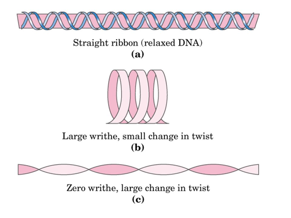
The Twist (T) and The Writhe of DNA
Twist is a measure of the helical winding of the DNA strands around each other. Given that DNA prefers to form B-type helix, the preferred twist = number of basepair/10; Writhe is a measure of the coiling of the axis of the double helix. A right-handed coil is assigned a negative number (negative supercoiling) and a left-handed coil is assigned a positive number (positive supercoiling). Topology theory tells us that the sum of T (or Tw)and W (or Wr)equals to linking number: L=T+W For example, in the circular DNA of 5400 basepairs, the linking number is 5400/10=540 If no supercoiling, then W=0, T=L=540; If positive supercoiling, W=+20, T=L-W=520;
 and a left-handed coil is assigned a positive number (positive supercoiling). Topology theory tells us that the sum of T (or Tw)and W (or Wr)equals to linking number: L=T+W. For example, in the circular DNA of 5400 basepairs, the linking number is 5400/10=540. If no supercoiling, then W=0, T=L=540; If positive supercoiling, W=+20, T=L-W=520;")
60
An Example of L, T, and W Calculation
A relaxed circular, double stranded DNA (1600 bps) is in a solution where conditions favor 10 bps per turn. What are the L, T, and W? During replication, part of this DNA unwinds (200 bps) while the rest of the DNA still favor 10 bps per turn. What are the new L, T, and W? 1400 bps 1600 bps 200 bps L=160 T=( )/10=140 W=L-T=+20 L=1600/10=160 W=0 (relaxed) T=L-W =160
 is in a solution where conditions favor 10 bps per turn. What are the L, T, and W During replication, part of this DNA unwinds (200 bps) while the rest of the DNA still favor 10 bps per turn. What are the new L, T, and W 1400 bps bps. 200 bps. L=160. T=( )/10=140. W=L-T=+20. L=1600/10=160. W=0 (relaxed) T=L-W =160.")
61
Wr=-2 Wr=2 Tw=200

62
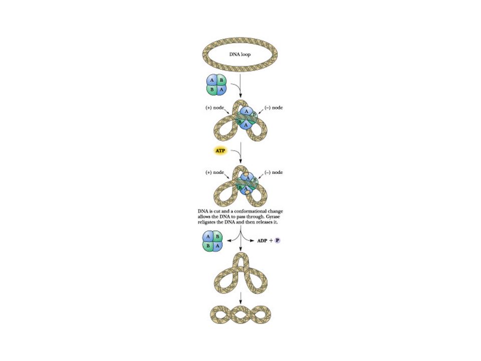
DNA Gyrase The bacterial enzyme DNA gyrase is a topoisomerase that introduces negative supercoils into DNA

64
Mechanism of topoisomerase II (DNA gyrase)
")
65
Topoisomerases catalyze changes in the linking number of DNA
Topoisomerase I treats in different length of time

66
Superhelix Density The superhelix density or specific linking difference is defined as L/L0 and is sometimes termed sigma, s . For our example, s = -4/40, or As a ratio, s is a measure of supercoiling that is independent of length. Its sign reflects whether the supercoiling tends to unwind (negative s ) or overwind (positive s ) the helix.
 or overwind (positive s ) the helix.")
67
Toroidal Supercoiled DNA
Negatively supercoiled DNA can arrange into a toroidal state. The toroidal state of negatively supercoiled DNA is stabilized by wrapping around proteins which serve as spools for the DNA “ribbon.” This toroidal conformation of DNA is found in protein: DNA interactions that are the basis of phenomena as diverse as chromosome structure and gene expression.

68
The toroidal state of negatively supercoiled DNA is stabilized by wrapping around proteins which serve as spools for the DNA “ribbon.”

69
palindrome is a word, phrase,or sentence that is spelled identically reading forward or backward; two examples are ROTATOR and NURSES RUN..

70
Such sequence are self-complementary within each strand and therefore have the potential to form hairpin or cruciform (cross-shaped) structures
 structures")
71
Cruciforms

72
DNA compaction requires a special form of supercoiling There are two types of supercoiling: plectonemic (from the Greek plektoe, “twisted”, and nema, “thread”) and solenoidal.
 and solenoidal.")
73
Plectonemic Supercoiling An electron micrograph of
plectonemically supercoiled plasmid DNA

74
(b) An interpretation of the observed structure
(b) An interpretation of the observed structure. The purple lines show the axis of the supercoil.
 An interpretation of the observed structure. The purple lines show the axis of the supercoil.")
75
(c) An indealized representation of this structure
 An indealized representation of this structure")
76
plectonemic and solenoidal supercoiling.

77
7.5 · Chromosome Structure
Human chromosomes

78
Chromatin and nucleoid structure
nucleosomes

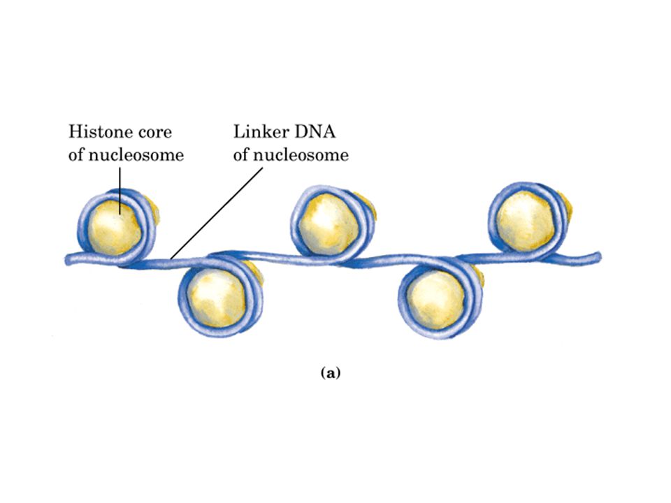
79
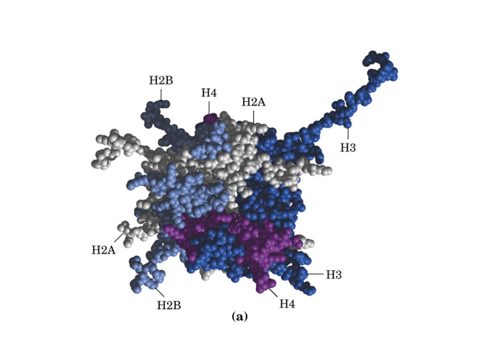
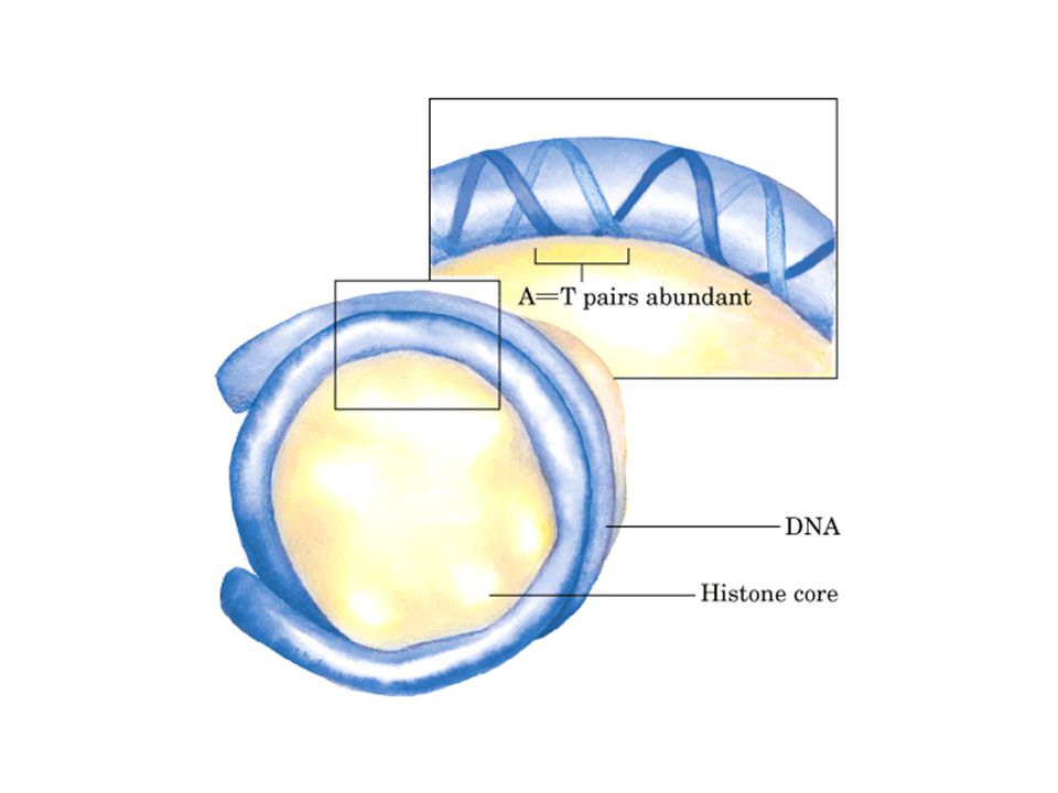
Nucleosomes Nucleosomes look like “beads on a string” under microscope. The beads contain a pair of four histone proteins, H2A, H2B, H3, and H4 (octamer). The string is double stranded DNA; The surface of the octamer contain features that guide the course of DNA such that DNA can wrap 1.65 turns around in a left-handed conformation. H1 proteins serves to seal the ends of the DNA and connects consecutive nucleosomes. nucleosomes
. The string is double stranded DNA; The surface of the octamer contain features that guide the course of DNA such that DNA can wrap 1.65 turns around in a left-handed conformation. H1 proteins serves to seal the ends of the DNA and connects consecutive nucleosomes. nucleosomes.")
82
Histones are small, basic proteins

83
Each of histones has variant forms because certain amino acid side chains are enzymatically modified by methylation, ADP-ribosylation, phosphorylation, or acetylation. Such modifications affact the net electric charge, shape, and other properties of histones, as well as the structural and functional properties of chromatin, and they play a role in the regulation of transcription.

84
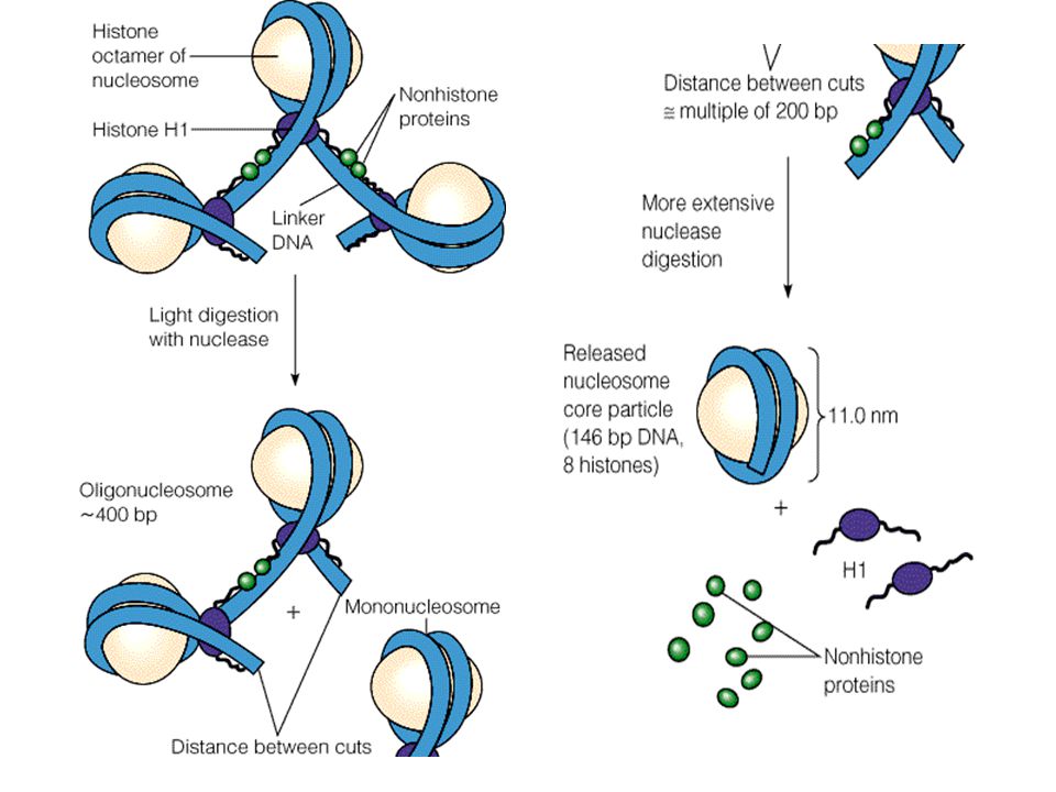
Nucleosome are the fundamental organizational units of chromatin

90
Chromatin assembly

92
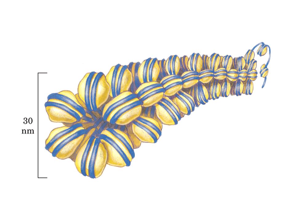
The 30 nm fiber, a higher-order organization of nucleosomes

95
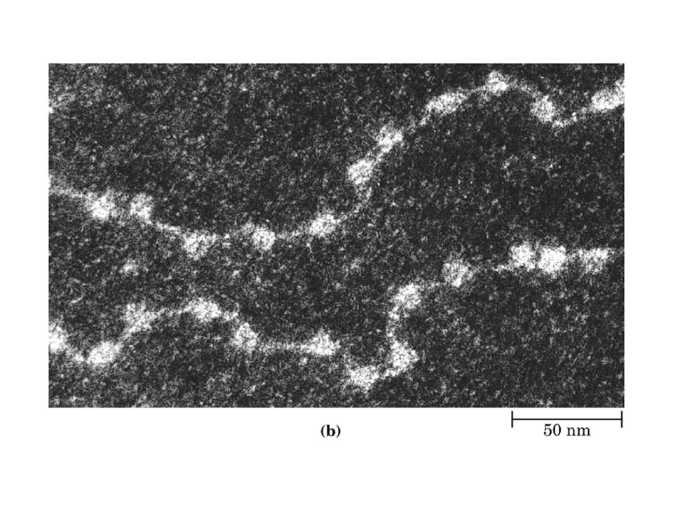
A partially unraveled human chromosome, revealing numerous loops of DNA attached to a scaffoldlike structure.

96
Loops of chromosomal DNA attached to a nuclear scaffold

97
Organization of Chromotin and Chromosomes:

98
7.6 · Chemical Synthesis of Nucleic Acids

99
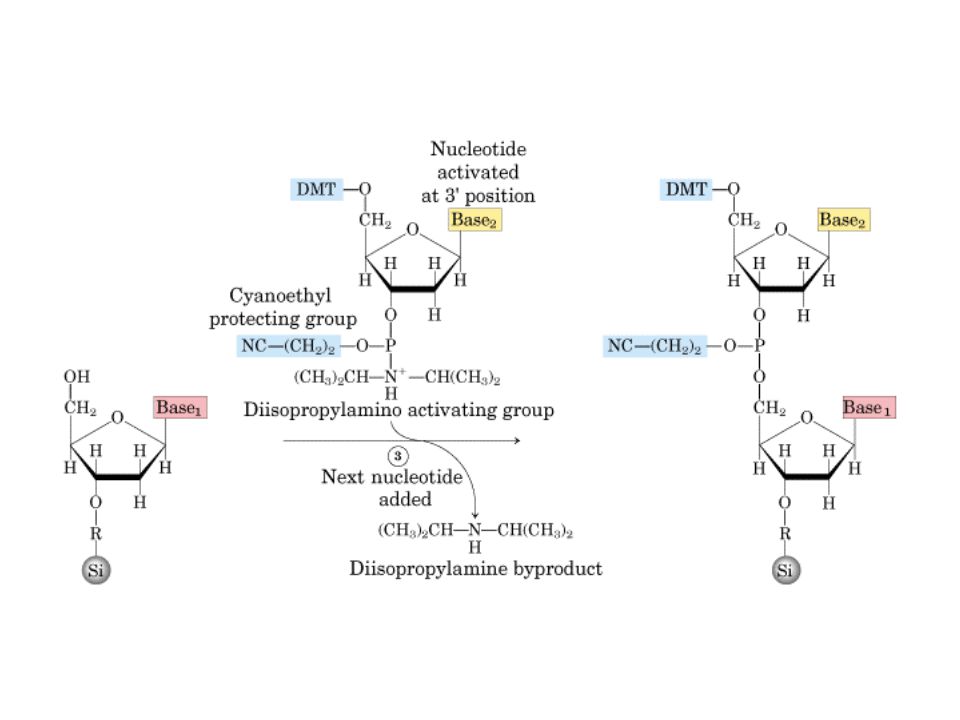
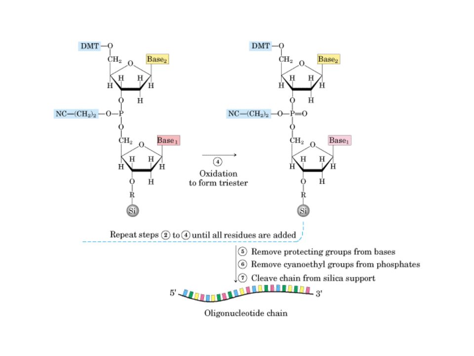
DNA synthesis DMT::二甲氧三苯甲基

100
DNA synthesis DMT::二甲氧三苯甲基

103
Commercially available automated instruments, called DNA synthesizers or “gene machines,” are capable of carrying out the synthesis of oligonucleotides of 150 bases or more.

104
7.7 · Secondary and Tertiary Structure of RNA
Types of RNAs: Transfer RNA (adaptor molecule) Messenger RNA (template for protein synthesis) Ribosomal RNA (protein synthesis) Small nuclear RNA (splicesomal RNA) Small nucleolar RNA (ribosomal RNA processing) Interference RNA (gene silencing) microRNA (translation regulation) Virus RNA (code virus genome) In comparison with DNA structures, much less is known about RNA structures. Most RNA are associated with proteins which facilitate their structural folding.
 Messenger RNA (template for protein synthesis) Ribosomal RNA (protein synthesis) Small nuclear RNA (splicesomal RNA) Small nucleolar RNA (ribosomal RNA processing) Interference RNA (gene silencing) microRNA (translation regulation) Virus RNA (code virus genome) In comparison with DNA structures, much less is known about RNA structures. Most RNA are associated with proteins which facilitate their structural folding.")
105
Transfer RNA Structures
Secondary Structure Of large ribosomal RNA Tertiary Structure Of large ribosome subunit TyC Loop Anticodon Stem V ariable loop D Loop Anticodon Loop

106
Ribosomal RNA Secondary Structure Of large ribosomal RNA
Tertiary Structure Of large ribosome subunit Ban et al., Science 289 ( ), 2000
,")
107
Catalytic RNA Secondary Structure Of large ribosomal RNA
Tertiary Structure Of large ribosome subunit

108
Objectives of Chapter 7 Describe DNA helix structure properties;
Recognize supercoiling in DNA and its handness; Calculate L, or T, or W when the other two parameters are known; Describe nucleosome and chromotin structure; Describe the structural elements in tRNA;

Similar presentations


 Recognize and apply the.>")

 Nucleic Acids. Information encoded in a DNA molecule is transcribed via synthesis of an RNA molecule The sequence of the RNA molecule.>")


, Thymine (T), Uracil (U, in RNA) Purine bases: Adenine (A), Guanine.>")
 Nucleic Acids. DNA 1 o Structure - Linear array of nucleotides 2 o Structure – double helix 3 o Structure - Super-coiling, stem- loop.>")
, Thymine (T), Uracil (U, in RNA) Purine bases: Adenine (A), Guanine.>")

 Dr. Sumbul Fatma>")


. The double helix Nitrogenous Bases and Pentose Sugars.>")