Download presentation
Presentation is loading. Please wait.

1
Complications of Cardiac Catheterization
Grossman’s cardiac catheterization, angiography, and intervention Chapter 3 CV R4 陳柏升 Supervisor: Prof. 蔡良敏

2
Introduction The risk of producing a major complication ― less than 1% during most procedure type currently The risks of complication for the individual patient Demographics (age, gender) The cardiac anatomy (left main CAD, AS, LV dysfunction) The clinical situation (ACS, shock, ARF) The type of procedure being performed (diagnosis, PCI)
 The cardiac anatomy (left main CAD, AS, LV dysfunction) The clinical situation (ACS, shock, ARF) The type of procedure being performed (diagnosis, PCI)")
3
Introduction Familiarity with those risks can be of immeasurable value in the following: Anticipating increased risk of complication Taking extra precautions to avoid them (TPM for PCI) Promptly recognizing complications when they occur Taking corrective and potentially life-saving action
 Promptly recognizing complications when they occur. Taking corrective and potentially life-saving action.")
4
Introduction Discussed with the patient and family
The detailed of the planned procedure and its anticipated risks Informed consent Documented in the patient’s chart Specify the type of procedure that is planed The potential major complications and their estimated risk of occurrence

5
Introduction Should be intimately knowledgeable about the potential complications of the procedures Collect information about the frequency of these complications on at least a yearly basis and review those data with physician staff

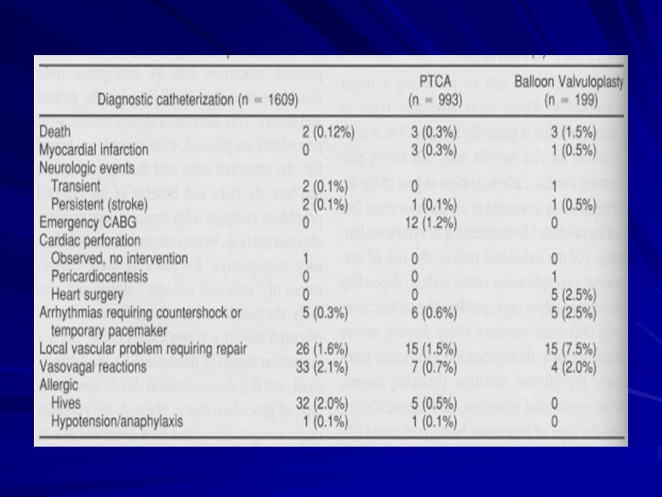
7
Death Diagnostic catheterization
Incidence The first Society for Cardiac Angiography registry from 1979 to 1981: 0.14% procedure-related mortality The second registry from 1984 to 1987: slightly more to 0.1% The third registry in 1990s: 0.08%, with a 1.5% incidence of any major complications High risk groups

9
Death Diagnostic catheterization
Severe left main disease More than 20 times higher than patients with 1-V-D Careful engaging and “puff” test in RAO and caudal angulation view to screen for left main disease If ostial left main stenosis is present, AP injection may be performed → RAO and cranial angulation to see LAD and diagonal branches More view offers little information and increase the risk fo triggering the vicious cycle to irreversible collapse IABP for unstable patient and arrange for bypass surgery

10
Death Diagnostic catheterization
Severe left ventricular dysfunction (EF < 30%) Seven-fold increased risk Particularly is associated with PCWP > 25mmHg and systolic BP < 100 mmHg An effort to make CHF under control before cath Right heart catheterization should always be performed to get more information PWCP > 30mmHg, every effort made to improve hemodynamic status MAP > 65 mmHg → diuretics, oxygen supply, vasodilator MAP < 65 mmHg → a positive inotrpic agent IABP Percutaneous cardiopulmonary bypass (CPS) low-osmolar-contrast agent to produce less myocardial depression
 Seven-fold increased risk. Particularly is associated with PCWP > 25mmHg and systolic BP < 100 mmHg. An effort to make CHF under control before cath. Right heart catheterization should always be performed to get more information. PWCP > 30mmHg, every effort made to improve hemodynamic status. MAP > 65 mmHg → diuretics, oxygen supply, vasodilator. MAP < 65 mmHg → a positive inotrpic agent. IABP. Percutaneous cardiopulmonary bypass (CPS) low-osmolar-contrast agent to produce less myocardial depression.")
11
Death Diagnostic catheterization
Patients who have previously undergone CABG Older, more diffuse coronary and generalized atherosclerosis, worse left ventricular function, more length and complex procedure The Post CABG trial: 2635 diagnostic angiogram; no death, 0.7% major complications Pediatric patients ― high risk group Potential explanations for declining mortality rate Improvement in catheter design, imaging systems, contrast agents High annual proceduce volume ( >1.5 million/year) Shorter procedure time Heparin use?
 Shorter procedure time. Heparin use")
12
Death Intervention procedures
More higher mortality in intervention because More aggressive catheters Superselective cannulation of diseased coronary arteries Brief interruption of coronary and systemic flow Roughly 10-fold higher than purely diagnostic procedure (1% : 0.1%) A wide variation in risk based on patient comorbidities, clinical condition, and procedure type
 A wide variation in risk based on patient comorbidities, clinical condition, and procedure type.")
13
Myocardial infarction ― Diagnostic catheterization
Transient myocardial ischemia is relatively common during diagnostic catheterization and coronary intervention Respond promptly to drug therapy Deflation of the angioplasty balloon

14
Myocardial infarction ― Diagnostic catheterization
Myocardial infarction is an uncommon but important complication of diagnostic cardiac catheterization Reduction progressively 0.25% in the late 1970s to 0.05% currently Causes of reduction Experience ― greater attention to catheter flushing, pressure damping Heparin use for angioplasty High risk groups ― patient-related and technique-related The extent of vessel diseases: 1-V-D : 3-V-D : left main → 0.06% : 0.08% : 0.17% The clinical indication (unstable angina or recent subendocardial infarction) IDDM “Angioplsty stand-by” for these unstable patients
 IDDM. Angioplsty stand-by for these unstable patients.")
15
Myocardial infarction ― Diagnostic catheterization
IABP or remaining intravenous heparinization for patients whose severe lesions are suitable for bypass surgery Unstable patient should not reversed heparinization with prostamine, absent a life-threatening bleeding complications Resume heparinization in 2 hours later after sheath is removed Post-procedure MI is rare in patients who complete their catheterization without unstable signs Should severe ischemic instability develop after the patient leaves the cath lab, aggressive therapy in indicated → coronary intervention or bypass surgery

16
Myocardial infarction ― Interventional procedure
Mechanisms Dissection abrupt vessel closure “snow-plow” occlusion of side branches Spasm or no-reflow Thrombosis distal embolization Incidence rate decreased progressive, the current experience suggested that emergent bypass surgery and Q-wave infarction rates is 1% Some elevation of CPK and CK-MB ― at least 20% of patients with otherwise successful intervention Low order (1 to 3 times the upper limit of normal) appears to have no short- or long-term consequences Only elevation 5 times above normal tends to adversely impact late survival ― views as a major complication, equivalent to Q-wave infarction
 appears to have no short- or long-term consequences. Only elevation 5 times above normal tends to adversely impact late survival ― views as a major complication, equivalent to Q-wave infarction.")
17
Cerebrovascular complications
Uncommon but potentially devastating complications Decrasing incidence: 0.23% (1973) → 0.07% (current) embolic in origin Most cases Many emboli are dislodged from unsuspected aortic plaque or diffuse atherosclerosis; emboli originate from cardiac chambers, thrombotic coronary arteries, or surface of cardiac valves Technical errors: sloppy catheter flushing, introducing of air bubbles during contrast injection, inadvertent placement of wires and catheters into arch vessels, prolonged (> 3mins) wire dwell time, etc. Avoid “catching” in aortic surface Dislodgeable mural thrombus Avoid transseptal catheterization or mitral valvuloplasty in patients with left atrial thrombus, which may increase the incidence of stroke
 → 0.07% (current) embolic in origin. Most cases. Many emboli are dislodged from unsuspected aortic plaque or diffuse atherosclerosis; emboli originate from cardiac chambers, thrombotic coronary arteries, or surface of cardiac valves. Technical errors: sloppy catheter flushing, introducing of air bubbles during contrast injection, inadvertent placement of wires and catheters into arch vessels, prolonged (> 3mins) wire dwell time, etc. Avoid. catching in aortic surface. Dislodgeable mural thrombus. Avoid transseptal catheterization or mitral valvuloplasty in patients with left atrial thrombus, which may increase the incidence of stroke.")
18
Cerebrovascular complications
embolic in origin (cont.) In patients with right-to-left shunt, paradoxical embolization may lead to stroke Active left side endocarditis (AV or MV) : left side cath does not increase the incidence of embolic events (0/35) AJC 1979;44:1306 Intracerebral hemorrhage Patients receiving Aggressive anticoagulation Antiplatelet Thrombolytic therapy Neurologic consultation, image study (CT, MRI) The distinction between embolic and hemorrhagic stroke is critical Transient neurologic deficits High-osmolar-contrast agents into the carotid and vertebral vessels
 In patients with right-to-left shunt, paradoxical embolization may lead to stroke. Active left side endocarditis (AV or MV) : left side cath does not increase the incidence of embolic events (0/35) AJC 1979;44:1306. Intracerebral hemorrhage. Patients receiving. Aggressive anticoagulation. Antiplatelet. Thrombolytic therapy. Neurologic consultation, image study (CT, MRI) The distinction between embolic and hemorrhagic stroke is critical. Transient neurologic deficits. High-osmolar-contrast agents into the carotid and vertebral vessels.")
19
Local vascular complications
One of the most common problems Problems including Vessel thrombosis Distal embolization Dissection Poorly controlled bleeding at the punctual site poorly placed puncture vessel laceration excessive anticoagulation poor technique in either suture closure or groin compression Hemorrhage and hematoma ― evident within 12 hours; false lumen ― evident for days or even several weeks later

20
Local vascular complications Diagnostic catheterization
The Society for Cardiac Angiography registries ― 0.5 to 0.6% in incidence Brachial approach Arterial thrombosis Causes Formation of a thrombus in the proximal arterial part and failure to remove prior to repair Secondary to an intimal flap within the arterial lumen Secondary to local spasm Preventions Meticulous attention to the details of arterial repair Adequate heparinization: systemic and local Treatment Fogarty catheter thrombectomy Percutaneous transluminal angioplasty

21
Local vascular complications Diagnostic catheterization
Brachial approach (cont.) Other complications Injury to median nerve Cutdown or compression by hematoma Mild case: numbness and weakness for 3 to 4 weeks and return to normal: occasionally up to 6 months Delayed dehiscence of arterial sutures with late arterial bleeding Bacterial arteritis Local cellulitis-phlebitis Extensive soft tissue is dissected Large vein are used and tied off The catheterization procedure is long Seroma and hematoma forms Nonviable tissue is left in the incision Poor surgical technique or violation of sterile procedure occurs
 Other complications. Injury to median nerve. Cutdown or compression by hematoma. Mild case: numbness and weakness for 3 to 4 weeks and return to normal: occasionally up to 6 months. Delayed dehiscence of arterial sutures with late arterial bleeding. Bacterial arteritis. Local cellulitis-phlebitis. Extensive soft tissue is dissected. Large vein are used and tied off. The catheterization procedure is long. Seroma and hematoma forms. Nonviable tissue is left in the incision. Poor surgical technique or violation of sterile procedure occurs.")
22
Local vascular complications Diagnostic catheterization
Femoral approach Thrombosis (femoral artery) Extremely rare, except a small femoral artery lumen (PAOD, DM, female), a large-diameter catheter or sheath (IABP) or long duration of catheter S/S: leg pain or numbness, diminished distal pulse Obstructive limb ischemia generally resolves and distal pulse returned when the sheath is removed Ongoing complaint and diminished or absent distal pulse with catheter removal → flow-obstructing dissection or thrombus → urgent vascular surgery → within 2 to 6 hours!! Results in extension of thrombosis into smaller distal branch and muscle necrosis if delayed Femoral venous thrombosis or pulmonary embolism Rare (multiple venous lines or compression by large arterial hematoma etc.) but may be underreported: up to 10% asymptomatic positive lung perfusion scan Continuous drip of heparinized saline t venous sidearm throughout the procedure to avoid this problem
 Extremely rare, except a small femoral artery lumen (PAOD, DM, female), a large-diameter catheter or sheath (IABP) or long duration of catheter. S/S: leg pain or numbness, diminished distal pulse. Obstructive limb ischemia generally resolves and distal pulse returned when the sheath is removed. Ongoing complaint and diminished or absent distal pulse with catheter removal → flow-obstructing dissection or thrombus → urgent vascular surgery → within 2 to 6 hours!! Results in extension of thrombosis into smaller distal branch and muscle necrosis if delayed. Femoral venous thrombosis or pulmonary embolism. Rare (multiple venous lines or compression by large arterial hematoma etc.) but may be underreported: up to 10% asymptomatic positive lung perfusion scan. Continuous drip of heparinized saline t venous sidearm throughout the procedure to avoid this problem.")
23
Local vascular complications Diagnostic catheterization
Femoral approach (cont.) Poorly controlled bleeding ― more common Suggest laceration of the femoral artery Try next-larger-diameter sheath or compressed manually until the procedure is completed Reverse heparin and control bleeding with prolonged cpompression Blood transfusion Hematoma formation usually resolve over 1 to 2 weeks S/S: femoral nerve compression → quadriceps, weakness → takes weeks even months to resolve; surgical repair is not required generally Hematoma may extend to retroperitoneal bleeding if puncture site is above inguinal ligament unexplained hypotension, decreased Hct, ipsilateral flank pain; response to fluid challenge best prevention
 Poorly controlled bleeding ― more common. Suggest laceration of the femoral artery. Try next-larger-diameter sheath or compressed manually until the procedure is completed. Reverse heparin and control bleeding with prolonged cpompression. Blood transfusion. Hematoma formation. usually resolve over 1 to 2 weeks. S/S: femoral nerve compression → quadriceps, weakness → takes weeks even months to resolve; surgical repair is not required generally. Hematoma may extend to retroperitoneal bleeding if puncture site is above inguinal ligament. unexplained hypotension, decreased Hct, ipsilateral flank pain; response to fluid challenge. best prevention.")
24
Local vascular complications Diagnostic catheterization
Femoral approach (cont.) Pseudoaneurysm Hematoma continuity with the arterial lumen Blood flow in and out of the arterial puncture, expanding the cavity pulsation, audible bruit, Duplex scan Therapy Surgical repair transducer compress the neck for 30 to 60 minutes procoagulant solutions or embolization coils with echo guiding Prevention: accurate puncture of the common femoral artery and effective initial control of bleeding A-V fistula Not be clinically evident for days after procedure Ongoing bleeding may decompress into the adjacent venous puncture site To and fro continuous bruit Surgical repair if fistula tends to enlarge with time or does not close within 2-4 weeks High risks: low puncture site (superficial or profunda femoral arteries)
 Pseudoaneurysm. Hematoma continuity with the arterial lumen. Blood flow in and out of the arterial puncture, expanding the cavity. pulsation, audible bruit, Duplex scan. Therapy. Surgical repair. transducer compress the neck for 30 to 60 minutes. procoagulant solutions or embolization coils with echo guiding. Prevention: accurate puncture of the common femoral artery and effective initial control of bleeding. A-V fistula. Not be clinically evident for days after procedure. Ongoing bleeding may decompress into the adjacent venous puncture site. To and fro continuous bruit. Surgical repair if fistula tends to enlarge with time or does not close within 2-4 weeks. High risks: low puncture site (superficial or profunda femoral arteries)")
26
Local vascular complications Interventional procedure
A significantly high incidence of local vascular complications than pure diagnostic procedure ― 1 to 2% Use of larger sheath The intensity and duration of anticoagulation Removal of the sheaths only after an overnight dwell Various approaches for collagen plugging or percutaneous suture-mediated closure have been used Avoid the discomfort of prolonged manual or mechanical compression Allow early even immediate ambulation Failed to demonstrate significant reduction of major vascular complications compared with compression

27
Arrhythmia or conduction disturbance
A variety of cardiac arrhythmia or conduction disturbance may occur ― VPCs, VT, VF, Af Monitor the surface ECG with physiologic monitor with the pressure tracing and alarm system The tools (defibrillator, pacemakers, ventilation support) and drugs to treat the rhythm disorder All cardiac catheterization personnel must be fully certified in ACLS
 and drugs to treat the rhythm disorder. All cardiac catheterization personnel must be fully certified in ACLS.")
28
Ventricular fibrillation
VPC and brief run VT(3-5 beats) are common During the passage of catheter into the right or left ventricle The offending catheter must be repositioned immediately Other causes inducing ventricular fibrillation Catheter transmission of “leakage” electrical current to the heart Eliminated by the adoption of standards for grounding system, less than 20 μA between any two exposed conduction source Intracoronary contrast injection Most commonly with injection of ionic (high-osmolar) contrast agent into the right coronary artery, especially the injection is prolonged or the catheter pressure is damped Change in injection technique and formulation of contrast agents reduce the incidence, 1.28%(1974) → <0.4%(1991)
 are common. During the passage of catheter into the right or left ventricle. The offending catheter must be repositioned immediately. Other causes inducing ventricular fibrillation. Catheter transmission of leakage electrical current to the heart. Eliminated by the adoption of standards for grounding system, less than 20 μA between any two exposed conduction source. Intracoronary contrast injection. Most commonly with injection of ionic (high-osmolar) contrast agent into the right coronary artery, especially the injection is prolonged or the catheter pressure is damped. Change in injection technique and formulation of contrast agents reduce the incidence, 1.28%(1974) → <0.4%(1991)")
29
Ventricular fibrillation
Other causes inducing ventricular fibrillation (cont.) Patients with baseline prolongation of the QT interval Some refractory ventricular ectopy is the setting of profound transmural ischemia or early myocardial infarction Therapy iv lidocaine (1.5 mg/kg over 1 minute, with a second bolus of 0.75 mg/kg 7 minutes later) procainamide (15 mg/kg over 20 minutes,↓BP ,↑QRS or QT) iv amiodarone (5 mg/kg over 20 minutes, 1 gm/24 hours)
 Patients with baseline prolongation of the QT interval. Some refractory ventricular ectopy is the setting of profound transmural ischemia or early myocardial infarction. Therapy. iv lidocaine (1.5 mg/kg over 1 minute, with a second bolus of 0.75 mg/kg 7 minutes later) procainamide (15 mg/kg over 20 minutes,↓BP ,↑QRS or QT) iv amiodarone (5 mg/kg over 20 minutes, 1 gm/24 hours)")
30
Atrial arrhythmia Atrial extrasystoles are common during
Catheter advancement from the right atrium to the SVC Looping of the catheter in the right atrium to facilitate passage in a patient with enlargement of the right-side heart chamber Usually subside once the catheter is repositioned But may go on to Af or AF in sensitive patients tends to revert spontaneously over a period of minutes to hours Require additional therapy if they produce ischemia or hemodynamic instability

31
Atrial arrhythmia Therapy
DC shock in both Af and AF with hemodynamic instability Atrial flutter ― a brief (15 seconds) but rapid ( bpm) right atrial pacing Ensue a stable atrial pacing location or triggering a VF since catheter migration into the ventricle Atrial fibrillation ― may cause S/S with RVR, hypotension in patient with MS, hypertrophic cardiomyopathy, or diastolic left ventricular dysfunction Rate control iv beta blockers (inderal : 1mg ; esmolol : loading 500μg/kg/min for 30 sec, followed by 50~250 μg/kg/min) iv CCB (verapamil 5mg) Rhythm conversion iv procainamide (15 mg/kg for 20 min) ibutilide (on other QT-prolonging drugs; ↓K or Mg; bradycardia; QTc > 440 ms)> 60 kg : 1mg over 10 min ; within 4 hours, no other class III agents!!
 but rapid ( bpm) right atrial pacing. Ensue a stable atrial pacing location or triggering a VF since catheter migration into the ventricle. Atrial fibrillation ― may cause S/S with RVR, hypotension in patient with MS, hypertrophic cardiomyopathy, or diastolic left ventricular dysfunction. Rate control. iv beta blockers (inderal : 1mg ; esmolol : loading 500μg/kg/min for 30 sec, followed by 50~250 μg/kg/min) iv CCB (verapamil 5mg) Rhythm conversion. iv procainamide (15 mg/kg for 20 min) ibutilide (on other QT-prolonging drugs; ↓K or Mg; bradycardia; QTc > 440 ms)> 60 kg : 1mg over 10 min ; within 4 hours, no other class III agents!!")
32
Bradycardia Occurs commonly during coronary angiography
At the end of a right coronary artery injection with ionic (high-osmolar) contrast agent Forceful coughing clear contrast from the coronaries support aortic pressure and cerebral perfusion during asystole restore normal cardiac rhythm Vasovagal reaction One of the most common complication (3% incidence) bradycardia with hypotension, nausea, yawning, and sweating triggered by pain and anxiety especially in hypovolemic status 80% occurs as vascular access and 16% occur during sheath removal adequate preprocedure sedation and local anesthetic
 contrast agent. Forceful coughing. clear contrast from the coronaries. support aortic pressure and cerebral perfusion during asystole. restore normal cardiac rhythm. Vasovagal reaction. One of the most common complication (3% incidence) bradycardia with hypotension, nausea, yawning, and sweating. triggered by pain and anxiety especially in hypovolemic status. 80% occurs as vascular access and 16% occur during sheath removal. adequate preprocedure sedation and local anesthetic.")
33
Bradycardia Vasovagal reaction (cont.) Conduction disturbance
Treatment Cessation of the painful stimulus Rapid volume administration Atropine Additional pressor support may require if hypotension persist May imply cardiac perforation when the pericardium is irritated by blood during catheter manipulation Conduction disturbance Uncommon but potentially serious cause of bradycardia during cardiac catheterization Require no treatment except in the patient with preexisting bundle branch block ― asystole or cardiovascular collapse except escape rhythm takes over

34
Bradycardia Conduction disturbance (cont.) Complete AV block
Occurs during Rotational atherectomy, especially in RCA or LCX Aortic valvuloplasty Treatment Atropine is rarely helpful but should be given anyway since it has few adverse effects Coughing Pacing marker insertion Prophylactic right-sided pacing catheters

35
Perforation of the heart and great vessel
Rare Heart perforation 0.8% in 1968, RA-RV-LA-LV; 0.006% of diagnostic cath and 0.08% of coronary angioplasties now Stiffer catheter and elderly women (>65 y/o) S/S Cardiac silhouette may enlarge Pulsation of the heart borders on fluoroscopy may become blunted Bradycardia and hypotension due to vagal stimulation Cardiac tamponade ― elevation of RA pressure with loss with y descent Paracentasis via subxyphoid approach and protamine infusion Emergent surgical intervention? ― most will seal without surgery
 S/S. Cardiac silhouette may enlarge. Pulsation of the heart borders on fluoroscopy may become blunted. Bradycardia and hypotension due to vagal stimulation. Cardiac tamponade ― elevation of RA pressure with loss with y descent. Paracentasis via subxyphoid approach and protamine infusion. Emergent surgical intervention ― most will seal without surgery.")
36
Perforation of the heart and great vessel
Vessels perforation Aorta: rare, except in the case of weakening by ascending aortic dissection or aneurysm Pulmonary arteries Rare; too stiff-tip guidewire or balloon inflated in a distal branch Typically develop hemoptysis → require tamponade of the proximal pulmonary artery, embolization of the bleeding branch, double-lumen endotracheal tube, emergent lobectomy or pnemonectomy Coronary artery Unheared of diagnostic cath; the incidence rises to 1% with the advent of more aggressive new techniques for coronary intervention Most are limited to deep injury to vessel wall; free perforation may leads to frank tamponade within seconds to minutes → seal the site of leakage by inflation of a balloon catheter, pericardiocentasis and protamine use if necessary, coil embolization, covered stent, emergent surgical intervention

37
Infection and pyogen reaction
Endocarditis prophylaxis and routinely antibiotics use are not recommended, except performing a delayed intervention by exchanging sheaths that were placed in a earlier diagnostic procedure Any break in sterile technique is suspected Repeat procedure within 2 weeks → use contra-lateral groin Increasing infection rate from the same groin Full sterile precautions before procedure Procedure from brachial approach From femoral approach when the procedure is prolonged The sheath was remain in place for any period A stent and permanent pacemaker is being implanted A vascular grafy is punctured Guidelines of the Occupational Safety and Health Administration (OSHA): Sterile precautions for any procedure
: Sterile precautions for any procedure.")
38
Infection and pyogen reaction
Laboratory personnel Vaccination for hepatitis B Prophylactic zidovudine (AZT) for personnel being stuck by needle tainted with blood from HIV-positive patient Avoid muitiuse drug vials and clean the room thoroughly between procedures Post-procedure fever Phlebitis May develop after brachial catheterization Low grade fever and a warm tender cord overlying the affected vein Pyogen reaction Shaking chills during or within the first hour Fever spike as high as 102℉ Caused by the presence of contaminating material that remain on incompletely cleaned catheter surfaces Morphine use for symptom relief
 for personnel being stuck by needle tainted with blood from HIV-positive patient. Avoid muitiuse drug vials and clean the room thoroughly between procedures. Post-procedure fever. Phlebitis. May develop after brachial catheterization. Low grade fever and a warm tender cord overlying the affected vein. Pyogen reaction. Shaking chills during or within the first hour. Fever spike as high as 102℉ Caused by the presence of contaminating material that remain on incompletely cleaned catheter surfaces. Morphine use for symptom relief.")
39
Allergic and anaphylactoid reaction
Materials may cause allergic or anaphylactoid reactions Local anesthesia Iodinated contrast agent Protamine Preservatives Skin testing with the intended agent at 1:1000 dilution if desired Most common in triggering allergic reactions ― up to 1% Anaphylactoid reaction ― involve degranulation of circulating basophils and tissue mast cells by direct complement activation Other clinical manifestations caused by histamine or other agents ― sneezing, urticaria, angioedema, bronchospasmwarm shock

40
Allergic and anaphylactoid reaction
Iodinated contrast agent (cont.) High risk groups Other atopic disorders Allergy to penicillin Allergy to seafood (contained organic iodine) Prior reaction to contrast (as high as 15% to 35%) Premedications ― reduce the incidence od a secone reaction to 5% to 10% and that of severe reaction to below 1% Prednisolone ― 20mg tid for 24 to 48 hours H1 antihistamine ― diphenhydramine 25mg tid H2 blocker ― cimetidine or ranitadine Nonionic contrast agent ― adds a further margin of safety Record aortic pressure first when a patient have a history of well-documented prior severe contrast reactions Emergent treatment ― intravenous injection of dilute epinephrine, 1:10,000 epinephrine 1mL/minute until arterial pressure is restored Avoid excessive doses ― may precipitate life-threatening hypertension, tachycardia or even VF
 High risk groups. Other atopic disorders. Allergy to penicillin. Allergy to seafood (contained organic iodine) Prior reaction to contrast (as high as 15% to 35%) Premedications ― reduce the incidence od a secone reaction to 5% to 10% and that of severe reaction to below 1% Prednisolone ― 20mg tid for 24 to 48 hours. H1 antihistamine ― diphenhydramine 25mg tid. H2 blocker ― cimetidine or ranitadine. Nonionic contrast agent ― adds a further margin of safety. Record aortic pressure first when a patient have a history of well-documented prior severe contrast reactions. Emergent treatment ― intravenous injection of dilute epinephrine, 1:10,000 epinephrine. 1mL/minute until arterial pressure is restored. Avoid excessive doses ― may precipitate life-threatening hypertension, tachycardia or even VF.")
41
Allergic and anaphylactoid reaction
Prostamine High risk groups IDDM patients with NPH control (which contained with protamine) Emergent treatment ― as previous described Heparin-induced thrombocytopenia (HIT) ― rare Definition a fall in platelet count by at least 50% Accomplished by a positive serologic test for the responsible antibody (usually IgG) Platelet Fc receptor + PF-4/antibody → platelet activation Onset is typically 7 to 10 days; bovine-derived more common than porcine-derived Alternative anticoagulant LMWH ― frequently cross-react with heparin antibody Heparinoid (Organan, danaproid) Direct antithrmbin compounds (hirudin, hirulog, or argatroban)
 Emergent treatment ― as previous described. Heparin-induced thrombocytopenia (HIT) ― rare. Definition. a fall in platelet count by at least 50% Accomplished by a positive serologic test for the responsible antibody (usually IgG) Platelet Fc receptor + PF-4/antibody → platelet activation. Onset is typically 7 to 10 days; bovine-derived more common than porcine-derived. Alternative anticoagulant. LMWH ― frequently cross-react with heparin antibody. Heparinoid (Organan, danaproid) Direct antithrmbin compounds (hirudin, hirulog, or argatroban)")
42
Renal dysfunction At least 5% of patients experience a transient rise in serum creatinine greater than 1mg/dL following cardiac angiography Precise mechanism of contrast-induced renal dysfunction ― not been established High risk groups ― up to 50% DM Multiple myeloma Volume depletion Preexisting renal dysfunction Patients who are receiving certain drugs(eg. GM, NSAID, ACE-I) Most are non-oliguria, peak within 1 to 2 days and return to baseline by 7 days
 Most are non-oliguria, peak within 1 to 2 days and return to baseline by 7 days.")
43
Renal dysfunction Fewer than 1% of patients go on to require chronic dialysis Prospective trials comparing high and low-osmolar contrast agents have failed to showed consistent benefit Main defense against contrast-induced nephropathy ― Limitation of total contrast volume to 3mL/kg Adequate prehydration for any patients with impaired baseline renal dysfunction ― 12 hours before and after the contrast procedure

44
Renal dysfunction Systemic cholesterol embolization
Another cause of renal failure following cardiac catheterization ― 0.15% of cathetherizations High risk groups Those with diffuse atherosclerosis In whom insertion of a guiding catheter will frequently produce a shower of glistering particles on the table drape Hallmarks of diagnosis ― evidence of peripheral embolization Episodic hypertension or systemic eosinophilia may be apparent well before the other manifestations develops Develops slowly ― over weeks to months Half of the patients progressed to frank renal failure Treatment ― purely supportive

45
Hypotension One of the most common problem Causes
Hypovolemia: inadequate hydration, blood loss, excessive contrast-induced diuresis Reduction in cardiac output: ischemia, tamponade, arrhythmia, valvular regurgitation Inappropriate systemic arteriolar vasodilation: vasovagal, excessive nitrate administration, response to contrast or inotrope-vasodilator drug Right side catheterization for diagnosis and treatment Initial empirical treatment and definite correction of hypotension and its causes before hypotension leads to secondary ischemia and irreversible spiral of left ventricular dysfunction

46
Hypotension Treatment Low filling pressure
Rapid volume administration Atropine use if combined with inappropriate bradycardia Look for potential site of blood loss High filling pressure ― suggest primary cardiac dysfunction: ischemia, tamponade, VHD Supported empirically by inotropic agents, vasopressor or circulatory devices Immediate intervention or surgical intervention Artificial pacing if bradycardia is present and is not response to atropine Adequate airway maintenance Coexistent sepsis, contrast reaction or and idiosyncratic vasodilator reaction to dopamine infusion

47
Volume Overload Prone to fluid overload due to Treatment
Hypertonic contrast agents Myocardial depression Ischemia induced by contrast Poor baseline left ventricular function Supine position Risk of contrast-induced renal dysfunction Treatment Optimizing volume status before or early in the procedure Use of low-osmolar contrast agents Supportive measures: inotropes, diuretics, vasodilators, IABP Even more aggressive treatment is warranted once pulmonary edema develops, eg. intubation

48
Anxiety/Pain Oral sedatives pretreatment(diazepam 5 to 10mg, diphenhydramine 25 to 50mg) and liberal use of local anesthesia at the insertion site To understand why the patient is having pain first and whether anything to do to reverse the problem The catecholamine surge associated with pain and anxiety may worsen the condition of a patient Small dose of morphine, fentanyl, midazolam Make the procedure more tolerable for both the patient and the staff Monitor blood pressure, respiratory rate, and pulse oximetry Antagonist drugs should also be stocked

49
Respiratory insufficiency
Not uncommon Pulmonary edema Baseline lung disease Allergic reaction Over sedation Baseline ABG after sheath insertion Low-flow supplemental oxygenation(2L/min) helps avoid episodes of desaturation If oxygen consumption is to be measured as part of a calculation of cardiac output by the Fick method, oxygen administration should not be begun after that measurement
 helps avoid episodes of desaturation. If oxygen consumption is to be measured as part of a calculation of cardiac output by the Fick method, oxygen administration should not be begun after that measurement.")
50
Retained equipment Devices knot, become entrapped, or leave fragment in the circulation ― most of these events are precipitated when such devices are stressed beyond their design parameters Operators should be familiar with device performance limits and be familiar with devices and techniques that can be used to recover the errant fragments, such as vascular snares, bioptomes, baskets, et.

51
Other important aspects of procedural complication
Caseload Inverse correlation between the caseload of a cardiac cathetrization lab and each operator and their incidence of major complications Greater caseload leads to greater skill and technical proficiency ACC/AHA guideline: minimal 300 cases/year for an adult catheterization lab; minimal 150cases/year for each physician The same relationship between caseload and major complications of coronary intervention <200 cases/year for each lab or < 75 cases/year for each operator have more than twice the mortality and emergent surgery rates of higher-volume institutes “Practice makes perfect”

52
Other important aspects of procedural complication
Speed Be widely regarded as one factor that determines the risk of complication Few data are available on this subject Shorter procedure times have correlated with decreased overall risk; low speed of performing a procedure does not necessarily carry an increased risk of a complication, unless the slow speed reflects lack of operator skill Duration of the procedure should be considered as an important independent risk factor only when Lack of skill or experience on the part of operator It leads to severe cardiac decompensation in a critically ill patient poorly prepared to spend more than the minimal time in the supine position

53
Other important aspects of procedural complication
Pseudo-complications Patients suffering from serious cardiac disease experience major cardiac events as part of the natural history of their disease

54
Percutaneous Approach, Including Transseptal and Apical Puncture
Grossman’s cardiac catheterization, angiography, and intervention Chapter 4 CV R4 陳柏升 Supervisor: Prof. 蔡良敏

55
Catheterization via the femoral artery and vein Patient preparation
Shave an area approximately 10 cm in diameter surrounding the point of femoral pulse; prepare both side routinely The shaved area are scrubbed with a povidone-iodine/detergent mixture and then painted with povidone-iodine solution

56
Catheterization via the femoral artery and vein Selection of puncture site
Perform the puncture at the correct level ― 1 or 2 cm below the inguinal ligament (runs from the anterior superior iliac spine to the pubic bone) Skin nick in reference to the skin crease may be misleading in obese patients Inferior border of femoral neck by fluoroscopy Most difficulties in entering the femoral artery and vein arise as a result of inadequate identification of these landmarks Puncture above inguinal ligament Catheter advancement difficult Predispose to inadequate compression Hematoma formation or retroperitoneal bleeding following catheter removal Puncture at more 3 cm below the inguinal ligament Failed to enter the vessel lumen Increase the risk of false aneurysm or thrombotic occlusion due to smaller caliber Excessive bleeding AV-fistula
 Skin nick in reference to the skin crease may be misleading in obese patients. Inferior border of femoral neck by fluoroscopy. Most difficulties in entering the femoral artery and vein arise as a result of inadequate identification of these landmarks. Puncture above inguinal ligament. Catheter advancement difficult. Predispose to inadequate compression. Hematoma formation or retroperitoneal bleeding following catheter removal. Puncture at more 3 cm below the inguinal ligament. Failed to enter the vessel lumen. Increase the risk of false aneurysm or thrombotic occlusion due to smaller caliber. Excessive bleeding. AV-fistula.")
58
Local anesthesia Adequate local anesthesia is essential
A linear intradermal wheal of 1% or 2% lidocaine is raised slowly along a course overlying both femoral artery and vein Anesthesia of deeper tissues Approximately 10 to 15 mL of 1% lidocaine administrated usually provide adequate anesthesia

59
Femoral vein puncture The femoral vein puncture is usually prior to femoral artery puncture Guidewire should slide through the needle and 30 cm into the vessel with no perceptible resistance Never be overcome by force Fluoroscopy to check the tip of guidewire if necessary If it is necessary to withdraw the wire, this should always be done gently because of snagging on the tip of the needle Valsalva maneuver may help by distending the femoral vein

60
0.038-inch, 145-cm J guidewire Thin-wall Needle Potts-Cournand Needle Seldinger Needle Doppler-guiding Smart Needle

61
The protruding wire is wiped with a moistened gauzed pad
Vascular sheaths are equipped with a backbleed valve and sidearm connector to control bleeding around the catheter shaft and to provide a means of administering drugs or extra iv fluids Burring at the end of the sheath, a new sheath should be obtained Infuse the sidearm of the venous sheath from a 1-L bag of NS Original sheath and dilator assembly Cordis sheath USCI Hemaquet

62
Catheterization the right heart from the femoral vein
Right heart catheterization Mean left heart filling pressure (PCWP) Pulmonary artery pressure Measure the cardiac output Detect left-to-right shunt Ongoing measure of changes in the hemodynamic state Fluid and contrast loading and medication loading Right heart catheterization is not performed routinely for primary diagnosis of CAD except S/S of CHF Noninvasive evidence of depressed left ventricular function Associated valvular disease Recent myocardial infarction
 Pulmonary artery pressure. Measure the cardiac output. Detect left-to-right shunt. Ongoing measure of changes in the hemodynamic state. Fluid and contrast loading and medication loading. Right heart catheterization is not performed routinely for primary diagnosis of CAD except. S/S of CHF. Noninvasive evidence of depressed left ventricular function. Associated valvular disease. Recent myocardial infarction.")
63
PWP pressure measurementcatheter bipolar pacing electrodes
Swan-Ganz COURNAND G-L Baim-Turi bipolar pacing electrodes

64
Balloon-wedge-pressure
Goodale-Lubin (USCI) Multi-Purpose (CORDIS) Cournand Swan-Ganz (ARROW) Balloon-wedge-pressure NIH Berman Side hole +(2) - +(8) +(6) End hole + Balloon Wire Fr (Inch) 0.035 0.025
 Multi-Purpose. (CORDIS) Cournand. Swan-Ganz. (ARROW) Balloon-wedge-pressure. NIH. Berman. Side hole. +(2) - +(8) +(6) End hole. + Balloon. Wire Fr. (Inch)")
66
Catheterization the right heart from the femoral vein
Attempts to perform right heart catheterization occasional result in entry into other structure Cross a patent foramen ovale Recognized by a change in the pressure waveform Position of the catheter tip across the spine Fully oxygenated blood Enter the ostium of the coronary sinus Right atrial waveform with far lower oxygen saturation (20% to 30%) than was present in the SVC Anatomic abnormalities can also be suspected when the catheter takes unusual position or course
 than was present in the SVC. Anatomic abnormalities can also be suspected when the catheter takes unusual position or course.")
67
Persistent left SVC PDA APVR

68
Femoral artery puncture
As venous puncture (Seldinger needle or single-wall-puncture needle) Smart needle which contains a Dopple crystal when the femoral pulse is difficult to palpate or numerous needle insertions have been fruitless Guidewire should move freely up If flow is not brisk or if the wire still cannot be advanced, the needle should be removed and the groin should be compressed for 5 minutes A third attempt on the same vessel is unwise!! Resistance (+) : extensive iliac disease or subintimal position of the wire → a small bolus of contrast injected gently under fluoroscepic monitoring Subintimal wire passage has occurred → cath should be relocated the other femoral artery of other approach Abdominal aortic aneurysm : favor to use soft-tip guidewire; avoid perforation or dislodgment of cavitary thrombus or debris
 Smart needle which contains a Dopple crystal when the femoral pulse is difficult to palpate or numerous needle insertions have been fruitless. Guidewire should move freely up. If flow is not brisk or if the wire still cannot be advanced, the needle should be removed and the groin should be compressed for 5 minutes. A third attempt on the same vessel is unwise!! Resistance (+) : extensive iliac disease or subintimal position of the wire → a small bolus of contrast injected gently under fluoroscepic monitoring. Subintimal wire passage has occurred → cath should be relocated the other femoral artery of other approach. Abdominal aortic aneurysm : favor to use soft-tip guidewire; avoid perforation or dislodgment of cavitary thrombus or debris.")
69
Femoral artery puncture
Prosthetic aortobifemoral graft : not ideal approach Frequently in an aging population with diffuse atherosclerotic disease the graft wall is tough, diffuse atherosclerotic or thrombotic debris, graft closure or infection The graft should be identified as a separate structure from the adjacent native femoral artery Avoid guidewire pass through the anastomosis and into the native lumen Prophylactic antibiotics (Kefzol 1gm q8h x 1 day)
")
70
Cathetrization the left heart from the femoral artery
15-cm length sheath with sidearm is commonly used via guidewire 23-cm length sheath is suitable in the presence of severe tortuosity Connected to pressured flush system to avoid clot formation in the sheath; sidearm is connected to manifold The sheath should be “power” flushed immediately after each catheter is introduced or withdrawn Catheters advance or withdraw via sheath with guidewire

71
Heparin Ideally, 5000 U iv heparinization immediately after left-sided cath, and lesser amount of heparin (2500 to 3000 U) in smaller patients If PCI is planned => U or 70 to 100 U/kg are required Keep ACT: 300 seconds or 250 to 275 seconds if Gp IIbIIIa is planned to use LMWH (enoxiparin) (clexane) Systemic heparinization is appropriate for : More prolonged or complex diagnostic catheterizations A guidewire will be required to cross a stenotic aortic valve All PCI Protamine : 1 mL = 10 mg of protamine for every 1000 IU of heparin Adverse reactions: hypotension and vascular collapse, especially in patients with IDDM or previous protamine exposure (high level of IgG, IgE anti-protamine antibodies)
 (clexane) Systemic heparinization is appropriate for : More prolonged or complex diagnostic catheterizations. A guidewire will be required to cross a stenotic aortic valve. All PCI. Protamine : 1 mL = 10 mg of protamine for every 1000 IU of heparin. Adverse reactions: hypotension and vascular collapse, especially in patients with IDDM or previous protamine exposure (high level of IgG, IgE anti-protamine antibodies)")
72
Catheter selection Pig tail 145° angled pigtail
Teflon Gensini catheter (not used) Pig tail
 Pig tail.")
73
Catheter selection Slightly delayed and accentuated of systolic peak but identical diastolic and mean pressure, except an extensively diseased iliac artery

74
Crossing the aortic valve
Normal Dilated root Small root Stenosis Judkins right coronary catheter Angled pigtail Left Amplatz catheter (260-cm-length guidewire) Straight guidewire
 Straight guidewire.")
75
Control of the puncture site following sheath removal
ACT < 160 sec → removing sheath Three fingers of the left hand that are positioned sequentially up the femoral artery beginning at the skin puncture (10 to 15 min) More prolonged compression (30 to 45 min) for larger arterial sheaths (balloon valvuloplasty) or performed in the setting of thrombolytic agents or Iib/IIIa receptor blockers Mechanical device (Compressar or FemoStop) to apply similar local pressure Adequate control of puncture site bleeding and not compromising distal perfusion Inspection for hematoma or active oozing and assessed distal pulse Bed rest for 4 to 6 hours with a sandbag (and pressure bandage in hypertension, obesity, or AR) in place over the puncture site Elevation of the head and chest to 30° to 45° Orthostatic hypotension → Lie completely flat Reinspection for recurrent bleeding, hematoma formation, a bruit of pseudoaneurysm or A-V fistula formation, or loss of distal pulses
 More prolonged compression (30 to 45 min) for larger arterial sheaths (balloon valvuloplasty) or performed in the setting of thrombolytic agents or Iib/IIIa receptor blockers. Mechanical device (Compressar or FemoStop) to apply similar local pressure. Adequate control of puncture site bleeding and not compromising distal perfusion. Inspection for hematoma or active oozing and assessed distal pulse. Bed rest for 4 to 6 hours with a sandbag (and pressure bandage in hypertension, obesity, or AR) in place over the puncture site. Elevation of the head and chest to 30° to 45° Orthostatic hypotension → Lie completely flat. Reinspection for recurrent bleeding, hematoma formation, a bruit of pseudoaneurysm or A-V fistula formation, or loss of distal pulses.")
76
Puncture closure device
Angioseal Vasaseal

77
Puncture closure device
Prostar suture device Duett device

78
Puncture closure device
Advantages Shortens the tine to hemostasis and ambulation Candidates Patient with increasing risk of bleeding with manual compression Other condition that make prolonged bedrest is undesirable (back pain, trouble voiding) Cost A single, accurate, frontal wall puncture and the favorable conditions prevail within the vessels and the surrounding soft tissues are necessary
 Cost. A single, accurate, frontal wall puncture and the favorable conditions prevail within the vessels and the surrounding soft tissues are necessary.")
79
Alternative sites for left heart catheterization
Axillary, brachial, radial arteries and lumbar aorta Transseptal from the right atrium to left atrium or direct puncture via left ventricular apex in certain cases The operator wishing to use one of the alternative access The local anatomy Detailed of maximal allowable catheter size Limitations on catheter selection Techniques for achieving postprocedure hemostasis Range of complications that may ensue from bleeding or thrombosis at that anatomic location

80
Percutaneous entry of the axillary artery
Axillary fossa exposure and local anesthesia, needles puncture and guidewire techiniques as prescribed earlier ― enter over the head of the humerus Left axillary artery is generally preferred to allow use of performed Judkins catheter and avoid the brachicephalic trunk Effective control of the puncture site after catheter removal is critical ― accumulation of even modest amounts of hematoma around the artery can cause nerve compression

81
Percutaneous entry of the brachial aetery
Surgical cut-down or using percutaneous techniques ― shorter procedure time and no increase in complications A 21-gauge arterial needle, a special heavy-duty guide-wire and a 5F or 6F sheath Compressed manually or proximal occlusion obtained by inflation of a blood pressure cuff for 20 to 25 minutes

82
Percutaneous entry of the radial artery
Adapted to the performance of diagnostic angiography and many types of percutaneous coronary intervention Liberal use of lidocaine, nitroglycerin, calcium channel blocker to control local spasm “wrist-band” compression devices to control bleeding Patients can get up and walk immediately Radial artery thrombosis ― 5% Allen test

83
Percutaneous entry of the radial artery
A preferred access in many lab Rapid ambulation Availability of stents or other devices that can be used through current large-lumen guiding catheter Paucity of entry-site complications

84
Percutaneous entry of the lumbar aorta
A technique of radiologists to study extensive vascular disease Adapted to the performance of coronary angiography and stent placement recently Prone position Complicates angiographic views Limits resuscitative efforts Inability to apply direct pressure over the arterial entry site ― limits aggressive anticoagulation

85
Transseptal puncture Indications Contraindications
Direct left atrial pressure recoding is desired (pulmonary venous disease) Differentiate true hypertrophic subaortic stenosis from catheter entrapment Retrograde left-sided heart catheterization had failed Be dangerous because of the presence of a certain type of mechanical prosthetic valve Contraindications Distorted anatomy due to congenital heart disease Marked left or right atrium enlargement Significant chest or spine deformity Inability to lie flat Ongoing anticoagulation Left atrial thrombud.tumor
 Differentiate true hypertrophic subaortic stenosis from catheter entrapment. Retrograde left-sided heart catheterization had failed. Be dangerous because of the presence of a certain type of mechanical prosthetic valve. Contraindications. Distorted anatomy due to congenital heart disease. Marked left or right atrium enlargement. Significant chest or spine deformity. Inability to lie flat. Ongoing anticoagulation. Left atrial thrombud.tumor.")
89
Transseptal puncture The goal of transseptal catheterization is to cross from the right atrium to the left atrium through the fossa ovalis in 10% of patients, this maneuver is performed inadvertently during right heart catheterization The location of fossa ovalis may change in patients with sever valvular disease Successful entry into the left atrium Left atrial pressure waveform Withdrawal of oxygenated blood The typical fluoroscopic appearance of the left atrium Major risk ― inadvertent puncture of adjacent structure (aortic root, coronary sinus, posterior free wall of the right atrium) General benign Major complications is infrequently in experienced hands “Needle tip” perforation: <3% Tamponade: < 1% Death: < 0.5%
 General benign. Major complications is infrequently in experienced hands. Needle tip perforation: <3% Tamponade: < 1% Death: < 0.5%")
90
Apical Left Ventricular Puncture
Historically, a variety of direct puncture techniques were used to enter the cardiac chambers, but only apical left ventricular survived Indication: measure left ventricular pressure in a patient where retrograde and transseptal catheterization of the LV are precluded by the presence of mechanical aortic and mitral prostheses

91
Apical Left Ventricular Puncture
Procedure Confirm the location of apex Local anesthesia Puncture with 18-gauge needle J-guidewire, 4F dilator followed by 4F pigtail catheter LV angiography Complications Major ― tamponade or pneumothorax: 3% Others ― hemothorax, intramyocardial injections, VT, pleuritic chest discomfort(10%), reflex hypotension due to vagal stimulation(5%)
, reflex hypotension due to vagal stimulation(5%)")
Similar presentations


 CAD is most common form of heart disease and causes premature death. In UK, 1 in 3 men and.>")


