Download presentation
Presentation is loading. Please wait.

1
CHAPTER 2 Water and Aqueous Solutions
Learning goals: to understand What kind of interactions occur between molecules Why water is a good medium for life Why nonpolar moieties aggregate in water How dissolved molecules alter properties of water How weak acids and bases behave in water How buffers work and why we need them How water participates in biochemical reactions

2
Biochemistry Part 2 Lehningers Biochemistry

3
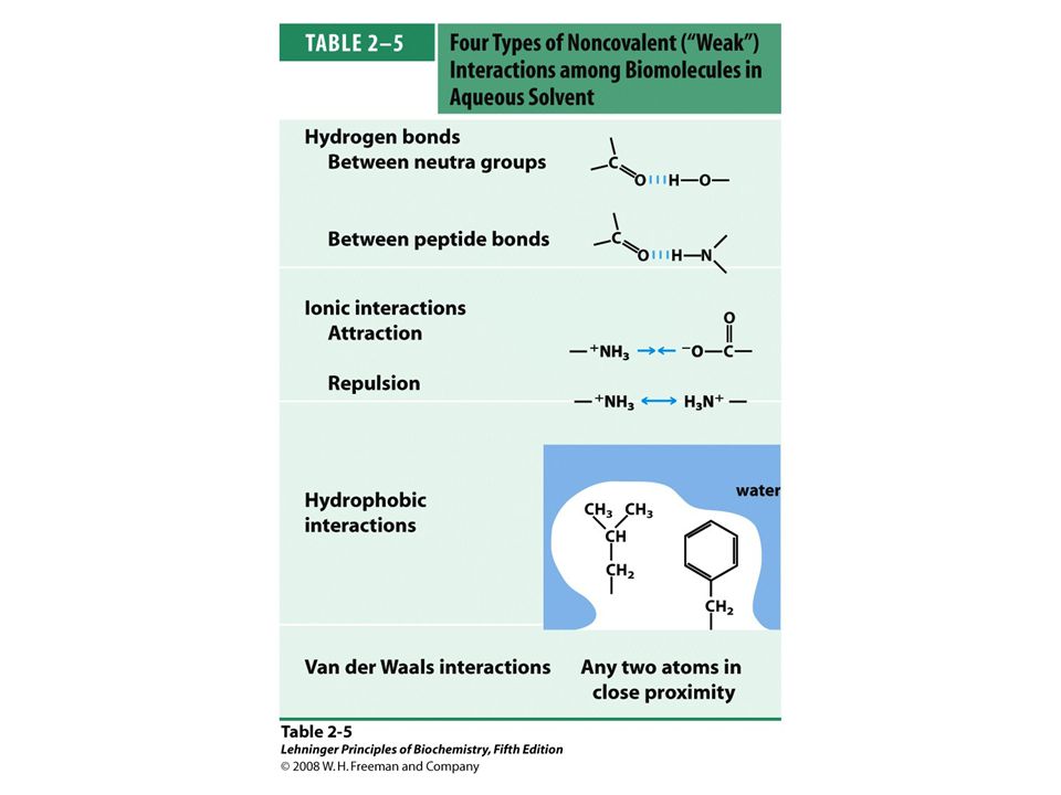
Physics of Non-covalent Interactions
Non-covalent interactions do not involve sharing a pair of electrons. Based on their physical origin, one can distinguish between Ionic (Coulombic) Interactions Electrostatic interactions between permanently charged species, or between the ion and a permanent dipole Dipole Interactions Electrostatic interactions between uncharged, but polar molecules Van der Waals Interactions Weak interactions between all atoms, regardless of polarity Attractive (dispersion) and repulsive (steric) component Hydrophobic Effect Complex phenomenon associated with the ordering of water molecules around non-polar substances
 Interactions. Electrostatic interactions between permanently charged species, or between the ion and a permanent dipole. Dipole Interactions. Electrostatic interactions between uncharged, but polar molecules. Van der Waals Interactions. Weak interactions between all atoms, regardless of polarity. Attractive (dispersion) and repulsive (steric) component. Hydrophobic Effect. Complex phenomenon associated with the ordering of water molecules around non-polar substances.")
4
Examples of Noncovalent Interactions

6
Hydrogen Bonds Strong dipole-dipole or charge-dipole interaction that arises between an acid (proton donor) and a base (proton acceptor) Typically 4-6 kJ/mol for bonds with neutral atoms, and 6-10 kJ/mol for bonds with one charged atom Typically involves two electronegative atoms (frequently nitrogen and oxygen) Hydrogen bonds are strongest when the bonded molecules are oriented to maximize electrostatic interaction. Ideally the three atoms involved are in a line
 Hydrogen bonds are strongest when. the bonded molecules are oriented to. maximize electrostatic interaction. Ideally the three atoms involved are in a line.")
7
FIGURE 2-5 Directionality of the hydrogen bond
FIGURE 2-5 Directionality of the hydrogen bond. The attraction between the partial electric charges (see Figure 2-1) is greatest when the three atoms involved in the bond (in this case O, H, and O) lie in a straight line. When the hydrogen-bonded moieties are structurally constrained (when they are parts of a single protein molecule, for example), this ideal geometry may not be possible and the resulting hydrogen bond is weaker.
 is greatest when the three atoms involved in the bond (in this case O, H, and O) lie in a straight line. When the hydrogen-bonded moieties are structurally constrained (when they are parts of a single protein molecule, for example), this ideal geometry may not be possible and the resulting hydrogen bond is weaker.")
8
Hydrogen Bonds: Examples

9
FIGURE 2-3 Common hydrogen bonds in biological systems
FIGURE 2-3 Common hydrogen bonds in biological systems. The hydrogen acceptor is usually oxygen or nitrogen; the hydrogen donor is another electronegative atom.

10
Importance of Hydrogen Bonds
Source of unique properties of water Structure and function of proteins Structure and function of DNA Structure and function of polysaccharides Binding of a substrates to enzymes Binding of hormones to receptors Matching of mRNA and tRNA

11
Biological Relevance of Hydrogen Bonds

12
FIGURE 2-4 Some biologically important hydrogen bonds.

13
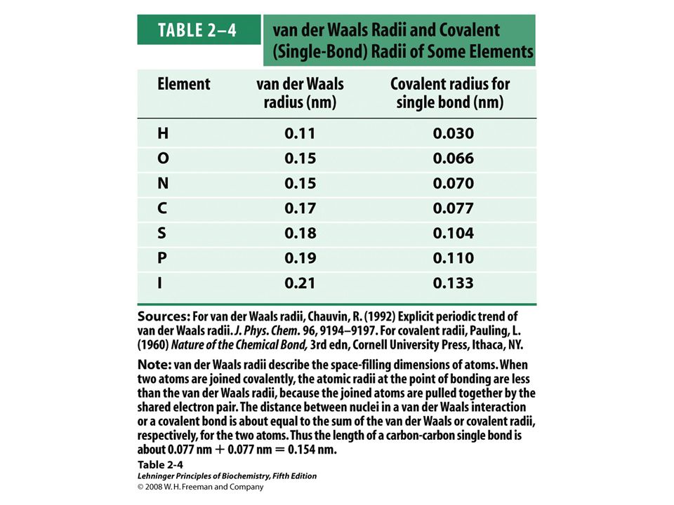
Van der Waals Interactions
Van der Waals interactions have two components: Attractive force (London dispersion) Depends on the polarizability Repulsive force (Steric repulsion) Depends on the size of atoms Attraction dominates at longer distances (typically nm) Repulsion dominates at very short distances There is a minimum energy distance (van der Waals contact distance)
 Depends on the polarizability. Repulsive force (Steric repulsion) Depends on the size of atoms. Attraction dominates at longer distances (typically nm) Repulsion dominates at very short distances. There is a minimum energy distance (van der Waals contact distance)")
15
Origin of the London Dispersion Force
Quantum mechanical origin Instantaneous polarization by fluctuating charge distributions Universal and always attractive Stronger in polarizable molecules Important only at a short range

16
Biochemical Significance of Van der Waals Interactions
Weak individually Easily broken, reversible Universal: Occur between any two atoms that are near each other Importance determines steric complementarity stabilizes biological macromolecules (stacking in DNA) facilitates binding of polarizable ligands
 facilitates binding of polarizable ligands.")
17
Water is the Medium for Life
Life evolved in water (UV protection) Organisms typically contain 70-90% water Chemical reactions occur in aqueous milieu Water is a critical determinant of the structure and function of proteins, nucleic acids, and membranes
 Organisms typically contain 70-90% water. Chemical reactions occur in aqueous milieu. Water is a critical determinant of the structure and function of proteins, nucleic acids, and membranes.")
18
Structure of the Water Molecule
Octet rule dictates that there are four electron pairs around an oxygen atom in water. These electrons are on four sp3 orbitals Two of these pairs covalently link two hydrogen atoms to a central oxygen atom. The two remaining pairs remain nonbonding (lone pairs) Water geometry is a distorted tetrahedron The electronegativity of the oxygen atom induces a net dipole moment Because of the dipole moment, water can serve as both a hydrogen bond donor and acceptor.
 Water geometry is a distorted tetrahedron. The electronegativity of the oxygen atom induces a net dipole moment. Because of the dipole moment, water can serve as both a hydrogen bond donor and acceptor.")
19
FIGURE 2-1a Structure of the water molecule
FIGURE 2-1a Structure of the water molecule. (a) The dipolar nature of the H2O molecule is shown in a ball-and-stick model; the dashed lines represent the nonbonding orbitals. There is a nearly tetrahedral arrangement of the outer-shell electron pairs around the oxygen atom; the two hydrogen atoms have localized partial positive charges (δ+) and the oxygen atom has a partial negative charge (δ–).
 The dipolar nature of the H2O molecule is shown in a ball-and-stick model; the dashed lines represent the nonbonding orbitals. There is a nearly tetrahedral arrangement of the outer-shell electron pairs around the oxygen atom; the two hydrogen atoms have localized partial positive charges (δ+) and the oxygen atom has a partial negative charge (δ–).")
20
Hydrogen Bonding in Water
Water can serve as both an H donor and an H acceptor Up to four H-bonds per water molecule gives water the anomalously high boiling point anomalously high melting point unusually large surface tension Hydrogen bonding in water is cooperative. Hydrogen bonds between neighboring molecules are weak (20 kJ/mole) relative to the H–O covalent bonds (420 kJ/mol)
 relative to the H–O covalent bonds (420 kJ/mol)")
21
FIGURE 2-1b Structure of the water molecule
FIGURE 2-1b Structure of the water molecule. (b) Two H2O molecules joined by a hydrogen bond (designated here, and throughout this book, by three blue lines) between the oxygen atom of the upper molecule and a hydrogen atom of the lower one. Hydrogen bonds are longer and weaker than covalent O—H bonds.
 Two H2O molecules joined by a hydrogen bond (designated here, and throughout this book, by three blue lines) between the oxygen atom of the upper molecule and a hydrogen atom of the lower one. Hydrogen bonds are longer and weaker than covalent O—H bonds.")
22
Water as a Solvent Water is a good solvent for charged and polar substances amino acids and peptides small alcohols carbohydrates Water is a poor solvent for nonpolar substances nonpolar gases aromatic moieties aliphatic chains

24
Water Dissolves Many Salts
High dielectric constant reduces attraction between oppositely charged ions in salt crystal, almost no attraction at large (> 40 nm) distance Strong electrostatic interactions between the solvated ions and water molecules lowers the energy of the system Entropy increases as ordered crystal lattice is dissolved
 distance. Strong electrostatic interactions between the solvated ions and water molecules lowers the energy of the system. Entropy increases as ordered crystal lattice is dissolved.")
25
FIGURE 2-6 Water as solvent
FIGURE 2-6 Water as solvent. Water dissolves many crystalline salts by hydrating their component ions. The NaCl crystal lattice is disrupted as water molecules cluster about the C– and Na+ ions. The ionic charges are partially neutralized, and the electrostatic attractions necessary for lattice formation are weakened.

26
Ice: Water in a Solid State
Water has many different crystal forms; the hexagonal ice is the most common Hexagonal ice forms a regular lattice, and thus has a low entropy Hexagonal ice has lower density than liquid water; ice floats

27
FIGURE 2-2 Hydrogen bonding in ice
FIGURE 2-2 Hydrogen bonding in ice. In ice, each water molecule forms four hydrogen bonds, the maximum possible for a water molecule, creating a regular crystal lattice. By contrast, in liquid water at room temperature and atmospheric pressure, each water molecule hydrogen-bonds with an average of 3.4 other water molecules. This crystal lattice structure makes ice less dense than liquid water, and thus ice floats on liquid water.

28
The Hydrophobic Effect
Refers to the association or folding of non-polar molecules in the aqueous solution Is one of the main factors behind: Protein folding Protein-protein association Formation of lipid micelles Binding of steroid hormones to their receptors Does not arise because of some attractive direct force between two non-polar molecules

29
Solubility of Polar and Non-polar Solutes
Why are non-polar molecules poorly soluble in water?

31
Low Solubility of Hydrophobic Solutes can be Explained by Entropy
Bulk water has little order: - high entropy Water near a hydrophobic solute is highly ordered: - low entropy Low entropy is thermodynamically unfavorable, thus hydrophobic solutes have low solubility

32
FIGURE 2-7a Amphipathic compounds in aqueous solution
FIGURE 2-7a Amphipathic compounds in aqueous solution. (a) Long-chain fatty acids have very hydrophobic alkyl chains, each of which is surrounded by a layer of highly ordered water molecules.
 Long-chain fatty acids have very hydrophobic alkyl chains, each of which is surrounded by a layer of highly ordered water molecules.")
33
Origin of the Hydrophobic Effect (1)
Consider amphipathic lipids in water Lipid molecules disperse in the solution; nonpolar tail of each lipid molecule is surrounded by ordered water molecules Entropy of the system decreases System is now in an unfavorable state

34
FIGURE 2-7b (part 1) Amphipathic compounds in aqueous solution
FIGURE 2-7b (part 1) Amphipathic compounds in aqueous solution. (b) By clustering together in micelles, the fatty acid molecules expose the smallest possible hydrophobic surface area to the water, and fewer water molecules are required in the shell of ordered water. The energy gained by freeing immobilized water molecules stabilizes the micelle.
 Amphipathic compounds in aqueous solution. (b) By clustering together in micelles, the fatty acid molecules expose the smallest possible hydrophobic surface area to the water, and fewer water molecules are required in the shell of ordered water. The energy gained by freeing immobilized water molecules stabilizes the micelle.")
35
Origin of the Hydrophobic effect (2)
Non-polar portions of the amphipathic molecule aggregate so that fewer water molecules are ordered. The released water molecules will be more random and the entropy increases. All non-polar groups are sequestered from water, and the released water molecules increase the entropy further. Only polar “head groups” are exposed and make energetically favorable H-bonds.

36
FIGURE 2-7b (part 2) Amphipathic compounds in aqueous solution
FIGURE 2-7b (part 2) Amphipathic compounds in aqueous solution. (b) By clustering together in micelles, the fatty acid molecules expose the smallest possible hydrophobic surface area to the water, and fewer water molecules are required in the shell of ordered water. The energy gained by freeing immobilized water molecules stabilizes the micelle.
 Amphipathic compounds in aqueous solution. (b) By clustering together in micelles, the fatty acid molecules expose the smallest possible hydrophobic surface area to the water, and fewer water molecules are required in the shell of ordered water. The energy gained by freeing immobilized water molecules stabilizes the micelle.")
37
FIGURE 2-7b (part 3) Amphipathic compounds in aqueous solution
FIGURE 2-7b (part 3) Amphipathic compounds in aqueous solution. (b) By clustering together in micelles, the fatty acid molecules expose the smallest possible hydrophobic surface area to the water, and fewer water molecules are required in the shell of ordered water. The energy gained by freeing immobilized water molecules stabilizes the micelle.
 Amphipathic compounds in aqueous solution. (b) By clustering together in micelles, the fatty acid molecules expose the smallest possible hydrophobic surface area to the water, and fewer water molecules are required in the shell of ordered water. The energy gained by freeing immobilized water molecules stabilizes the micelle.")
38
Hydrophobic Effect Favors Ligand Binding
Binding sites in enzymes and receptors are often hydrophobic Such sites can bind hydrophobic substrates and ligands such as steroid hormones Many drugs are designed to take advantage of the hydrophobic effect

39
FIGURE 2-8 Release of ordered water favors formation of an enzyme-substrate complex. While separate, both enzyme and substrate force neighboring water molecules into an ordered shell. Binding of substrate to enzyme releases some of the ordered water, and the resulting increase in entropy provides a thermodynamic push toward formation of the enzyme-substrate complex (see p. 192).
..")
40
Colligative Properties
Some properties of solution — boiling point, melting point, and osmolarity — do not depend strongly on the nature of the dissolved substance. These are called colligative properties Other properties — viscosity, surface tension, taste, and color, among other — depend strongly on the chemical nature of the solute. These are non-colligative properties. Cytoplasm of cells are highly concentrated solutions and have high osmotic pressure

41
FIGURE 2-11 Osmosis and the measurement of osmotic pressure
FIGURE 2-11 Osmosis and the measurement of osmotic pressure. (a) The initial state. The tube contains an aqueous solution, the beaker contains pure water, and the semipermeable membrane allows the passage of water but not solute. Water flows from the beaker into the tube to equalize its concentration across the membrane. (b) The final state. Water has moved into the solution of the nonpermeant compound, diluting it and raising the column of water within the tube. At equilibrium, the force of gravity operating on the solution in the tube exactly balances the tendency of water to move into the tube, where its concentration is lower. (c) Osmotic pressure (Π) is measured as the force that must be applied to return the solution in the tube to the level of that in the beaker. This force is proportional to the height, h, of the column in (b).
 The initial state. The tube contains an aqueous solution, the beaker contains pure water, and the semipermeable membrane allows the passage of water but not solute. Water flows from the beaker into the tube to equalize its concentration across the membrane. (b) The final state. Water has moved into the solution of the nonpermeant compound, diluting it and raising the column of water within the tube. At equilibrium, the force of gravity operating on the solution in the tube exactly balances the tendency of water to move into the tube, where its concentration is lower. (c) Osmotic pressure (Π) is measured as the force that must be applied to return the solution in the tube to the level of that in the beaker. This force is proportional to the height, h, of the column in (b).")
42
Effect of Extracellular Osmolarity

43
FIGURE 2-12 Effect of extracellular osmolarity on water movement across a plasma membrane. When a cell in osmotic balance with its surrounding medium—that is, a cell in (a) an isotonic medium—is transferred into (b) a hypertonic solution or (c) a hypotonic solution, water moves across the plasma membrane in the direction that tends to equalize osmolarity outside and inside the cell.
 an isotonic medium—is transferred into (b) a hypertonic solution or (c) a hypotonic solution, water moves across the plasma membrane in the direction that tends to equalize osmolarity outside and inside the cell..")
44
Ionization of Water
H2O H+ + OH- O-H bonds are polar and can dissociate heterolytically Products are a proton (H+) and a hydroxide ion (OH-) Dissociation of water is a rapid reversible process Most water molecules remain un-ionized, thus pure water has very low electrical conductivity (resistance: 18 M•cm) The equilibrium H2O H+ + OH- is strongly to the left Extent of dissociation depends on the temperature
 and a hydroxide ion (OH-) Dissociation of water is a rapid reversible process. Most water molecules remain un-ionized, thus pure water. has very low electrical conductivity (resistance: 18 M•cm) The equilibrium H2O H+ + OH- is strongly to the left. Extent of dissociation depends on the temperature. ")
45
Proton Hydration Protons do not exist free in solution.
They are immediately hydrated to form hydronium (oxonium) ions A hydronium ion is a water molecule with a proton associated with one of the non-bonding electron pairs Hydronium ions are solvated by nearby water molecules The covalent and hydrogen bonds are interchangeable. This allows for an extremely fast mobility of protons in water via “proton hopping”
 ions. A hydronium ion is a water molecule with a proton associated with one of the non-bonding electron pairs. Hydronium ions are solvated by nearby water molecules. The covalent and hydrogen bonds are interchangeable. This allows for an extremely fast mobility of protons in water via proton hopping")
46
Proton Hopping

47
FIGURE 2-13 Proton hopping
FIGURE 2-13 Proton hopping. Short “hops” of protons between a series of hydrogen-bonded water molecules result in an extremely rapid net movement of a proton over a long distance. As a hydronium ion (upper left) gives up a proton, a water molecule some distance away (lower right) acquires one, becoming a hydronium ion. Proton hopping is much faster than true diffusion and explains the remarkably high ionic mobility of H+ ions compared with other monovalent cations such as Na+ and K+.
 gives up a proton, a water molecule some distance away (lower right) acquires one, becoming a hydronium ion. Proton hopping is much faster than true diffusion and explains the remarkably high ionic mobility of H+ ions compared with other monovalent cations such as Na+ and K+.")
48
Ionization of Water: Quantitative Treatment
Concentrations of participating species in an equilibrium process are not independent but are related via the equilibrium constant [H+]•[OH-] H2O H+ + OH- Keq = ———— [H2O] Keq can be determined experimentally, it is 1.8•10-16 M at 25 °C [H2O] can be determined from water density, it is 55.5 M Ionic product of water: In pure water [H+] = [OH-] = 10-7 M

49
What is pH? pH is defined as the negative logarithm of the hydrogen ion concentration. Simplifies equations The pH and pOH must always add to 14 pH can be negative ([H+] = 6 M) In neutral solution, [H+] = [OH-] and the pH is 7 pH = -log[H+]
 In neutral solution, [H+] = [OH-] and the pH is 7. pH = -log[H+]")
50
pH Scale: 1 unit = 10-fold

52
FIGURE 2-14 The pH of some aqueous fluids.

53
Dissociation of Weak Electrolytes: Principle
Weak electrolytes dissociate only partially in water Extent of dissociation is determined by the acid dissociation constant Ka We can calculate the pH if the Ka is known. But some algebra is needed!

54
pKa = -log Ka (strong acid large Ka small pKa)
pKa measures acidity pKa = -log Ka (strong acid large Ka small pKa)
")
55
FIGURE 2-15 Conjugate acid-base pairs consist of a proton donor and a proton acceptor. Some compounds, such as acetic acid and ammonium ion, are monoprotic; they can give up only one proton. Others are diprotic (carbonic acid and glycine) or triprotic (phosphoric acid). The dissociation reactions for each pair are shown where they occur along a pH gradient. The equilibrium or dissociation constant (Ka) and its negative logarithm, the pKa, are shown for each reaction. *For an explanation of apparent discrepancies in pKa values for carbonic acid (H2CO3), see p. 63.
 or triprotic (phosphoric acid). The dissociation reactions for each pair are shown where they occur along a pH gradient. The equilibrium or dissociation constant (Ka) and its negative logarithm, the pKa, are shown for each reaction. *For an explanation of apparent discrepancies in pKa values for carbonic acid (H2CO3), see p")
56
Buffers are mixtures of weak acids and their anions
Buffers resist change in pH At pH = pKa, there is a 50:50 mixture of acid and anion forms of the compound Buffering capacity of acid/anion system is greatest at pH = pKa Buffering capacity is lost when the pH differs from pKa by more than 1 pH unit

57
FIGURE 2-16 The titration curve of acetic acid
FIGURE 2-16 The titration curve of acetic acid. After addition of each increment of NaOH to the acetic acid solution, the pH of the mixture is measured. This value is plotted against the amount of NaOH added, expressed as a fraction of the total NaOH required to convert all the acetic acid (CH3COOH) to its deprotonated form, acetate (CH3COO–). The points so obtained yield the titration curve. Shown in the boxes are the predominant ionic forms at the points designated. At the midpoint of the titration, the concentrations of the proton donor and proton acceptor are equal, and the pH is numerically equal to the pKa. The shaded zone is the useful region of buffering power, generally between 10% and 90% titration of the weak acid.
 to its deprotonated form, acetate (CH3COO–). The points so obtained yield the titration curve. Shown in the boxes are the predominant ionic forms at the points designated. At the midpoint of the titration, the concentrations of the proton donor and proton acceptor are equal, and the pH is numerically equal to the pKa. The shaded zone is the useful region of buffering power, generally between 10% and 90% titration of the weak acid.")
58
Henderson–Hasselbalch Equation: Derivation
HA H+ + A-

59
Biological Buffer Systems
Maintenance of intracellular pH is vital to all cells Enzyme-catalyzed reactions have optimal pH Solubility of polar molecules depends on H-bond donors and acceptors Equilibrium between CO2 gas and dissolved HCO3- depends on pH Buffer systems in vivo are mainly based on phosphate, concentration in millimolar range bicarbonate, important for blood plasma histidine, efficient buffer at neutral pH Buffer systems in vitro are often based on sulfonic acids of cyclic amines HEPES PIPES CHES

60
Water as a reactant in biochemistry

61
Bound Water in Proteins

62
FIGURE 2-9 Water binding in hemoglobin
FIGURE 2-9 Water binding in hemoglobin. (PDB ID 1A3N) The crystal structure of hemoglobin, shown (a) with bound water molecules (red spheres) and (b) without the water molecules. The water molecules are so firmly bound to the protein that they affect the x-ray diffraction pattern as though they were fixed parts of the crystal. The two α subunits of hemoglobin are shown in gray, the two β subunits in blue. Each subunit has a bound heme group (red stick structure), visible only in the β subunits in this view. The structure and function of hemoglobin are discussed in detail in Chapter 5.
 The crystal structure of hemoglobin, shown (a) with bound water molecules (red spheres) and (b) without the water molecules. The water molecules are so firmly bound to the protein that they affect the x-ray diffraction pattern as though they were fixed parts of the crystal. The two α subunits of hemoglobin are shown in gray, the two β subunits in blue. Each subunit has a bound heme group (red stick structure), visible only in the β subunits in this view. The structure and function of hemoglobin are discussed in detail in Chapter 5.")
63
FIGURE 2-10 Water chain in cytochrome f
FIGURE 2-10 Water chain in cytochrome f. Water is bound in a proton channel of the membrane protein cytochrome f, which is part of the energy-trapping machinery of photosynthesis in chloroplasts (see Figure 19-64). Five water molecules are hydrogen-bonded to each other and to functional groups of the protein: the peptide backbone atoms of valine, proline, arginine, and alanine residues, and the side chains of three asparagine and two glutamine residues. The protein has a bound heme (see Figure 5-1), its iron ion facilitating electron flow during photosynthesis. Electron flow is coupled to the movement of protons across the membrane, which probably involves “proton hopping” (see Figure 2-13) through this chain of bound water molecules.
. Five water molecules are hydrogen-bonded to each other and to functional groups of the protein: the peptide backbone atoms of valine, proline, arginine, and alanine residues, and the side chains of three asparagine and two glutamine residues. The protein has a bound heme (see Figure 5-1), its iron ion facilitating electron flow during photosynthesis. Electron flow is coupled to the movement of protons across the membrane, which probably involves proton hopping (see Figure 2-13) through this chain of bound water molecules.")
64
Summary The nature of intermolecular forces
The properties and structure of liquid water The behavior of weak acids and bases in water The way water can participate in biochemical reactions

Similar presentations


 Continued>")
 Topics Weak interactions in aqueous systems>")


 What holds atoms together? Ionic bonds Attraction between oppositely charged ions (atoms or molecules) Weak.>")