Download presentation
Presentation is loading. Please wait.

1
Page 863 Figure 23-25The pentose phosphate pathway.

7
FIGURE 14-27 Role of NADPH in regulating the partitioning of glucose 6- phosphate between glycolysis and the pentose phosphate pathway. When NADPH is forming faster than it is being used for biosynthesis and glutathione reduction (see Figure 14- 20), [NADPH] rises and inhibits the first enzyme in the pentose phosphate pathway. As a result, more glucose 6-phosphate is available for glycolysis.
, [NADPH] rises and inhibits the first enzyme in the pentose phosphate pathway. As a result, more glucose 6-phosphate is available for glycolysis..")
8
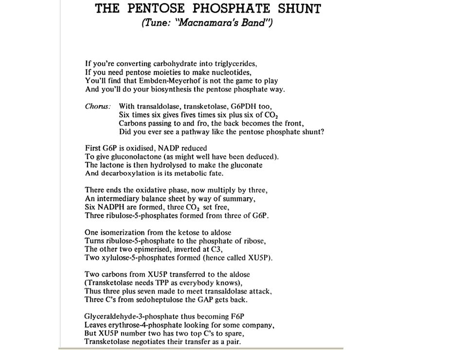
PPP Song

9
Lyrics http://books.google.com/books?id=oq9EN yL_d9YC&lpg=PP1&pg=PA1#v=onepage &q&f=false

12
LEHNINGER PRINCIPLES OF BIOCHEMISTRY Fifth Edition David L. Nelson and Michael M. Cox © 2008 W. H. Freeman and Company CHAPTER 15 Principles of Metabolic Regulation

23
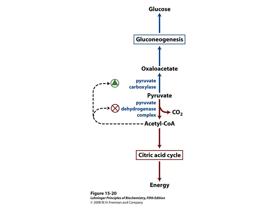
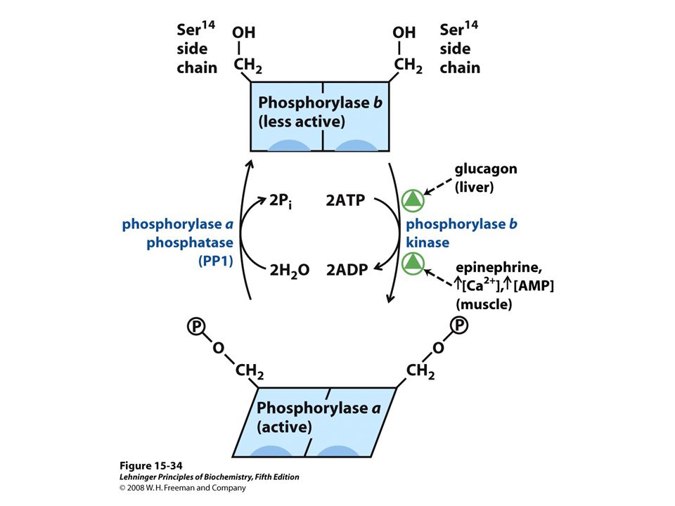
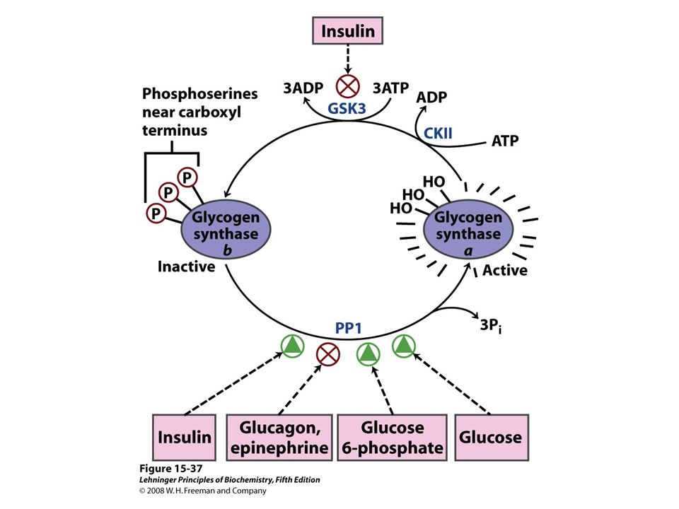
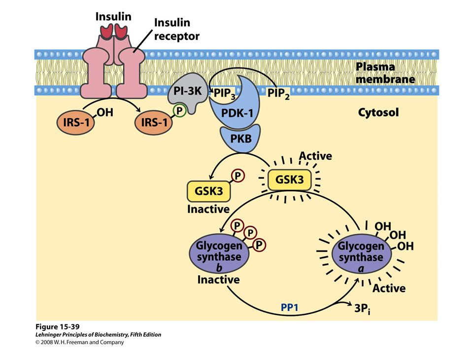
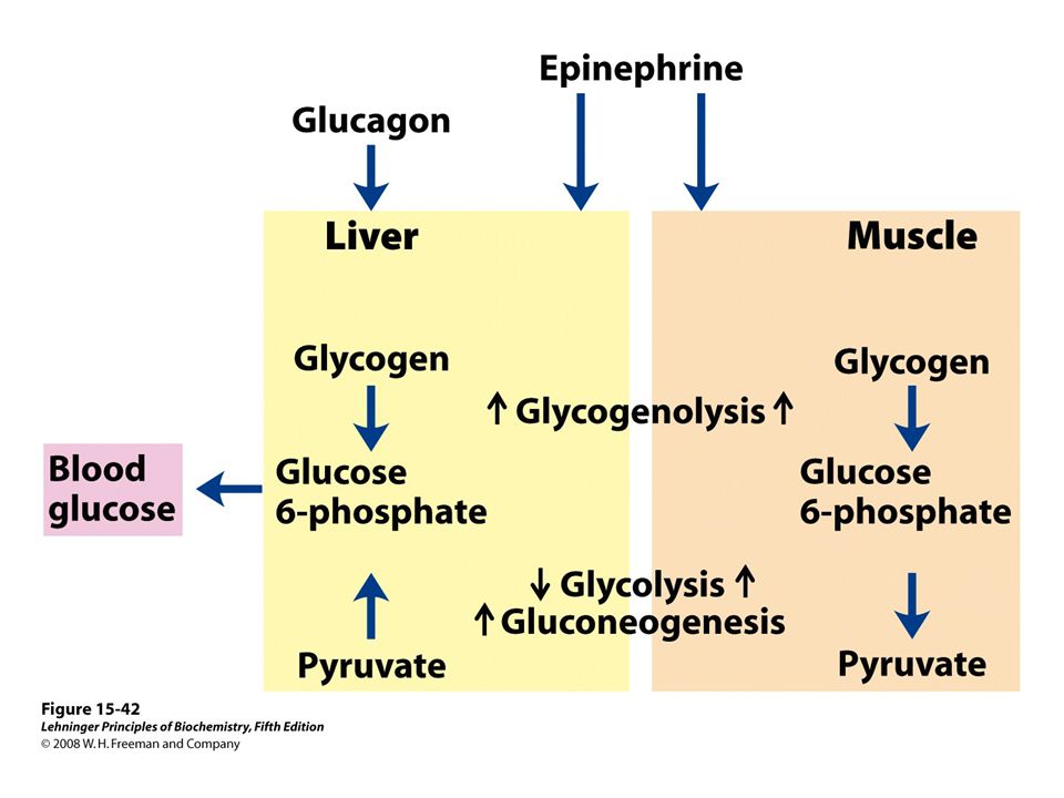
Insulin’s effects on transport and hexokinase activity, not the change in glycogen synthase activity, increase the flux toward glycogen synthesis.

24
Describe these differences! When blood glucose rises above 5 mM, hexokinase IV activity increases, but hexokinase I is already operating near Vmax and cannot respond to an increase in glucose concentration.

25
The protein inhibitor of hexokinase IV is a nuclear binding protein that draws hexokinase IV into the nucleus when the fructose 6-phosphate concentration in liver is high, and releases it to the cytosol when the glucose concentration is high.

26
FIGURE 15-14a Phosphofructokinase-1 (PFK-1) and its regulation. Ribbon diagram of E. coli PFK-1, showing two of its four identical subunits (PDB ID 1PFK). Each subunit has its own catalytic site, where the products ADP and fructose 1,6-bisphosphate are almost in contact, and its own binding sites for the allosteric regulator ADP (blue), located at the interface between subunits.
. Each subunit has its own catalytic site, where the products ADP and fructose 1,6-bisphosphate are almost in contact, and its own binding sites for the allosteric regulator ADP (blue), located at the interface between subunits..")
31
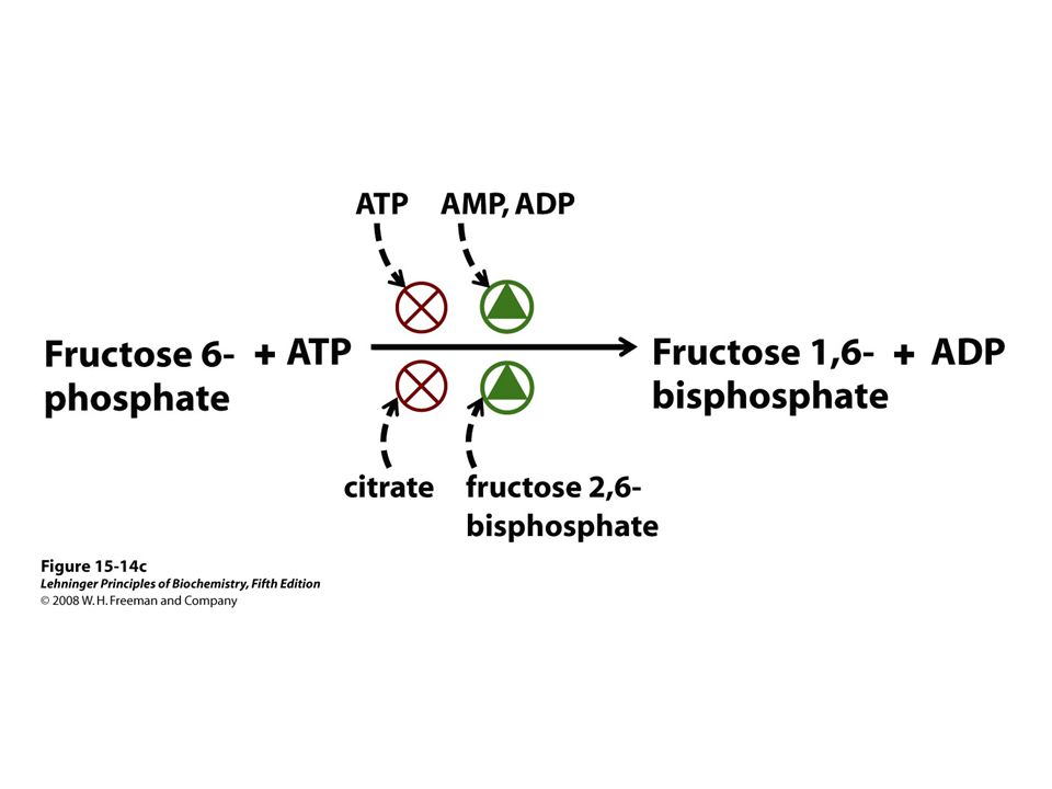
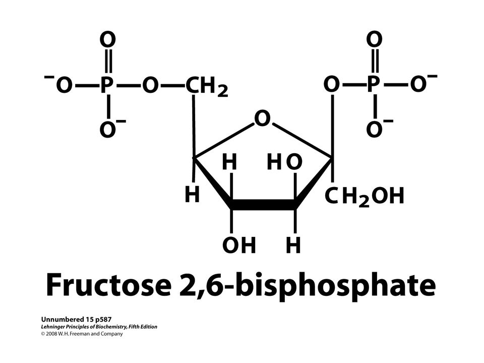
Fructose 2,6-bisphosphate (F26BP) has opposite effects on the enzymatic activities of phosphofructokinase-1 and fructose 1,6-bisphosphatase. (a) PFK-1 activity in the absence of F26BP is half-maximal when the concentration of fructose 6-phosphate is 2 mM. When 0.13 μM F26BP is present, the K0.5 for fructose 6-phosphate is only 0.08 mM. (b) FBPase-1 activity is inhibited by as little as 1 μM F26BP and is strongly inhibited by 25 μM. In the absence of this inhibitor the K0.5 for fructose 1,6- bisphosphate is 5 μM, but in the presence of 25 μM F26BP the K0.5 is >70 μM. Fructose 2,6-bisphosphate also makes FBPase-1 more sensitive to inhibition by another allosteric regulator, AMP.
 PFK-1 activity in the absence of F26BP is half-maximal when the concentration of fructose 6-phosphate is 2 mM. When 0.13 μM F26BP is present, the K0.5 for fructose 6-phosphate is only 0.08 mM. (b) FBPase-1 activity is inhibited by as little as 1 μM F26BP and is strongly inhibited by 25 μM. In the absence of this inhibitor the K0.5 for fructose 1,6- bisphosphate is 5 μM, but in the presence of 25 μM F26BP the K0.5 is >70 μM. Fructose 2,6-bisphosphate also makes FBPase-1 more sensitive to inhibition by another allosteric regulator, AMP..")
33
Both enzyme activities are part of the same polypeptide chain, and they are reciprocally regulated by insulin and glucagon.

34
3D structure of one enzyme subunit of the testis PFK- 2/FBPase-2 isoenzyme The co-ordinates were retrieved from the PDB database (accession code 1BIF) containing ATPγS in the PFK-2 domain. In the upper right hand PFK-2 domain, ATP is on the left and Fru-6-P is on the right. In the lower left hand FBPase-2 domain, two inorganic phosphates indicate the position of the Fru-2,6-P2-binding site. The PFK-2 domain is composed of a β-sheet surrounded by α- helices. Two subdomains, composed of α-helices (above) form a flexible cover and are involved in Fru-6-P binding and catalysis (Biochem J. 2004 August 1; 381(Pt 3): 561–579.Biochem J. 2004 August 1; 381(Pt 3): 561–579.
 form a flexible cover and are involved in Fru-6-P binding and catalysis (Biochem J August 1; 381(Pt 3): 561–579.Biochem J August 1; 381(Pt 3): 561–579..")
35
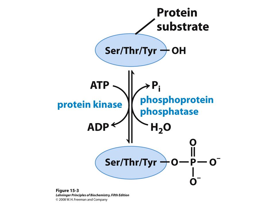
Phosphoprotein phosphatase

39
The PEP carboxykinase promoter region, showing the complexity of regulatory input to this gene.

40
Glycogen granules in a hepatocyte

49
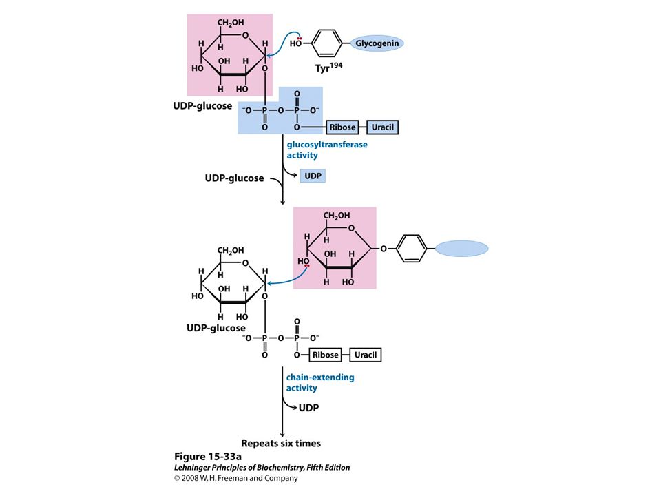
Glycogenin structure. Muscle glycogenin (Mr 37,000) forms dimers in solution. The substrate, UDP-glucose, is bound to a Rossmann fold near the amino terminus and is some distance from the Tyr194 residues—15 Å from the Tyr in the same monomer, 12 Å from the Tyr in the dimeric partner. Each UDP-glucose is bound through its phosphates to a Mn2+ ion that is essential to catalysis. Mn2+ is believed to function as an electron-pair acceptor to stabilize the leaving group, UDP. The glycosidic bond in the product has the same configuration about the C-1 of glucose as the substrate UDP-glucose, suggesting that the transfer of glucose from UDP to Tyr194 occurs in two steps. The first step is probably a nucleophilic attack by Asp162, forming a temporary intermediate with inverted configuration. A second nucleophilic attack by Tyr194 then restores the starting configuration

64
“Alfonse, Biochemistry makes my head hurt!!” \

Similar presentations


 2-phosphoglycerate. B) oxaloacetate. C)>")


 Molecular formula. (b) Schematic diagram illustrating its branched structure. Page 627.>")


