Download presentation
Presentation is loading. Please wait.

1
Lecture 11: GPCR pathways Fain Chapter 4 10/7/09

3
Kao - high transmission fiber optic cables from pure materials

4
Fiber optic networks Current amount of fiber goes around world 25,000 times

5
Boyle and Smith - CCD

8
Central dogma DNA mRNA protein

9
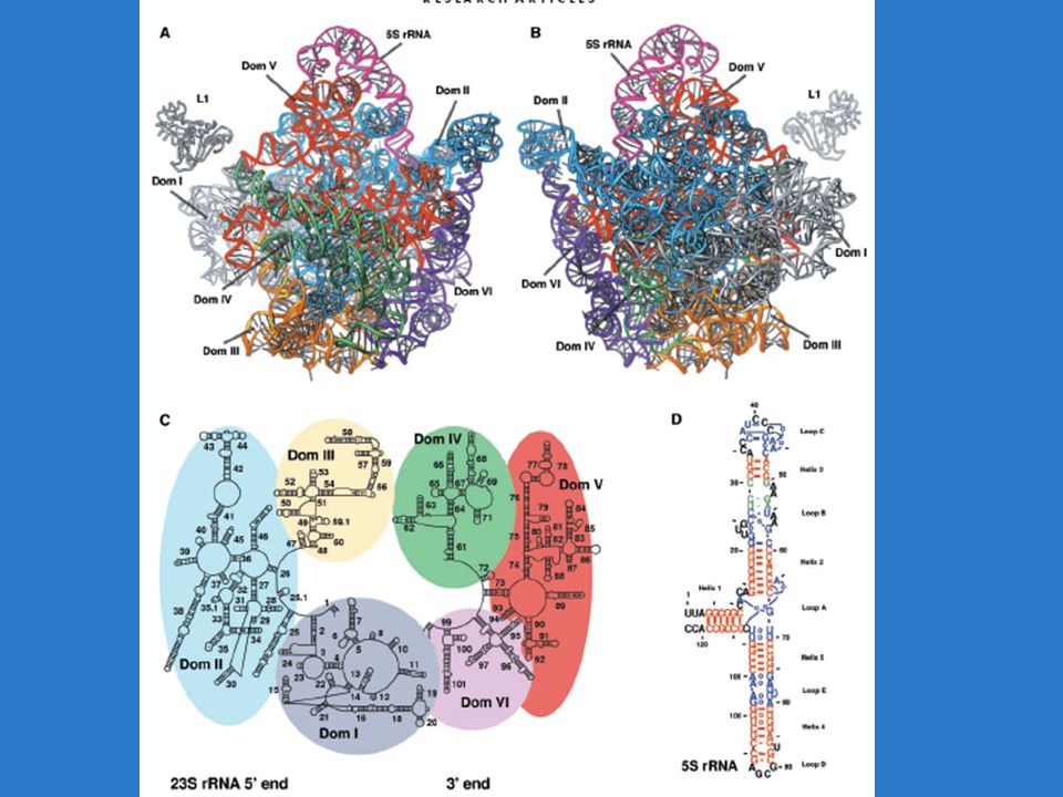
The amazing ribosome creates proteins

10
Ada Yonath crystallizes ribosome Small subunit - 32 proteins Large subunit - 46 proteins Geobacillus stearothermophilus - hot springs Haloarcula marismortui - Dead Sea

17
Homework #5 Gene duplication Build a tree (Pasteur) Think about gene function (OMIM) Locate genes (Map viewer)
 Think about gene function (OMIM) Locate genes (Map viewer)")
18
Homework #1: GNA trees GNAI2 GNAI3 GNAI1 GNAT1 GNAT2 GNAT3Taste Cone Rod

19
Search for gene of interest

20
Link to chromosome location Click here

21
Can control which maps are shown

22
Can remove those you don’t want (rna maps) Highlight items and then click remove Then update with OK
 Highlight items and then click remove Then update with OK")
23
Find chromosomal location of gene - many links

24
You can zoom in or out to see more detail

25
Location of GNAT and GNAI GNAT2 GNAI3 1p13 GNAT1 GNAI2 3p21 GNAT3 GNAI1 7q21

26
Location of GNAI and GNAT

27
Rest of semester Individual senses Fain chapters Primary literature Midterm project Trp channel analysis - next week Individual project topics Diversity of one sense across organisms Signal transduction Sensory diversity within one organism

28
The wonders of G protein signaling Signal amplification Control, regulation and specificity Evolution of diversity Gene expression

29
Ch4: Metabotropic signal transduction Indirect link from receptor to channel Use messenger system Receptor G protein Effector 2nd messenger Channel Neural signal Receptors are G protein coupled Similar to hormone and neurotransmitter signal transduction mechanisms

30
Metabotropic sensory transduction Figure 4.1 Channel

31
G proteins activate effectors Adenylyl cyclase = makes cAMP Guanyl cyclase = makes cGMP Phospholipase C = makes DAG and IP 3 Phospholipase A = makes arachidonic acid Phosphodiesterase = hydrolyzes cAMP or cGMP 2nd messengers open/close channels change ion concentration and membrane potential

32
Diversity of GPCRs Human genome 1500-2000 GPCRs (3-5% of genome) Kinds Hormone receptors FSH, Oxytocin, Vasopressin Synaptic transmitters Dopamine, opiates, glutamate Sensory receptors Olfactory receptors Visual pigments Taste receptors for bitter, sweet and AA
 Kinds Hormone receptors FSH, Oxytocin, Vasopressin Synaptic transmitters Dopamine, opiates, glutamate Sensory receptors Olfactory receptors Visual pigments Taste receptors for bitter, sweet and AA")
33
Basic GPCR structure 7 TM regions Phosphorylation sites on C terminus G protein binds to C terminus and intracellular loops 2 and 3 Figure 4.3

34
Basic GPCR structure 7 TM regions Phosphorylation sites on C terminus G protein binds to C terminus and intracellular loops 2 and 3 Ligand binds either - in membrane - norepinephrine - olfaction -extracellular site - glutamate - GABA Figure 4.3

35
Xray crystal structure of GPCR Palczewski et al 2000 11-cis retinal Rhodopsin

36
Yokoyama and Starmer 1996 GPCR phylogeny N=neurotransmitter P=peptides S=sensory

37
G proteins Ones that interact with GPCR are trimeric - and Act like switch Binding site on for GDP If exchange GDP for GTP, becomes activated Dissociates from

38
G protein= Numbers of different versions of subunits in human genome 20-30 G 5 G 12 G

39
1994 Nobel prize in medicine

40
GGGG GPA Ancient G proteins

41
GGGG G s stimulates adenylate cyclase includes olfactory

42
GGGG G s stimulates adenylate cyclase includes olfactory G i /G o Inhibitory and other Includes vision and taste Transducin gustducin

43
GGGG G s stimulates adenylate cyclase includes olfactory G i /G o inhibits Includes vision and taste G q Activates PLC

44
G and tethered to membrane tethered by geranyl geranyl (gg) tethered by palmitoyl (p)
 tethered by palmitoyl (p)")
45
G bound to GDP is inactive

46
Activated GPCR activates G protein GDP GPCR

47
GTP binding activates G GDP GTP + Get dissociation of G and G GPCR *

48
GTP binding activates G + Both G and G can activate effector molecules Effector AMP cAMP

49
GTP hydrolysis inactivates G Recombines with G + Hydrolysis to GDP + G will hydrolyze its own GTP slowly GTPase activating proteins speed hydrolysis Regulator of G protein signaling (RGS)
")
50
Lichtarge wanted to explain properties of G proteins How are they kept inactive? G -G binding How do they interact with receptors? GPCR binding How are they activated? GDP-GTP binding pocket How do they interact with effectors? How are they inactivated?

51
Evolutionary trace analysis

52
Evolutionary trace (ET) method ET is a way to compare proteins and identify conserved functional regions Ask evolution where these regions are? Hypothesis: Selection acts on AA sequence Parts of molecule which are critical for function will be highly conserved Parts of molecule which vary are not important

53
ET Compare proteins with same function Sites which are fixed are key to function Ignore variable sites Compare proteins with different function Invariant sites - same for both functions Class specific - fixed within function and different between functions

54
ET method 1.Gather protein homologs and align sequences 2.Use phylogenetic methods to group them into functional groups Determine fixed sites in each group 3.Compare fixed sites between groups: Class-specific sites - distinguish function Invariant sites 4.Map functionally important sites onto 3D structure If they cluster, this is likely an active site

55
Lichtarge compared 120 sequences for G G s stimulates adenylate cyclase (AC) G t stimulates PDE G i inhibits AC G o G q activates PLC Also compare 20 G and 16 G
 G t stimulates PDE G i inhibits AC G o G q activates PLC Also compare 20 G and 16 G ")
56
ET method

57
Key changes in function occur along nodes to groups

58
GTP binding activates G GDP GTP + Get dissociation of G and G GPCR *

59
Conserved sites mapped onto Xray crystal struture G A1 binds to receptor along with C terminal tail GTP is shown in blue

60
Conserved sites mapped onto Xray crystal struture 9 of 17 sites in A1 class specific A1 likely interacts with effectors too

61
Effector can bind to site A2 also 14 of 32 sites in A2 are class specific and so specific to effector

62
Remove subunit to find conserved binding region Conserved sites identified by ET comparisons

63
Remove G to conserved binding site A2 is binding site of to

64
Test importance of site for function Mutate sites in G sequence by replacing them one by one with alanine Express mutant G and combine with G and rhodopsin Add light If G is working: R+hv R * + G G -GTP-S 35 Measure S 35 to quantify amount of G t activation

65
Onrust et al. 1997

66
ET sites agree with those found by site directed mutagenesis Sites important for interactions receptor Sites important for receptor binding

67
Biochemists can study protein function Change one site at time and see what happens Or just let evolution do the tests See what sites are important!

68
Homework Q1 - about Nobel prizes awarded this week Q2 - about past Nobel prize lecture If you can’t think of one to do, watch Roderick Mackinnon talk about ion channels Lots of good ones!

Similar presentations


 Cell Signaling & Signal Transduction Steroid hormones (also thyroid hormone) enter cells to regulate gene expression. Signal.>")


 Membranes. Membrane transport Membranes are selectively permeable barriers Hydrophobic uncharged small molecules can freely diffuse.>")


