Download presentation
Presentation is loading. Please wait.

1
Islamic University _Gaza Department of Biotechnology
Lab #3 Primary Culture Islamic University _Gaza Faculty of science Department of Biotechnology By: Mahmoud W EL-Hindi

2
Four stages to consider:
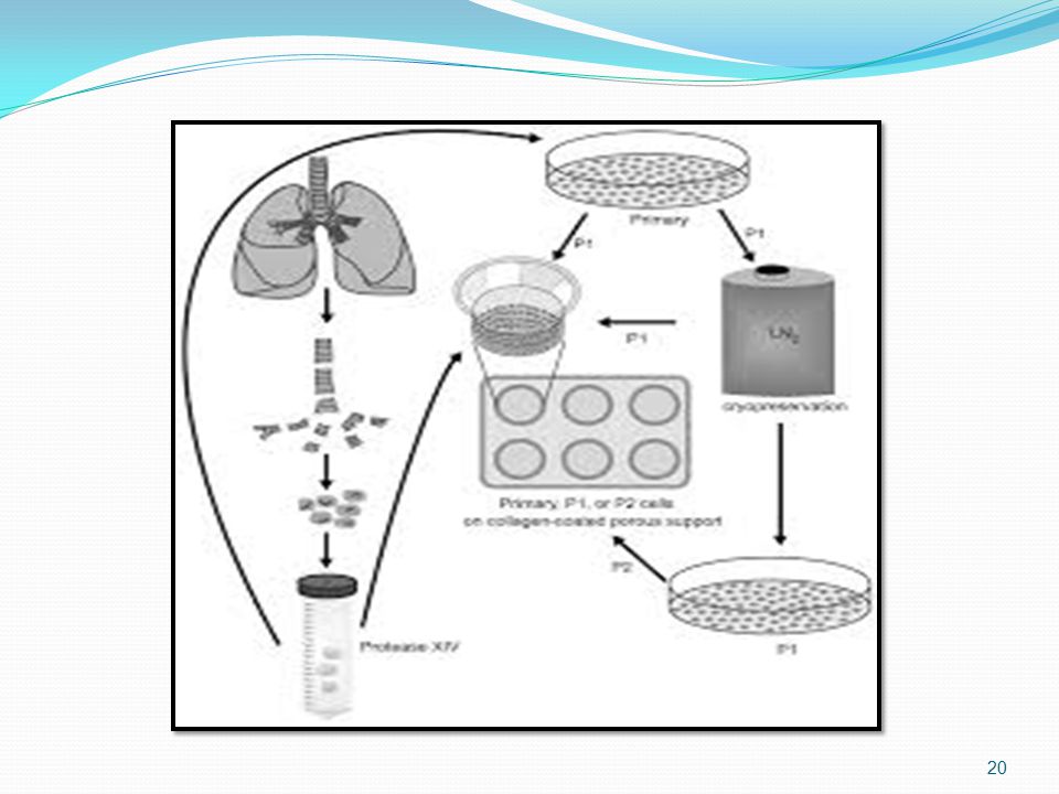
Obtained of the sample. Isolation of the tissue . Dissection and/or disaggregation . Culture after seeding into vessel culture .

3
Cont. After isolation, a primary cell culture may be obtained either by allowing cells to migrate out from fragments of tissue adhering to a suitable substrate or by disaggregation the tissue mechanically or enzymatically to produce a suspension of cells, some of which will ultimately attach to the substrate .

4
Cont. It appears to be essential for most normal untransformed cells, with the exception of hematopoietic cells, to attach to a flat surface in order to survive and proliferate with maximum efficiency . Transformed cells on the other hand, particularly cells from transplantable animal tumors, are often able to proliferate in suspension .

5
Cont. The enzymes used most frequently for tissue disaggregation are crude preparations of trypsin, collagenase, elastase, pronase, dispase, DNase, and hyaluronidase, alone or in various combination.

6
Cont. Such as : Elastase & Dnase for type II alveolar cell isolation collagenase with Dispase and collagenase with hyaluronidase . There are other, non mammalian enzymes, such as trypsin, TrypLE(Invitrogen), recombinant microbial, and Accutase and Accumax (Innovative Cell Technologies), also available for primary disaggregation.
, recombinant microbial, and Accutase and Accumax (Innovative Cell Technologies), also available for primary disaggregation.")
7
Cont. Crude preparations are often more successful than purified enzyme preparations, because the former contain other proteases as contaminations, although the latter are generally less toxic and more specific in there action. Trypsin and pronase give the most complete disaggregation, but may damage the cells. Collagenase and dispase, on the other hand, give incomplete disaggregation, but are less harmful.

8
Cont. Hyaluronidase can be used in conjunction with collagenase to digest the intracellular matrix, and Dnase is used to disperse DNA released from lysed cells; DNA tends to impair proteolysis and promote reaggregation.

9
Requirements & conditions of Tissues:.
(1) Fat and necrotic tissue is best removed during dissection . (2) The tissue should be chopped finely with sharp instruments to cause minimum damage . (3) Enzymes used for disaggregation should be removed subsequently by gentle centrifugation . (4) The concentration of cells in the primary culture should be much higher than that normally used for subculture, because the proportion of cells form the tissue that survives in primary culture may be quite low.
 Fat and necrotic tissue is best removed during dissection . (2) The tissue should be chopped finely with sharp instruments to cause minimum damage . (3) Enzymes used for disaggregation should be removed subsequently by gentle centrifugation . (4) The concentration of cells in the primary culture should be much higher than that normally used for subculture, because the proportion of cells form the tissue that survives in primary culture may be quite low.")
10
Cont. (5) A rich medium, such as Ham’s F12, is preferable to a simple medium, such as Eagle’s MEM, and, if serum is required, fetal bovine often gives better survival than does calf or horse. Isolation of specific cell types will probably require selective media. (6) Embryonic tissue disaggregates more readily, yields more viable cells, and proliferates more rapidly in primary culture than does adult tissue.
 A rich medium, such as Ham’s F12, is preferable to a simple medium, such as Eagle’s MEM, and, if serum is required, fetal bovine often gives better survival than does calf or horse. Isolation of specific cell types will probably require selective media. (6) Embryonic tissue disaggregates more readily, yields more viable cells, and proliferates more rapidly in primary culture than does adult tissue.")
11
Materials & Solutions :
Rabbits . Dissecting tools . Petri dishes . Pipettes . Tips . Dissecting stage . Incubator . Culture flasks . Centrifuge tubes .

12
Cont. Ethanol 70% . Phosphate Buffer Saline (PBS ) sterile.
Hanks BSS . DMEM media . Cups .

13
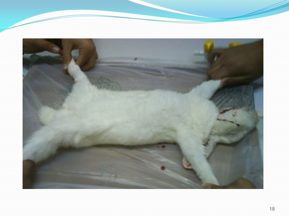
Protocol (1) Transfer tissue to fresh, sterile DBSS, and rinse .
(2) Transfer tissue to a second dish; dissect off unwanted tissue, such as fat or necrotic material, and transfer to a third dish . (3) Chop finely with crossed scalpels into about 1-mm cubes. (4) Transfer by pipette to a sterile centrifuge tube . (5) Allow the pieces to settle.
 Transfer tissue to a second dish; dissect off unwanted tissue, such as fat or necrotic material, and transfer to a third dish . (3) Chop finely with crossed scalpels into about 1-mm cubes. (4) Transfer by pipette to a sterile centrifuge tube . (5) Allow the pieces to settle.")
14
Cont. (6) Wash by resuspending the pieces in DBSS, allowing the pieces to settle, and removing the supernatant fluid. Repeat this step tow more times. (7) Transfer the pieces to a culture flask “ 20_30” pieces. (8) Remove most of the fluid, and add 1ml growth medium. (9) Cap the flask, and place it in an incubator 37C for 18 _ 24 h .
 Wash by resuspending the pieces in DBSS, allowing the pieces to settle, and removing the supernatant fluid. Repeat this step tow more times. (7) Transfer the pieces to a culture flask 20_30 pieces. (8) Remove most of the fluid, and add 1ml growth medium. (9) Cap the flask, and place it in an incubator 37C for 18 _ 24 h .")
15
Cont. (10) If the pieces have adhered, then the medium volume may be made up gradually over the next 3 _5 days to 10ml and then changed weekly. (11) When observed the growth in culture you must transfer the tissue into fresh culture vessel . ( 12 ) Subculture if necessary .
 When observed the growth in culture you must transfer the tissue into fresh culture vessel . ( 12 ) Subculture if necessary .")
21
Good Luck Girls

Similar presentations