Download presentation
Presentation is loading. Please wait.

1
Chapter 10: Anatomy of the Muscular System

2
INTRODUCTION The body contains more than 600 skeletal muscles (Figures 10-5 and 10-6) From 40% to 50% of body weight is skeletal muscle Muscles, along with the skeleton, determine the form and contour of the body

3
Connective tissue components
Cells are surrounded and bundled by connective tissue Endomysium—encloses a single muscle fiber Perimysium—wraps around a fascicle (bundle) of muscle fibers Epimysium—covers the entire skeletal muscle All 3 connective tissue layers and continuous with tendons or aponeurosis Fascia surrounds epimysium and other organs within the body
 of muscle fibers. Epimysium—covers the entire skeletal muscle. All 3 connective tissue layers and continuous with tendons or aponeurosis. Fascia surrounds epimysium and other organs within the body.")
5
SKELETAL MUSCLE STRUCTURE (cont.)
Size, shape, and fiber arrangement (Figure 10-2) Skeletal muscles vary considerably in size, shape, and fiber arrangement Size: ranges from extremely small to large masses Shape: variety of shapes, such as broad, narrow, long, tapering, short, blunt, triangular, quadrilateral, irregular, flat sheets, or bulky masses Arrangement: variety of arrangements, such as parallel to a long axis, converging to a narrow attachment, oblique, pennate, bipennate, or curved; the direction of fibers is significant because of its relation to function
 Skeletal muscles vary considerably in size, shape, and fiber arrangement. Size: ranges from extremely small to large masses. Shape: variety of shapes, such as broad, narrow, long, tapering, short, blunt, triangular, quadrilateral, irregular, flat sheets, or bulky masses. Arrangement: variety of arrangements, such as parallel to a long axis, converging to a narrow attachment, oblique, pennate, bipennate, or curved; the direction of fibers is significant because of its relation to function.")
7
SKELETAL MUSCLE STRUCTURE (cont.)
Attachment of muscles (Figure 10-3) Origin: point of attachment that does not move when the muscle contracts Insertion: point of attachment that moves when the muscle contracts
 Origin: point of attachment that does not move when the muscle contracts. Insertion: point of attachment that moves when the muscle contracts.")
9
SKELETAL MUSCLE STRUCTURE (cont.)
Muscle actions (Figure 10-4) Most movements are produced by the coordinated action of several muscles; some muscles in the group contract while others relax Prime mover: a muscle that directly performs a specific movement Agonists: any “mover” muscle that directly performs a movement, including the prime mover Antagonist: muscles that, when contracting, directly oppose prime movers; relax while the prime mover (agonist) is contracting to produce movement; provide precision and control during contraction of prime movers Synergists: muscles that contract at the same time as the prime movers; facilitate prime mover actions to produce a more efficient movement Fixator muscles: joint stabilizers (type of synergist)
 Most movements are produced by the coordinated action of several muscles; some muscles in the group contract while others relax. Prime mover: a muscle that directly performs a specific movement. Agonists: any mover muscle that directly performs a movement, including the prime mover. Antagonist: muscles that, when contracting, directly oppose prime movers; relax while the prime mover (agonist) is contracting to produce movement; provide precision and control during contraction of prime movers. Synergists: muscles that contract at the same time as the prime movers; facilitate prime mover actions to produce a more efficient movement. Fixator muscles: joint stabilizers (type of synergist)")
11
SKELETAL MUSCLE STRUCTURE (cont.)
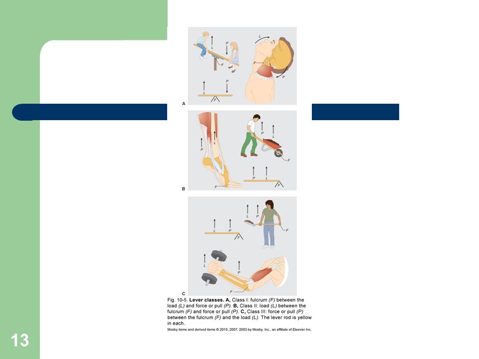
Lever systems In the human body, bones serve as levers and joints serve as fulcrums; contracting muscle applies a pulling force on a bone lever at the point of the muscle’s attachment to the bone, which causes the insertion bone to move about its joint fulcrum Composed of four component parts (Figure 10-5) Rigid bar (bone) Fulcrum around which the rod moves (joint) Load that is moved Pull that produces movement (muscle contraction) First-class levers Fulcrum lies between the pull and the load Not abundant in the human body; serve as levers of stability
 Rigid bar (bone) Fulcrum around which the rod moves (joint) Load that is moved. Pull that produces movement (muscle contraction) First-class levers. Fulcrum lies between the pull and the load. Not abundant in the human body; serve as levers of stability.")
12
SKELETAL MUSCLE STRUCTURE (cont.)
Lever systems (cont.) Second-class levers Load lies between the fulcrum and the joint at which the pull is exerted Presence of these levers in the human body is a controversial issue Third-class levers Pull is exerted between the fulcrum and load Permit rapid and extensive movement Most common type of lever found in the body
 Second-class levers. Load lies between the fulcrum and the joint at which the pull is exerted. Presence of these levers in the human body is a controversial issue. Third-class levers. Pull is exerted between the fulcrum and load. Permit rapid and extensive movement. Most common type of lever found in the body.")
14
HOW MUSCLES ARE NAMED Muscle names can be in Latin or English (this text uses English) Muscles are named according to one or more of the following features: Location, function, shape (Tables 10-2 to 10-4) Direction of fibers: named according to fiber orientation (Table 10-5) Number of heads or divisions (Table 10-5) Points of attachment: origin and insertion points Relative size: small, medium, or large (Table 10-6) Hints on how to deduce muscle actions
 Direction of fibers: named according to fiber orientation (Table 10-5) Number of heads or divisions (Table 10-5) Points of attachment: origin and insertion points. Relative size: small, medium, or large (Table 10-6) Hints on how to deduce muscle actions.")
16
IMPORTANT SKELETAL MUSCLES
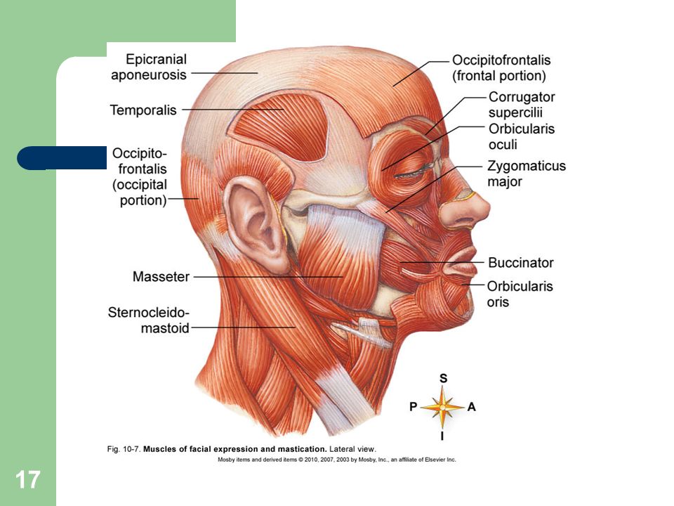
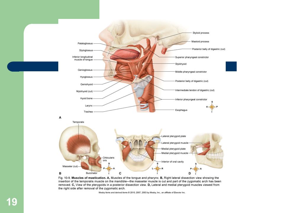
Muscles of facial expression: unique in that at least one point of attachment is to the deep layers of the skin over the face or neck (Figures 10-6 to 10-8; Table 10-7) Muscles of mastication: responsible for chewing movements (Figure 10-9; Table 10-7) Muscles that move the head: paired muscles on either side of the neck are responsible for head movements (Figure 10-10; Table 10-8)
 Muscles of mastication: responsible for chewing movements (Figure 10-9; Table 10-7) Muscles that move the head: paired muscles on either side of the neck are responsible for head movements (Figure 10-10; Table 10-8)")
21
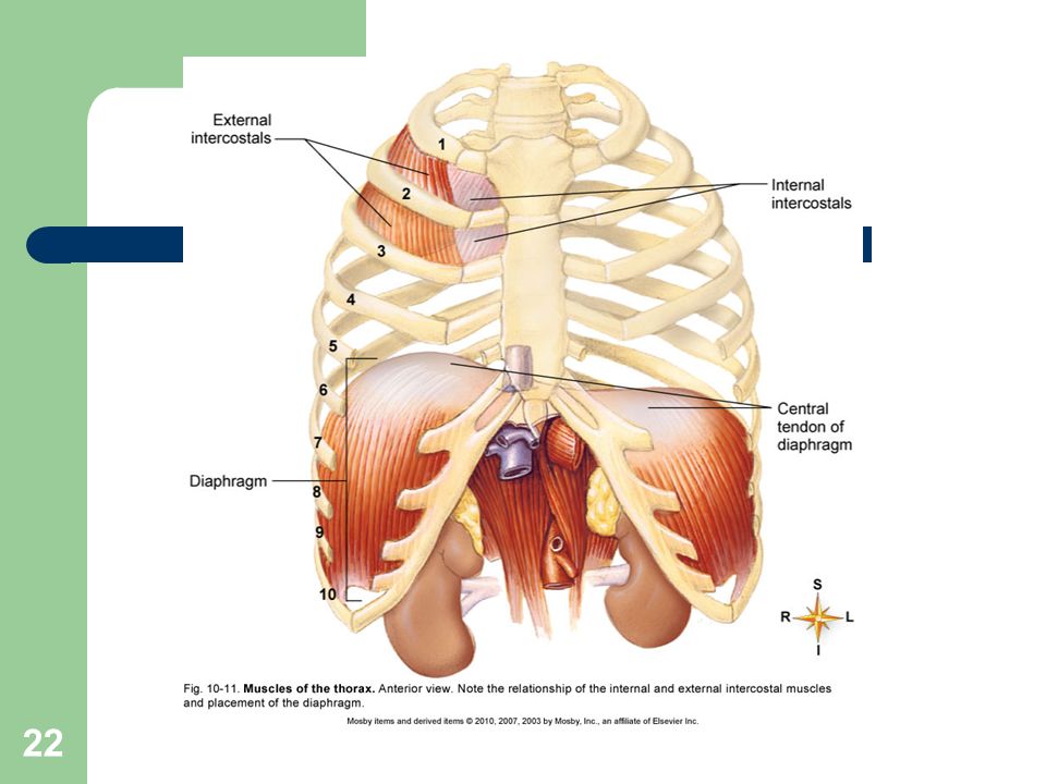
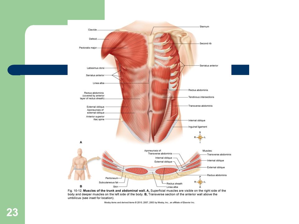
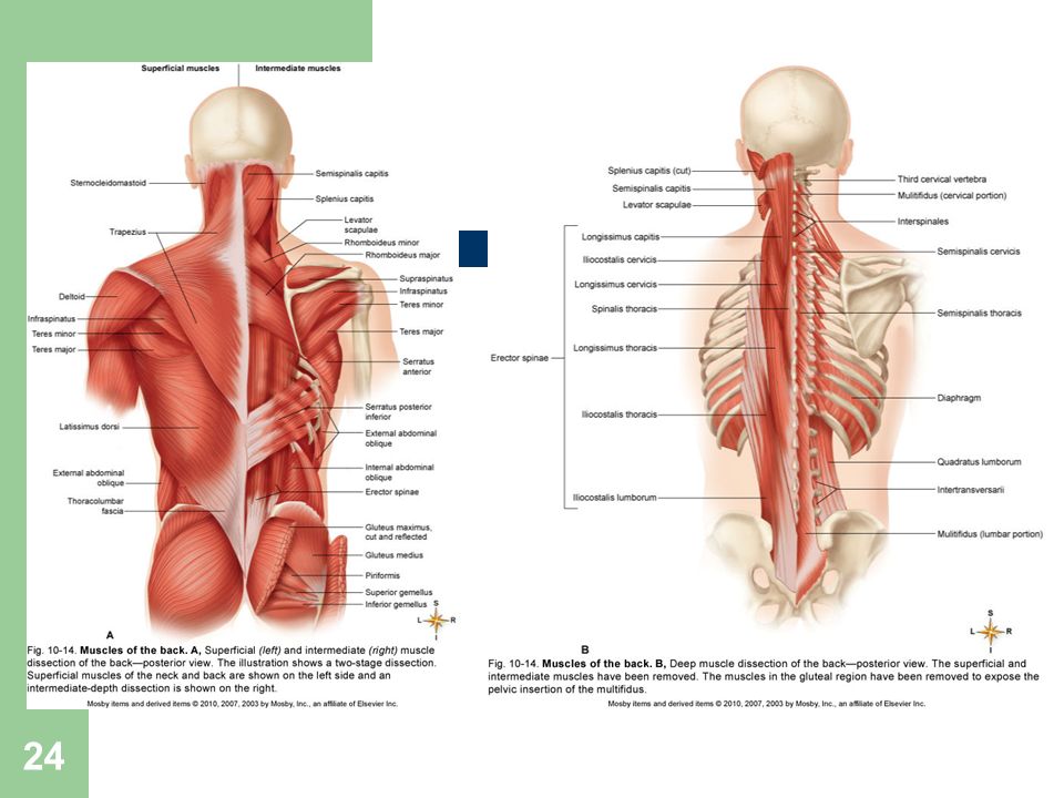
TRUNK MUSCLES Muscles of the thorax: critically important in respiration (Figure 10-11; Table 10-9) Muscles of the abdominal wall: arranged in three layers, with fibers in each layer running in different directions to increase strength (Figure 10-12; Table 10-10) Muscles of the back: bend or stabilize the back (Figure 10-14; Table 10-11) Muscles of the pelvic floor: support the structures in the pelvic cavity (Figure 10-15; Table 10-12)
 Muscles of the back: bend or stabilize the back (Figure 10-14; Table 10-11) Muscles of the pelvic floor: support the structures in the pelvic cavity (Figure 10-15; Table 10-12)")
26
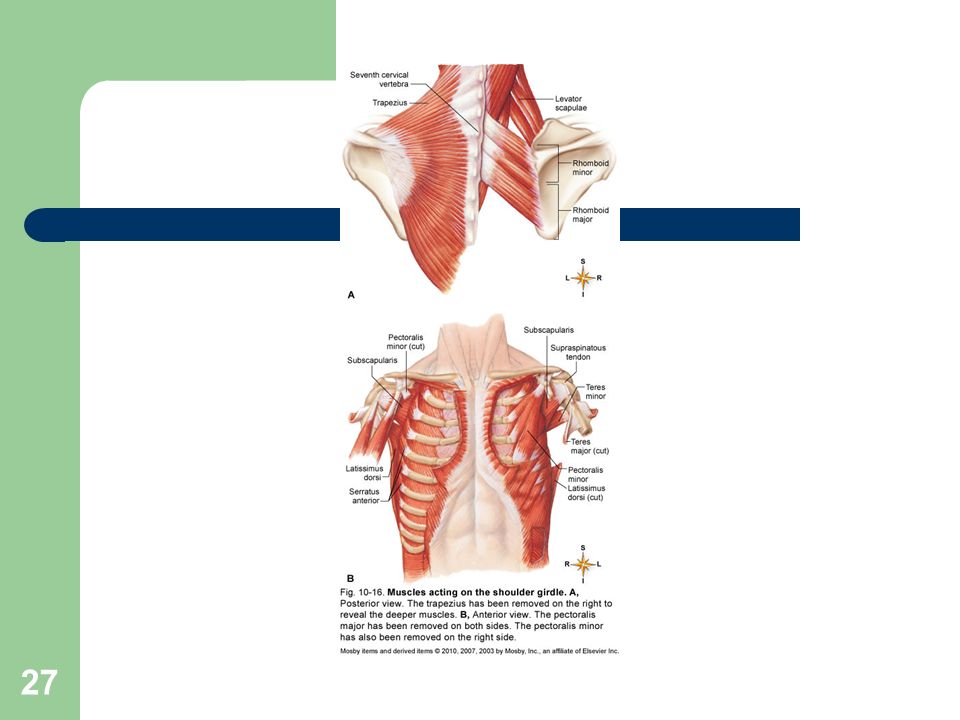
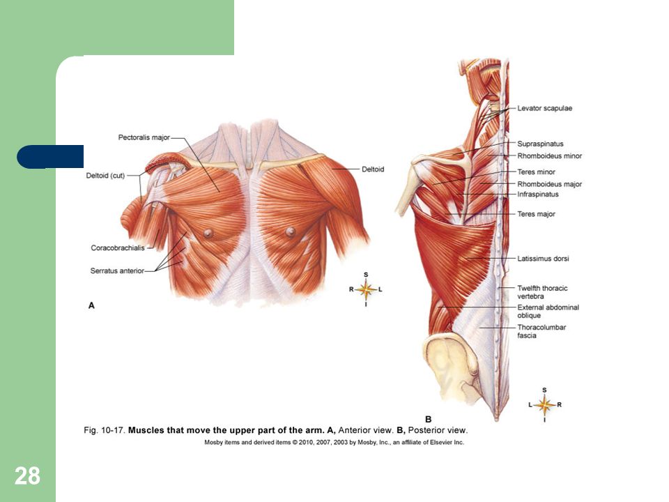
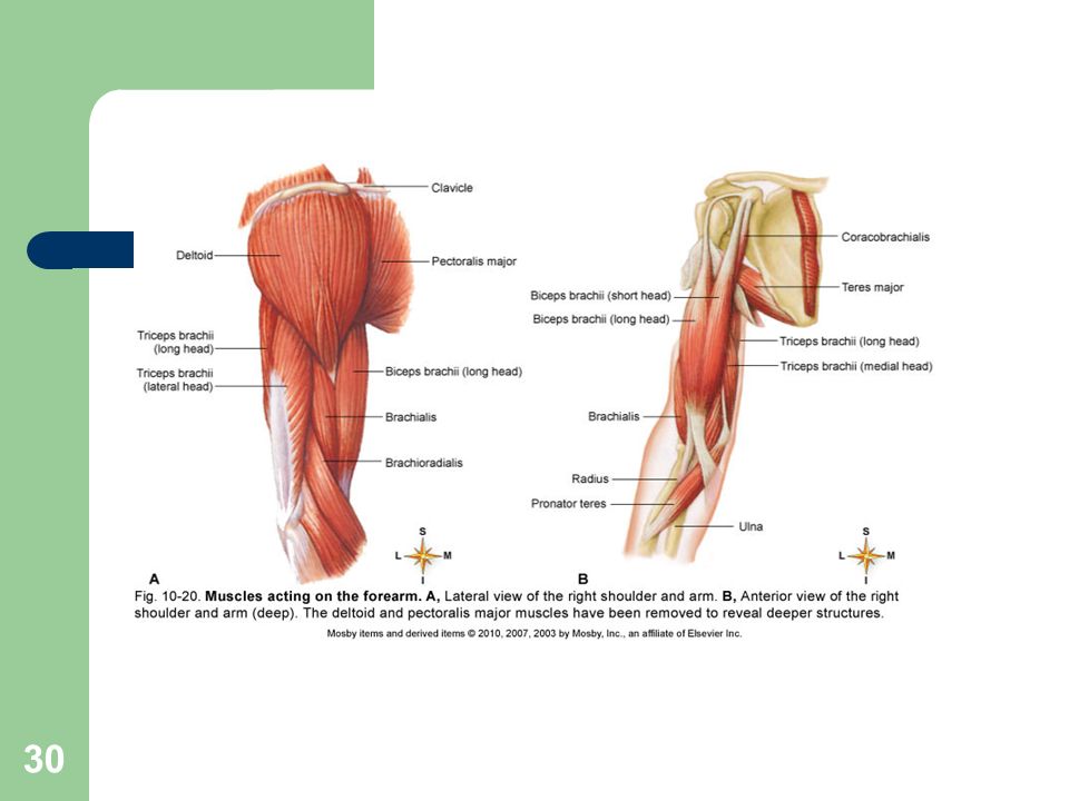
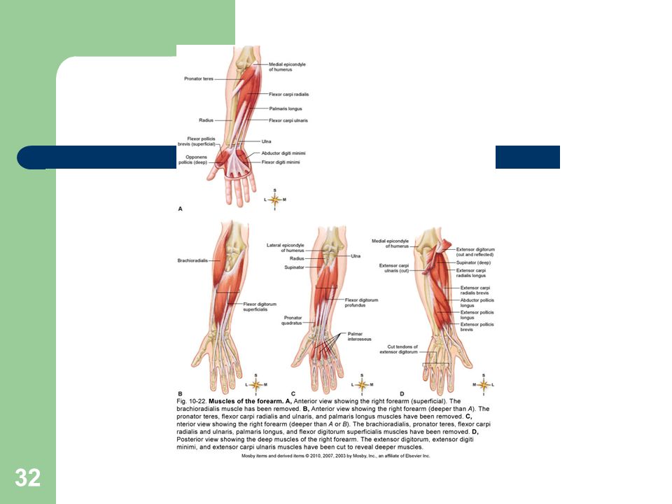
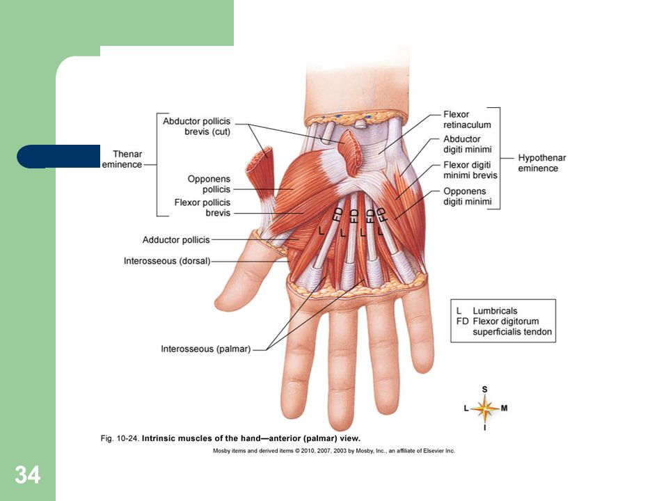
UPPER LIMB MUSCLES Muscles acting on the shoulder girdle: muscles that attach the upper extremity to the torso are located anteriorly (chest) or posteriorly (back and neck); these muscles also allow extensive movement (Figure 10-16; Table 10-13) Muscles that move the upper part of the arm: the shoulder is a synovial joint allowing extensive movement in every plane of motion (Figure 10-17; Table 10-14) Muscles that move the forearm: found proximal to the elbow and attach to the ulna and radius (Figures to 10-21; Table 10-15) Muscles that move the wrist, hand, and fingers: located on the anterior or posterior surfaces of the forearm (Figures to 10-24; Table 10-16)
 or posteriorly (back and neck); these muscles also allow extensive movement (Figure 10-16; Table 10-13) Muscles that move the upper part of the arm: the shoulder is a synovial joint allowing extensive movement in every plane of motion (Figure 10-17; Table 10-14) Muscles that move the forearm: found proximal to the elbow and attach to the ulna and radius (Figures to 10-21; Table 10-15) Muscles that move the wrist, hand, and fingers: located on the anterior or posterior surfaces of the forearm (Figures to 10-24; Table 10-16)")
35
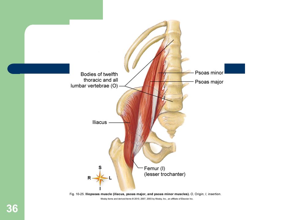
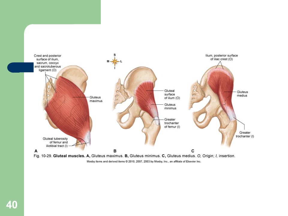
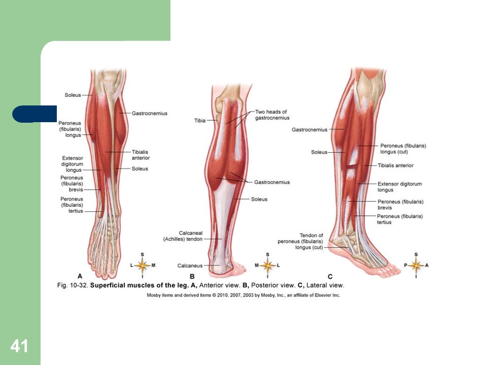
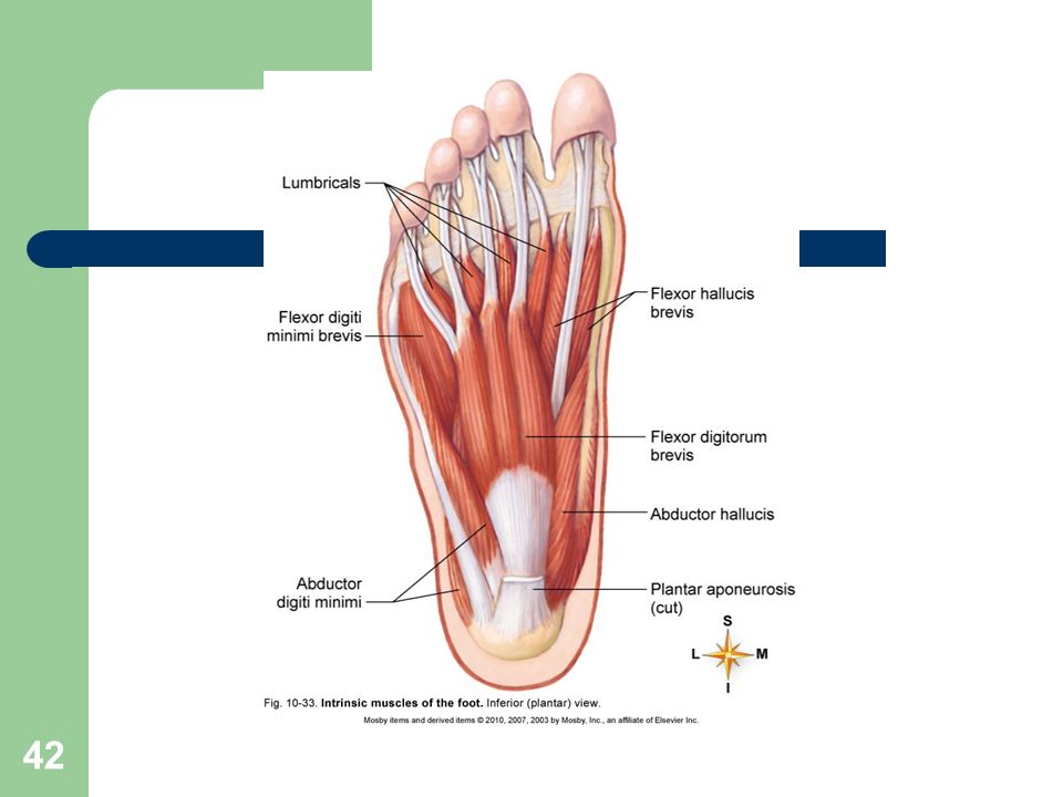
LOWER LIMB MUSCLES The pelvic girdle and lower extremity function in locomotion and maintenance of stability Muscles that move the thigh and lower part of the leg (Figures 10-6 and to 10-29; Tables and 10-18) Muscles that move the ankle and foot (Figures and 10-33; Table 10-19) Extrinsic foot muscles are located in the leg and exert their actions by pulling on tendons that insert on bones in the ankle and foot; responsible for dorsiflexion, plantar flexion, inversion, and eversion Intrinsic foot muscles are located within the foot; responsible for flexion, extension, abduction, and adduction of the toes
 Muscles that move the ankle and foot (Figures and 10-33; Table 10-19) Extrinsic foot muscles are located in the leg and exert their actions by pulling on tendons that insert on bones in the ankle and foot; responsible for dorsiflexion, plantar flexion, inversion, and eversion. Intrinsic foot muscles are located within the foot; responsible for flexion, extension, abduction, and adduction of the toes.")
43
POSTURE Maintaining the posture of the body is a major role of muscles
Good posture: body alignment that most favors function; achieved by keeping the body’s center of gravity over its base; requires the least muscular work to maintain How posture is maintained Muscles exert a continual pull on bones in the opposite direction from gravity Other structures have a role in maintaining posture Nervous system: responsible for the existence of muscle tone and regulation and coordination of the amount of pull exerted by individual muscles Respiratory, digestive, excretory, and endocrine systems all contribute to maintain posture

Similar presentations