Download presentation
Presentation is loading. Please wait.

1
NEW INSTRUMENTS/CASE DISCUSSION
Speaker M Jayasree Moderators J Manju Anju

2
Contents Optical Coherence Tomography Microperimetry
Multifocal Electroretinogram Glaucoma Diagnostics GDx HRT GLAUCOMA OCT HVF

3
OCULAR COHERENCE TOMOGRAPHY
PRINCIPLE Optical involves light and optics Used for Optical biopsy of retina. Coherence – two light beams of same wave length in phase. Uses principle of low coherence interferometry. Tomography Tome/Tomo – Greek for section /cutting

4
Determining and visualizing structure that absorb and scatter light
SDOCT Resolution – 6 microns Wavelength – 840 nm 3-D imaging No movement artifacts 25,000 A-scans/second Determining and visualizing structure that absorb and scatter light Noncontact Noninvasive

5
Indications Retinal- macula and Optic nerve.
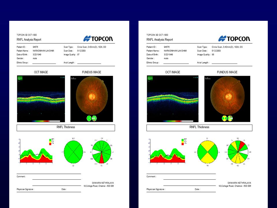
Inconclusive FFAs- done together. Quantify extent of lesions ex-CNVM, thickness of macula. Quantify RNFL thickness around Optic nerve head.

7
Normal Retina RNFL - high reflective (red)
IPL & OPL - moderate back scattering(yellow) Nuclear layers - min.back scattering (green) Photoreceptors - dark layer RPE & choriocapillaries - high reflective(red) Choroid & sclera - low reflectivity
 Nuclear layers - min.back scattering (green) Photoreceptors - dark layer. RPE & choriocapillaries - high reflective(red) Choroid & sclera - low reflectivity.")
8
Hyper reflectivity Hypo reflectivity
Above the NFL Posterior Hyaloid ,Epiretinal Membrane,Blood In the NFL Inflammation, Cotton Wool Spots, Blood Vessels, Flame Shaped Hemorrhages In the nuclear and plexiform layers Hard Exudates, Dot Blot Hemorrhages In the RPE CC layer Hyperpigmentation, CNV In the sub RPE layer Drusens, Blood, Fibrosis In the choroids Scar, Transmission Absence Edema Cystoid spaces Fluid – serous

9
NORMAL VITREO RETINAL INTERFACE
NORMAL FOVEAL CONTOUR WITH FOVEAL DIP INNER RETINAL RETINAL LAYERS ARE NORMAL NORMAL RPE CC COMPLEX FOVEAL THICKNESS

10
NORMAL VITREO RETINAL INTERFACE
FOVEAL CONTOUR ALTERED THICKENED HYPERREFLECTIVE INNER LAYERS E/O SRF , SUGGESTIVE OF ACTIVE CNVM WITH CYSTIC SPACES IN THE INNER RETINAL LAYERS

11
THICK ERM NOTED ALTERED FOVEAL CONTOUR LARGE CNVM, NO E/O SRF SUGGESTIVE OF SCARRED CNVM

12
THIN ERM ELEVATED FOVEAL CONTOUR THICKENED AND HIGH REFLECTIVE MEMBRANE NOTED SUBFOVEALLY NO E/O SRF,SUGGESTIVE OF SCARRED CNVM SMALL DRUSENOID PED

13
WRINKLED ILM ALTERED FOVEAL CONTOUR INNER RETINAL LAYERS ARE THICK AND CYSTIC SPACES THICKENED AND HIGH REFLECTIVE RPE-CC COMPLEX WITH MULTIPLE PED

14
NORMAL VITREO RETINAL INTERFACE
FOVEAL CONTOUR ELEVATED AND THINNED HYPER REFLECTIVE INNER LAYES CSR SEROUS PED

15
NORMAL VITREO RETINAL INTERFACE
FOVEAL CONTOUR ALTERED HEMORRHAGIC PED

16
WRINLED ILM FOVEAL CONTOUR ALTERED CYSTIC SPACES IN THE INNER RETINAL LAYERS SEROUS AND FIBROUS PED

17
INCOMPLETE POSTERIOR VITREOUS DETACHMENT
FOVEAL CONTOUR ALTERED FIBROVASCULAR PED

18
INCOMPLETE POSTERIOR VITREOUS DETACHMENT
FOVEAL CONTOUR ALTERED DRUSENOID PED

19
NORMAL VITREO RETINAL INTERFACE
FOVEAL CONTOUR ALTERED HARD EXUDATES IN THE INNER RETINAL LAYERS NORMAL RPE CC COMPLEX

20
NORMAL VITREO RETINAL INTERFACE
FOVEAL CONTOUR ALTERED DIFFUSE HARD EXUDATES IN THE INNER RETINAL LAYERS NORMAL RPE CC COMPLEX

21
NORMAL VITREO RETINAL INTERFACE
CSME CME FOVEOLAR DETACHMENT THICKENED HYPER REFLECTIVE CYSTIC SPACED INNER LAYERS NORMAL RPE-CC COMPLEX

22
NORMAL VITREO RETNAL INTERFACE
FOVEAL CONTOUR ELEVATED/THINNING NEURO SENSORY DETACHMENT SRF, SUGGESTIVE OF CSR NORMAL RPE COMPLEX

23
ERM WITH ILM FOLDS FOVEAL CONTOUR ALTERED INNER RETINAL THICKENED NORMAL RPE CC COMPLEX

24
ERM WITH WRINKLING ILM LAMELLAR MACULAR HOLE THIN INNER RETINAL LAYERS NORMAL RPE COMPLEX

25
FULL THICKNESS MACULAR HOLE WITH OPERCULUM

26
COMPLETE POSTERIOR VITREOUS DETACHMENT
VITREOUS TRACTION RELEASED

27
NORMAL VITREO RETINAL INTERFACE
FOVEAL SCHISIS THINNED CYTIC SPACES INNER LAYERS RPE ALTERED

28
FOVEAL CONTOUR ALTERED
OPTIC DISC PIT

29
OPTIC DISC PIT MACULOPATHY

30
VITREO MACULAR TRACTION

31
Case 1 PRE TREATMENT DATE OF VISIT 22/08/08 POST TREATMENT
BCVA OS 6/36, N36 SL OS WNL FUNDUS OS CNVM, SUB RETINAL HEAMORRHAGE OCT OS E/O SRF , SUGGESTIVE OF ACTIVE CNVM WITH CYSTIC SPACES IN THE INNER RETINAL LAYERS OS INJ AVASTIN GIVEN THRICE POST TREATMENT DATE OF VISIT 22/11/08 BCVA OS 6/12, N8 SL OS WNL FUNDUS OS SCARREDCNVM, OCT OS NO E/O SRF , SUGGESTIVE OF SCARRED CNVM WITH CYSTIC SPACES IN THE INNER RETINAL LAYERS

32
PROGNOSIS HOLE FORM FACTOR
HFF > % PRIMARY CLOSURE HFF = % PRIMARY CLOSURE HFF < Poor closure rates HIGHER HFF - BETTER POST OP VA

33
CASE 2 PRE TREATMENT POST TREATMENT S/P VIT+C3F8 UNDER GA
DATE OF VISIT (25/09/08) BCVA OS 6/48(ECC VIEW), N24 S/L OS NO ABNORMALITY FUNDUS OS FTMH OCT OS FULL THICKNESS MACULAR HOLE WITH COMPLETE PVD POST TREATMENT S/P VIT+C3F8 UNDER GA DATE OF VISIT (6/12/08) BCVA OS 6/12+, N8 S/L OS WNL FUNDUS OS OCT OS
 BCVA OS 6/48(ECC VIEW), N24. S/L OS NO ABNORMALITY. FUNDUS OS FTMH. OCT OS FULL THICKNESS MACULAR HOLE WITH COMPLETE PVD. POST TREATMENT. S/P VIT+C3F8 UNDER GA. DATE OF VISIT (6/12/08) BCVA OS 6/12+, N8. S/L OS WNL. FUNDUS OS. OCT OS.")
34
Multifocal Electroretinogram (mfERG) in Retinal Disorders
 in Retinal Disorders")
35
Multifocal electroretinogram
Simultaneous recording of focal retinal responses Offers direct, objective and topographical mapping of central 36-40º of retinal function Cone driven responses Fovea, Para fovea and near peripheral photopic retina function can be evaluated.

36
mfERG Stimulus Display contains array of hexagons-
Commonly used 103 hexagonal array Scaling basis - Photoreceptor density Produce local retinal responses of equal amplitude Flickers according to pseudorandom binary m-sequence

37
Waveforms grouping according to different retinal eccentricities
Subtends 35° horizontally and 31° vertically – viewing distance 53cm The responses obtained from six zones 1.6 ° 1.6°- 6° 6°-11.4° 11.4°-18.2° 18.2°-26.2° 26.2°-35°

38
First order kernel responses- Interpretation
Multiplot Trace Array

39
mfERG Components Parameters N1 – First negative trough
P1- First positive Peak Origins Cone Photoreceptors Dominated by on and off bipolar cells P1-IT P1- AMP N1- IT N1-AMP

40
Interpretation Larger delay in implicit times
Degenerative photoreceptor disease Larger delay in implicit times Local lesions damaging INL Larger reduction in amplitudes Damage to NFL or GCL No reduction or delay

41
Clinical applications
To distinguish retinal diseases from optic nerve disease Details extent of lesion Sensitive indicator for retinal drug toxicity Post-operative management following V-R surgery Assess sub-clinical retinal changes in DR

42
Case 1 Stargardt’s disease Few yellowish flecks at the macula

43
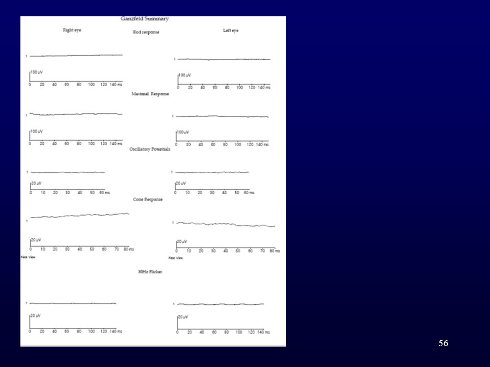
Fullfield ERG

44
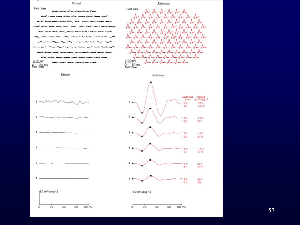
Multifocal ERG – Trace array

45
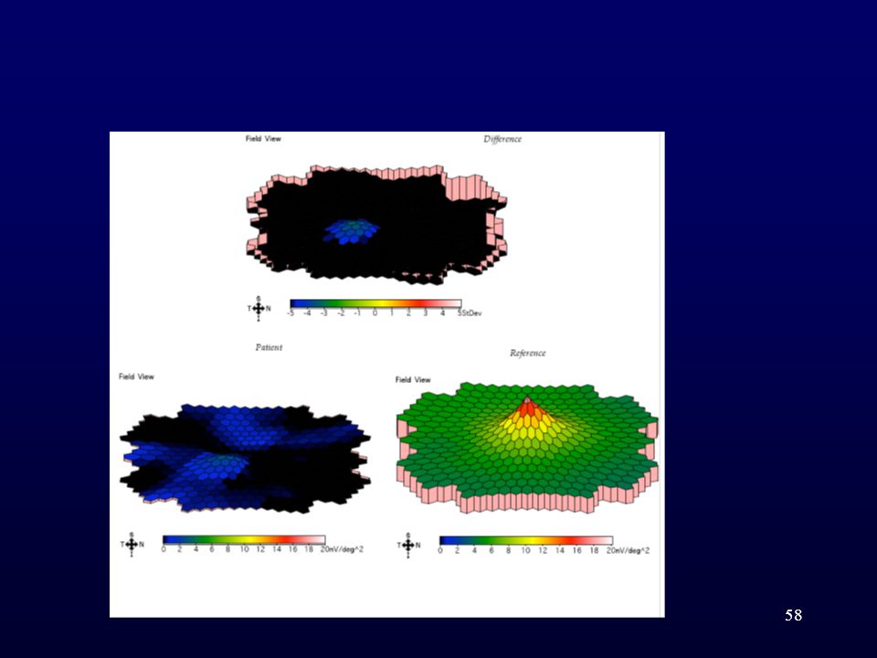
Crater like defect - MfERG delineates the macular pathology
Multiplot Crater like defect - MfERG delineates the macular pathology

46
Case 2 Cone Dystrophy

47
Fullfield ERG

48
Multifocal ERG – Trace array

49
Multiplot

50
Case 3

51
Fullfield ERG Retinitis Pigmentosa

52
Multifocal ERG – Trace array

53
MultiPlot

54
Case 4 JUVENILE RETINOSCHISIS

55
Case 5 Fine RPE stippling in normal looking fundus –
Chloroquine toxicity

59
MICROPERIMETRY It is an instrument for fundus related perimetry - assess the central visual field and correlates the findings directly to the fundus location Captures patient’s retina and simultaneously projects light stimuli on to the retina Stimuli have been projected and the operator customizes the pattern of stimuli according to the retinal disease Easily adapted to the different diseases of macula Assess also the patient’s fixation site and stability of fixation over time as stable, relatively unstable and unstable With or without pupillary dilation (if undilated pupil size min 4mm required) Persons who have low vision or fixation problems, the stimulus can be made thicker or larger – also the fixation target may be chosen as either single cross, four crosses or circle, any color , any size
 Persons who have low vision or fixation problems, the stimulus can be made thicker or larger – also the fixation target may be chosen as either single cross, four crosses or circle, any color , any size.")
60
Comparison with HVF HVF
MICROPERIMETRY Even a small central, paracenral scotoma can be detected, understand and assess macular disease Fixation location is assessed Direct corelation between threshold values and retinal sensitivity in decibels Operator decides the stimuli location and time interval between the stimuli Gives best results especially in cases of vitreo retinal surgeries(pre and post), before and after PDT THERAPHY, easily detects the defect is in the retina or visual pathway HVF Macular program available but relatively small central scotoma may be missed Cannot be assessed Similar but logarithmic values in apostils Cannot be done
, before and after PDT THERAPHY, easily detects the defect is in the retina or visual pathway. HVF. Macular program available but relatively small central scotoma may be missed. Cannot be assessed. Similar but logarithmic values in apostils. Cannot be done.")
61
Fixation cross, at the macula
Retinal sensitivity in decibels

62
MAP REPRESENTATION Fixation target four crosses Filled green square better sensitivity Filled yellow square moderate sensitivity Filled red square poor retinal sensitivity Empty square stimulus not seen

63
operator decides when and where to project each stimulus on the tracked retinal image and its corresponding initial intensity in manual micro perimetric exam In a semi-automatic exam the operator shall define a polygonal area on the tracked image and the number of stimuli inside the selected area. In an automatic exam the stimuli are proposed following a pattern chosen from among the pre-determined or customized by the operator.

64
Clinical applications of micro perimetry
ARMD/AMD(atrophic and neovascular) EARLY AMD DIABETIC MACULAR EDEMA VITREO RETINAL INTERFACE DISORDER LOW VISION PATIENT MACULAR HOLE CYST TOXIC MACULOPATHIES CSR HEREDITORY RETINAL DYSTROPIES(RP, STARGARDT’S DISEASE,BEST’S DISEASE)
 EARLY AMD. DIABETIC MACULAR EDEMA. VITREO RETINAL INTERFACE DISORDER. LOW VISION PATIENT. MACULAR HOLE CYST. TOXIC MACULOPATHIES. CSR. HEREDITORY RETINAL DYSTROPIES(RP, STARGARDT’S DISEASE,BEST’S DISEASE)")
65
Thank You

66
PRE POST MICROPERIMETRIC IMAGE
YET TO BE PASTED

Similar presentations


 Waxman MD PhD>")


 pigment.>")
 1)Noninvasive 2) non-contact imaging 3)Millimeter penetration Aproximately 2-3 mm in tissue with micrometer scale (axial.>")


>")

 A presentation courtesy of Zeiss.>")


