Download presentation
Presentation is loading. Please wait.

1
Case 2

2
Clinical data 30-year-old male 30-year-old male Tumorous mass in the rete testis/epididymis region. Duration 6 months Tumorous mass in the rete testis/epididymis region. Duration 6 months Excision Excision

3
1,3 x1,2 x0,8 cm 1,3 x1,2 x0,8 cm Whitish on cut surphace Whitish on cut surphace Immunohistochemistry: Immunohistochemistry: EMA, AE1-AE3: positive CEA, CD 34: negative

4
????
5
Caterpillar of moth Actias cf. selenae

8
Our diagnosis Adenomatoid tumor

9
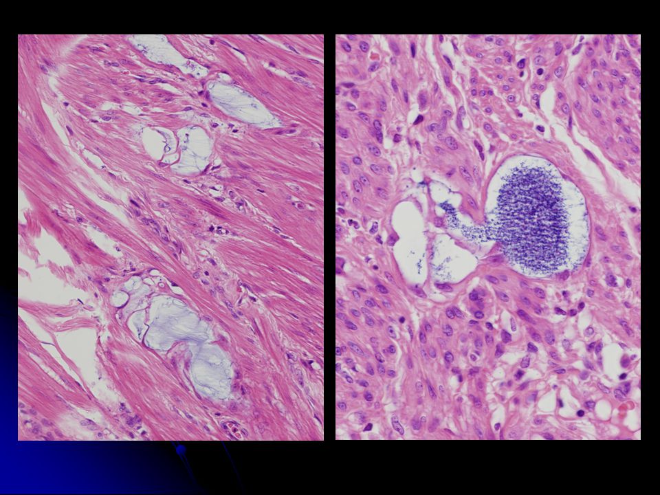
A very peculiar and constant feature is presence of thin intraluminal bridging strands A very peculiar and constant feature is presence of thin intraluminal bridging strands Bridging strands are so thin that resembled lipocytes of adipose tissue Bridging strands are so thin that resembled lipocytes of adipose tissue Strands bridged lumina of numerous tubular and slit-like structures and formed fine inconspicuous network in some cases. Strands bridged lumina of numerous tubular and slit-like structures and formed fine inconspicuous network in some cases. Bridging strands are present constantly in ATs. Bridging strands are present constantly in ATs.

10
Ultrastructure Ultrastructurally, bridges are formed by apposition of attenuated cytoplasm of two adjacent cells. Ultrastructurally, bridges are formed by apposition of attenuated cytoplasm of two adjacent cells. Luminal surfaces are covered by short, narrow, occasionally branched or bushy microvilli confirming their mesothelial origin Luminal surfaces are covered by short, narrow, occasionally branched or bushy microvilli confirming their mesothelial origin

11
Ultrastructure

Similar presentations


 Histopathologic findings Immunopathologic findings Immunohistochemistry on paraffin sections.>")


>")


