Download presentation
Presentation is loading. Please wait.

1
Chapter 6 The Muscular System

2
The essential function of a muscle is to shorten or contract
As a result of this ability, muscles are responsible for almost all body movement and can be viewed as the “machines of the body”

3
Overview of Muscle Tissues

4
Muscle Types - Three Skeletal Cardiac Smooth

5
The 3 Types of Muscle Tissue differ in:
Cell structure Body location How they are stimulated to contract

6
The three types of muscle tissue are similar because:
All muscle cells are elongated – called muscle fibers The ability of muscle to shorten or contract depends on 2 types of myofilaments (part of the cytoskeleton)
")
7
Skeletal Muscle Fibers are packaged into skeletal muscles that attach to the skeleton Cigar-shaped Multinucleate (many nuclei) Largest of the muscle fibers
 Largest of the muscle fibers.")
8
Striated muscle (appear striped)
Voluntary muscle (conscious control) Form smoother contours of the body
 Form smoother contours of the body.")
9
Key words to think of for skeletal muscle:
Skeletal, striated, voluntary

10
Skeletal muscles are very fragile, but they are capable of exerting tremendous power.
They are able to do this because: thousands of fibers are bundled together with connective tissue – these bundles are then bundled together

11
Tendons Attach muscle to bone

12
Figure 09.02

13
Functions of the tendons
Anchor muscles Provide durability and conserve space Crossover bony projections

14
Smooth Muscle No striations Involuntary
Found in the walls of hollow digestive organs Propels substances along a definite path

15
Spindle shaped Single nucleus Arranged in sheets or layers Muscle contraction is slow and sustained

16
Key words: Visceral, non-striated, involuntary

17
Cardiac Muscle Found only the heart Striated and involuntary

18
Key Words: Cardiac Striated involuntary

19
Muscle Functions Muscles play four important roles in the body.

20
1. Produce movement Moves the body

21
2. Maintain posture Allow you to remain in an erect or seated posture despite gravity

22
3. Stabilize Joints Muscle tendons are extremely important in reinforcing and stabilizing joints

23
4. Generating Heat Heat is a by product of muscle activity
ATP used as power – ¾ escapes as heat

24
Microscopic Anatomy of Skeletal Muscles
Skeletal muscle contains both actin and mysosin filaments The overlapping pattern of thick and thin filaments is responsible for the light and dark bands seen in skeletal striated muscle

25
The thick filaments are made up of a protein called myosin
The thin filaments are made of a protein called actin. Sarcomere: contractile unit

26
Skeletal Muscle Activity

27
The 2 special functional properties of muscles:
Irritability – ability to receive and respond to a stimuli Contractility – ability to shorten with adequate stimuli Skeletal muscles must be stimulated by nerve impulses to contract.

28
Motor Unit One neuron (nerve cell) and all the skeletal muscles it stimulates
 and all the skeletal muscles it stimulates")
29
Figure 09.09

30
How a muscle contracts A nerve impulse reaches the end of the nerve a neurotransmitter is released. The neurotransmitter that stimulates skeletal muscle is acetylcholine (Ach) When enough acetylcholine is released, sodium ions (Na+) will rush into the muscle. This rush of ions creates an electrical current known as the action potential. The action potential travels over the entire muscle causing it to contract.
 When enough acetylcholine is released, sodium ions (Na+) will rush into the muscle. This rush of ions creates an electrical current known as the action potential. The action potential travels over the entire muscle causing it to contract.")
31
The events that return a muscle to its resting state:
Diffusion of K+ (potassium) out of the cell Activation of the Na+/K+ pump
 out of the cell. Activation of the Na+/K+ pump.")
33
The Sliding Filament Theory
Muscle fibers contract when the sarcomere shortens. The sarcomere shortens when the actin fibers slide past the myosin filaments Myosin moves the actin.

34
The sliding filament theory refers to the movement of actin in relation to myosin.
ATP supplies the energy for muscle contraction. Myosin filaments do all the work. The actin filaments just sit there.

37
Figure 09.04

38
Myosin filaments breakdown ATP and have crossbridges that pull the actin filaments toward the center of the sarcomere.

39
Contraction of Skeletal Muscle as a whole

40
Figure 09.06

41
Graded Responses Different degrees of shortening
Different numbers of muscles contract

42
Graded muscle contractions can be produced in two ways:
By changing the speed of muscle stimulation By changing the number of muscle cells being stimulated

43
Providing Energy for Muscle Contraction

44
1. Direct phosphorylation of ADP by creatine phosphate (CP)
CP gives a phosphate to ADP to make ATP ATP is regenerated in a fraction of a second CP supplied energy used in 20 seconds

45
2. Aerobic Respiration – ATP is made by aerobic respiration
1 glucose – 36 ATP Fairly slow – needs continuous supply of oxygen Adenosine Triphosphate

46
3. Anaerobic Respiration and Lactic Acid Formation
No oxygen 2 ATP per glucose Lactic acid is made and builds up in muscles 5X faster than aerobic 30-40 seconds of strenuous exercise Problems: needs lots of glucose Small amount of ATP produced per glucose Lactic acid

47
3. Anaerobic Respiration and Lactic Acid Formation
No oxygen 2 ATP per glucose Lactic acid is made and builds up in muscles 5X faster than aerobic 30-40 seconds of strenuous exercise Problems: needs lots of glucose Small amount of ATP produced per glucose Lactic acid

48
Muscle Fatigue and Oxygen Debt

49
Muscle Fatigue Occurs when muscles are exercised strenuously

50
Fatigued When a muscle is unable to contract even though it is being stimulated

51
Oxygen Debt The volume of oxygen needed after exercise to get rid of the lactic acid formed during exercise

52
The major factor that effects the work that a muscle can do and how long it can do work without becoming fatigued is: How good the blood supply is

53
When muscles lack oxygen:
Lactic acid builds up Muscles also run out of ATP So lack of ATP and lactic acid build up cause a muscle to stop contracting

54
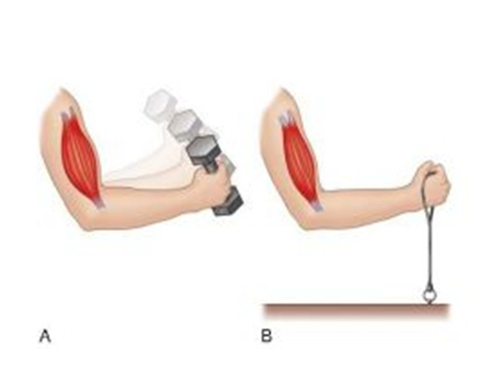
The Two Types of Muscle Contractions

55
Isotonic contractions
When the force of the muscle contraction is greater than the force that is resisting the contraction Ex. Weight is lifted

56
Isometric Contractions
When the resistance to the contraction is equal to the force generated in the muscle tissue (muscle do not contract or only a little) Pulling on a stationary bar
 Pulling on a stationary bar.")
58
Effect of Exercise on Muscles

59
Muscle inactivity always leads to: muscle weakening and wasting away
Regular exercise increases muscle: size, strength and endurance

60
Atrophy Diminish in size and become weaker result of no exercise

61
Hypertrophy Increase in size and strength Result of exercise

62
Anabolic steroids are sometimes taken by athletes to promote muscle growth

63
Undesirable effects of anabolic steroid use:
Cardiovascular disease Liver and kidney dysfunction Impotency Sterility Increase in rash behavior “roid rage”

64
Slow Twitch Muscle Fibers - Legs
Steadier tug More endurance Aerobic energy production – tire when fuel is gone Sports useful in: long distance running, biking, jogging, swimming Color: dark Many mitochondria, dense capillary beds which draw more blood and oxygen

65
Fast Twitch Muscle Fibers - Breasts
Anaerobic – tire quick because of lactic acid build up Designed for strength Explosions of energy Sports useful in: sprinting, weight lifting Color: white Few mitochondria, few blood cells

66
Aerobic or Endurance Excercise
Stronger more flexible muscles with greater resistance to fatigue Blood supply increases as cells form more mitochondria and store more oxygen

67
Body Benefits Overall body metabolism is more efficient
Improves digestion Skeleton stronger Heart pumps more blood with each beat Fat deposits cleared Lungs are more efficient at gas exchange

68
Resistance or Isometric Exercise
A few minutes every other day Muscles increase in size because muscle cells increase in size (not increase in number of cells) Amount of reinforcing connective tissue also increases
 Amount of reinforcing connective tissue also increases.")
69
Types of Muscles Muscles cannot push. They can only pull.
So most body movements are a result of the activity of pairs or teams of muscles acting together or against each other Muscle groups are arranged on the skeleton so that whatever one muscle can do, another group of muscles can do in reverse

70
Naming Skeletal Muscles

71
Skeletal muscles are given names based on:
Size – gluteus maximus Shape – deltoid Location Direction of fibers – rectus – up and down – transverse – horizontal Number of attachments – biceps – 2 attachments Action

72
Figure 09.22

73
Figure 09.23

74
Figure 09.24

75
Figure 09.25a

76
Figure 09.27b

77
Figure 09.27c

78
Figure 09.27d

79
Figure 09.28

80
Figure 09.29d

81
Figure 09.31b

82
Figure 09.34

83
Figure 09.35a

84
Figure 09.37a

85
Figure 09.37b

86
Figure 09.37c

87
Figure 09.38c

88
Figure 09.39a

89
Figure 09.39a

90
Figure 09.39b

91
Figure 09.40

92
The End

Similar presentations


 Generate heat - body temp 3 types: Skeletal - moves bone, voluntary Smooth.>")


