Download presentation
Presentation is loading. Please wait.

1
Cell Junctions structure & role presented by Suman.Thota M.Pharm I I-Semester Department of Industrial Pharmacy UNIVERSITY COLLEGE OF PHARMACEUTICAL SCIENCES KAKATIYA UNIVERSITY WARANGAL

2
Contents Plasma membrane its structure & components Extracellular matrix & its role Types of cell junctions Tight junctions molecular structure & role - Blood brain barrier Adherens junctions structure & role Desmosomes structure & role Hemidesmosomes Focal adhesions Gap junctions structure & function Conclusion References

3
Plasma Membrane Definition: The Plasma membrane is a thin bi- layered structure which surrounds each cell, consists of lipids (phospholipids 75%, cholesterol 20%,glycolipids 5%), proteins (partially or completely embedded), carbohydrates etc., ~6-10 nm thick. Plasma membrane is asymmetrical

4
Plasma membrane structure

6
EXTRACELLULAR ENVIRONMENT (cytoskeletal proteins beneath the plasma membrane) ADHESION PROTEIN oligosaccharide groups phospholipids cholesterol LIPID BILAYER RECOGNITION PROTEIN RECEPTOR PROTEIN CYTOPLASM PLASMA MEMBRANE (area of enlargment) TRANSPORT PROTEINS open channel protein gated channel proten (open) active transport protein gated channel proten (closed)
 ADHESION PROTEIN oligosaccharide groups phospholipids cholesterol LIPID BILAYER RECOGNITION PROTEIN RECEPTOR PROTEIN CYTOPLASM PLASMA MEMBRANE (area of enlargment) TRANSPORT PROTEINS open channel protein gated channel proten (open) active transport protein gated channel proten (closed)")
7
Many animal cells are intrinsically linked to other cells and to the extracellular matrix (ECM). The ECM fills the spaces between cells and tissues together. Bone and cartilage are mostly ECM plus a very few cells. ECM is most abundant in Connective tissue, that surrounds glands and blood vessels, is a gelatinous matrix containing many fibroblast cells. The ECM contains three classes of molecules: 1) Structural proteins (collagens and elastins); 2) Protein-polysachharide complexes (proteoglycans) 3) Adhesive glycoproteins to attach cells to matrix (fibronectins and laminins), Extracellular matrix
 Structural proteins (collagens and elastins); 2) Protein-polysachharide complexes (proteoglycans) 3) Adhesive glycoproteins to attach cells to matrix (fibronectins and laminins), Extracellular matrix.")
8
Many types of glycoproteins Form branched complexes Fibronectins act as docking site Bound to Integrins Integrins( transmembrane proteins) and are bound to cytoskeletal filaments
 and are bound to cytoskeletal filaments")
9
Role of ECM Gives tissue stability by linking areas of cells together Allows communication between cells Fibronectin and integrins are involved in signal sending and receiving

10
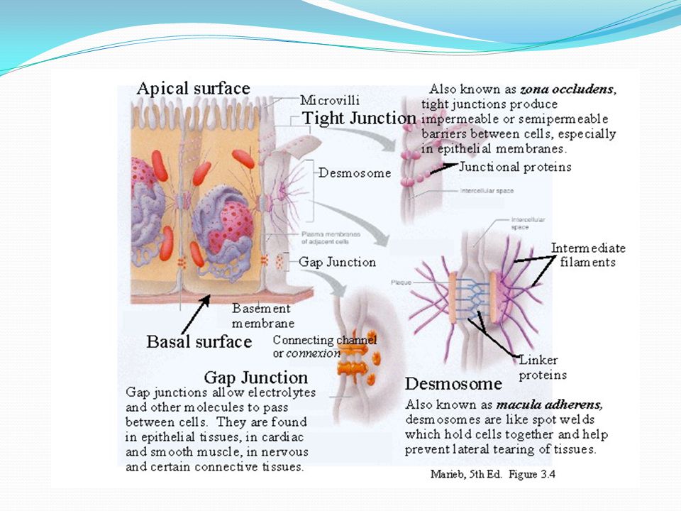
Cell junctions Definition “ Cell junctions are the contact points between the plasma membranes of tissue cells” i.e. Plasma membrane areas are specialized to provide contact between cells. Dense clusters of cell adhesion molecules (CAMs) on the outside linked to cytoskeleton on the inside through adapter proteins.
 on the outside linked to cytoskeleton on the inside through adapter proteins..")
13
Transport across epithelial cells Paracellular Pathway Transport is only passive driven by the gradients This physiological barrier exists to provide protection from the entry of toxins, bacteria,and viruses from the apical side to the basolateral side, and it allows the passage of selective molecules. Transcellular Pathway Transport is both active & passive Transport is directional, energy dependent, and governed by the cell- specific profile of transporters & channels positioned on the apical & basolateral cell membranes.

14
Between cells - Tight junctions - Adherens junctions - Desmosomes - Gap junctions Between cells and matrix - Hemidesmosomes - Focal adhesions Cell junctions (based on localization)
")
16
Cell junctions (based on function) Occluding junctions -Tight junctions -Adhering junctions - Adherens junctions - Desmosomes - Hemidesmosomes Communicating junctions - Gap junctions
 Occluding junctions -Tight junctions -Adhering junctions - Adherens junctions - Desmosomes - Hemidesmosomes Communicating junctions - Gap junctions")
17
Functions of cell junctions

18
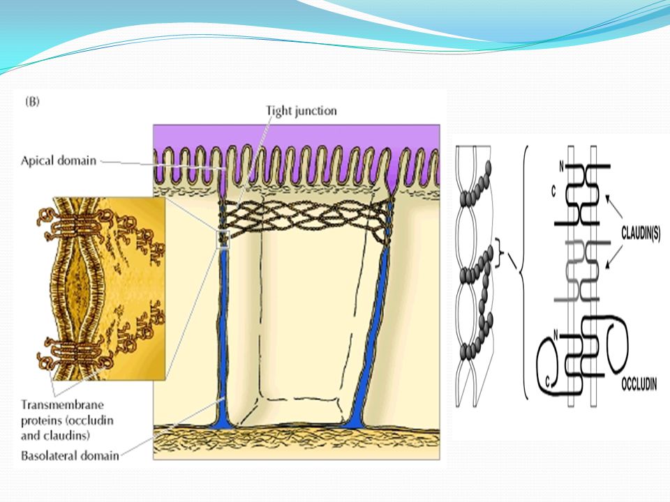
Tight junctions consists of web like strands of transmembrane proteins (occludin and claudins) that fuse the outer surfaces of adjacent plasma membranes together. i.e. Belts of transmembrane proteins that close extracellular space between cells Prevent passage of water and water-soluble substances Account for electrical resistance across epithelia Isolate parts of plasma membrane (Apical and Basolateral domains) Tight junctions (Zonula occludens)
 Tight junctions (Zonula occludens).")
20
E.g. Cells of epithelial tissues that line the STOMACH INTESTINES & URINARY BLADDER have many tight junctions to retard the passage of substances between cells & prevent the contents of these organs from leaking in to the blood and surrounding tissues Tight junctions cause cell surface polarity that produces the fence function and restricts free diffusion of lipids and proteins from the apical plasma membrane to the basolateral surface Thus, paracellular permeation of drug through the intercellular junctions depends on the pore size of the tight junctions This barrier is variable and physiologically regulated, and its disruption contributes to human diseases

21
Claudins and occludins (membrane proteins) zip two membranes together Stabilized by spectrin Connected to spectrin by adapter proteins ZO1 and ZO2 Molecular structure of tight junc tions
 zip two membranes together Stabilized by spectrin Connected to spectrin by adapter proteins ZO1 and ZO2 Molecular structure of tight junc tions")
22
The “tightness” varies according to the barrier needs. Leaky epithelia where there is need for some traffic. - Hormones - Vasopressin - Cytokines - Lack of ATP causes “leak” -Extravasating leukocytes open tight junctions Regulation of tight junctions

23
Leukocytes leave the circulation at sites of tissue inflammation by interacting with the endothelial cells of capillary walls

24
Role : Tight junctions prevent the passage of water and solutes between the cells and are common between epithelial cells exposed to harsh chemicals / powerful enzymes E.g.: Tight junctions between epithelial cells lining the digestive tract keep digestive enzymes, stomach acids, or waste products from damaging underlying tissues Tight junctions constitute the main barrier to paracellular diffusion. The diameters of the tight junction pores are approximately 4-8 Å and 10-15 Å in humans and animals, respectively. Because in humans the paracellular route will not allow the passage of molecules with diameters greater than ~8 Å, this route is unlikely to play an important role in the absorption of most compounds of pharmaceutical interest.

25
In intestinal epithelial cells transport of glucose from the intestinal lumen through the cell to the blood stream requires the uptake of glucose through apical surface sodium/glucose symport proteins and export by glucose transport proteins on the basolateral surface and tight junctions prevent the lateral movement of these transport proteins.

26
tight junctionstight junctions form seals that prevent the free passage of molecules (including ions) between the cells of epithelial sheets. tight junctionstight junctions separate the apical and basolateral domains of the plasma membrane by preventing the free diffusion of lipids and membrane proteins between thembasolateral domainsplasma membranelipidsproteins Consequently, specialized transport systems in the apical and basolateral domains are able to control the traffic of molecules between distinct extracellular compartments, such as the transport of glucose between the intestinal lumen and to the blood supply.basolateral domains

27
The BBB is the major barrier to the passage of active molecules from the blood compartment to the brain. The BBB, which segregates the brain interstitial fluid (ISF) from the circulating blood, is located at the level of the brain capillaries, where there is a convergence of different cell types: endothelial cells, pericytes, astrocytes and microglia’s (perivascular macrophages) The brain micro vessel endothelial cells (BMEC) that form the BBB, display important morphological characteristics such as the presence of tight junctions between the cells, the absence of fenestrations and a diminished pinocytic activity, that together help to restrict the passage of compounds from the blood into the extracellular environment of the brain. Blood brain barrier
 from the circulating blood, is located at the level of the brain capillaries, where there is a convergence of different cell types: endothelial cells, pericytes, astrocytes and microglia’s (perivascular macrophages) The brain micro vessel endothelial cells (BMEC) that form the BBB, display important morphological characteristics such as the presence of tight junctions between the cells, the absence of fenestrations and a diminished pinocytic activity, that together help to restrict the passage of compounds from the blood into the extracellular environment of the brain. Blood brain barrier.")
28
The two main barriers in the CNS: blood-brain barrier (A) and blood cerebrospinal fluid barrier (B).
 and blood cerebrospinal fluid barrier (B).")
29
Tight junctions provide significant Trans endothelial electrical resistance (TEER) to BMEC and impede the penetration of potential therapeutic agents such as oligonucleotides, antibodies, peptides and proteins. Under normal conditions the BBB acts as a barrier to toxic agents and safeguards the integrity of the brain. The BCSFB separates the blood from the cerebrospinal fluid (CSF) that runs in the subarachnoid space surrounding the brain. This barrier is located at the choroid plexus, and it is formed by epithelial cells held together at their apices by tight junctions, which limit paracellular flux.
 that runs in the subarachnoid space surrounding the brain. This barrier is located at the choroid plexus, and it is formed by epithelial cells held together at their apices by tight junctions, which limit paracellular flux..")
30
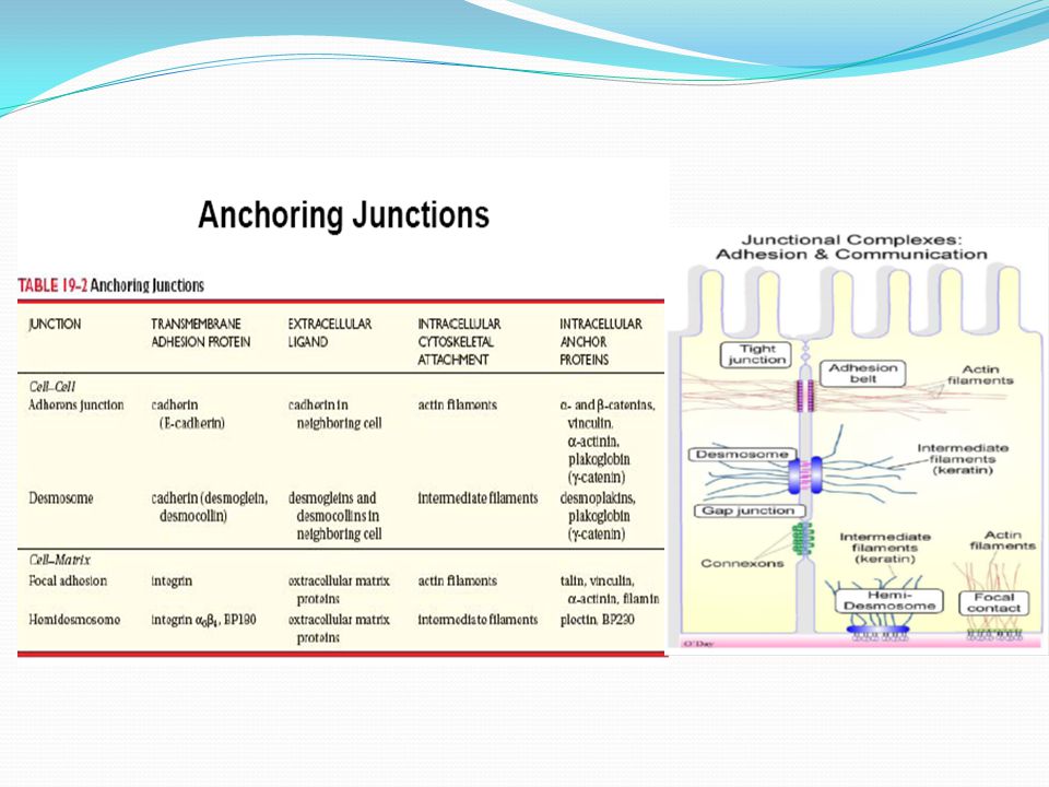
Adhesive junctions (Adherens junction, desmosomes & hemidesmosomes ) link adjoining cell to each other and to the ECM. Although adhesive junction types are similar in structure and function, they contain distinct 1) intracellular attachment proteins and 2) transmembrane linker proteins. Adhesive/Anchoring junctions
 intracellular attachment proteins and 2) transmembrane linker proteins. Adhesive/Anchoring junctions.")
32
The intracellular attachment proteins form a thick layer of fibrous material on the cytoplasmic side of the plasma membrane called a plaque which binds actin microfilaments in adherens junctions and intermediate filaments in desmosomes and hemidesmosomes. The transmembrane linker proteins are anchored to the plaque by the cytoplasmic domain and binds the ECM or to the same proteins on other cells.

33
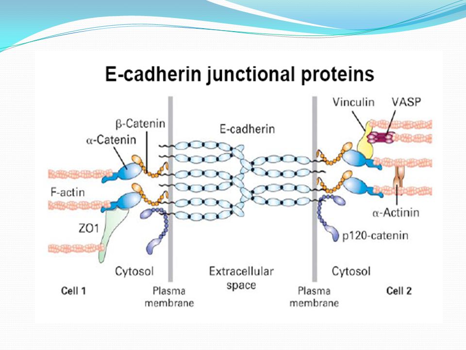
ADHERENS JUNCTIONS Adherens junctions resemble desmosomes except two adjoining cells are separated by a thin space of 20-25 nm and connect to actin microfilaments in the cytoplasm. Contain plaque- a dense layer of proteins on the inside of plasma membranes that attaches to both membrane proteins & to microfilaments of the cytoskeleton

34
Actually transmembrane glycoproteins called cadherins that join the cells. Each cadherin inserts in to the plaque form the opposite side of the plasma membrane, partially crosses the inter cellular space & connects to cadherins of an adjacent cell. In epitelial cells adherens junction often form extensive zones called Adhesion belts

36
Role : help epithelial surfaces resist separation during various contractile activities, as when food moves through the intestines. Hold cells tightly together Confer mechanical strength

37
Molecular structure of Adherens junctions Belt like junctions located just below tight junction. Homophilic pairing of E-cadherins, Adapter proteins (plakoglobin and α and β catenins) link cadherins to the belt of actin filaments.
 link cadherins to the belt of actin filaments..")
39
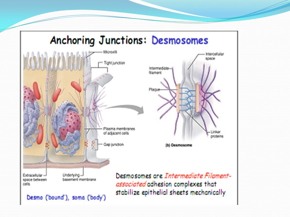
Desmosomes form strong points of adhesion between cells in a tissue such that two adjoining cells are separated by a thin space of 25-35 nm, the desmosome core, in which cadherin molecules mediate cell-cell adhesion Like adherent junctions desmosomes contain plaque & transmembrane glycoproteins(CADHERINS ) that extend in to the inter-cellular space between adjacent cell membranes & attach cells to one another Unlike adherens junction, the plaque of desmosomes does not attach to micro filaments. Instead, a desmosome plaque attachs to intermediate filaments of the cytoskeleton that consist of the protein keratin Desmosomes

40
Figure 12.64

42
Intermediate filaments extend from desmosomes on one side of the cell across the cytosol to desmosomes on the opposite side of the cell. This structural arrangement contributes to the stability of the cells & tissues. These Button like /spot-weld-like junctions are common among the cells that make up the epidermis, cardiac muscle cells in the heart Role :-Desmosomes prevent epidermal cells from separating under tension & cardiac muscle cells from pulling apart during contractions.

43
Two cadherins Desmoglein Desmocollin Adapter proteins Plakoglobin and desmoplakin Linked to epidermal keratins Cadherins bind the membranes of adjacent cells in a way that gives strength and rigidity to the entire tissue. Molecular structure of Desmosomes

44
The plaques on the inner surfaces of cells joined by desmosomes have a mixture of intracellular attachment proteins (desmoplakins and plakoglobin) which interact with the tonofilament intermediate filaments(Keratins). Desmosomes are very strong, and the connection can resist stretching & twisting In the skin, these links are so strong that dead cells are usually shed in thick sheets, rather than individually.

45
Similar to desmosomes but they do not link adjacent cells, name arises from the fact that they look like half of a desmosome Has totally different molecular structure, because the transmembrane glycoproteins in hemidesmosomes are INTEGRINS rather than cadherins On the inside of the plasma membrane, integrins attach to intermediate filaments made of the protein keratin Hemidesmosomes

46
Desmosome (B) Hemidesmosome ( c)
 Hemidesmosome ( c)")
47
On the outside of the plasma membrane, integrins attach to the protein laminin which is present in the basement membrane. Thus hemidesmosomes ANCHOR cells not to each other but to the basement membrane. i.e. Cell-matrix adhesion

48
Composed of integrins (outside) that bind to collagen and laminin-5( present in ECM) Cytosolic side consist of a plaque composed of adapter proteins (plectin) attaching integrins to keratin filaments Molecular structure of Hemidesmosomes
 that bind to collagen and laminin-5( present in ECM) Cytosolic side consist of a plaque composed of adapter proteins (plectin) attaching integrins to keratin filaments Molecular structure of Hemidesmosomes")
49
Focal adhesions It is the cell to matrix junctions, which connects the actin filaments of the cell to the ECM. The transmembrane proteins, which hold the cell membrane and the matrix, are called INTEGRINS ROLE:- Attach cells to 1) basal lamina 2) extracellular matrix Example; different epithelial cells
 basal lamina 2) extracellular matrix Example; different epithelial cells.")
50
At gap junctions, membrane proteins called connexins form tiny fluid filled tunnels called connexons that connect neighboring cells Plasma membranes of gap junctions are not fused together as tight junctions but are seperated by a very narrow inter cellular gap( space ) It has a diameter in the range of 2- 4 nm Hence forms Junctions that provide direct connections (door) between cells. Forms Channels or pores through the membranes of two cells and across the intercellular space Gap junctions

52
These channels are usually in an open state but will close when the cells become leaky or when the metabolic rate is depressed. Form electrical synapses - Direct transmission of action potential without transmitter, receptors etc. Allow the passage of small molecules such as metabolites & inorganic ions. The transfer of nutrients & perhaps wastes, takes place through gap junctions in avascular tissues such as lens & cornea of the eye.

53
Integrate the metabolism of the cells They allow cells in a tissue to communicate with one another In developing embryo, some of the chemical & electrical signals that regulate growth & cell differentiation travel via gap junctions. Gap junctions also enable nerve/muscle impulses to spread rapidly among cells- A process that is crucial for the normal operation of some parts of the nervous system & for the contraction of muscles in the heart, GIT & uterus.

54
Molecular Structure of Gap junctions A ring of 6 membrane proteins called connexins - connexons Two connexons on neighboring membranes form a transmembrane channel that interconnects the cytoplasms of two cells Connexons are size filters

55
Gap junctionsGap junctions separate cells by 2-3 nm and allow direct electrical and chemical communication. Connexons are tightly packed 7 nm wide hollow cylinders in two adjacent cell membranes that form a 3 nm thin hydrophilic channel that allows the passage small molecules and ions.

57
Regulation of Gap junctions Cytoplasmic levels of Ca 2 (increased levels) P H (acidification) Phosphorylation of the channel Oleamide – closes gap junctions and induces sleep
 P H (acidification) Phosphorylation of the channel Oleamide – closes gap junctions and induces sleep")
58
Cells that use Gap junctions Skin epithelium Endocrine glands GI epithelium Smooth muscle Cardiac muscle Osteocytes Glial cells

60
They provide open channels through the plasma membrane, allowing ions and small molecules (less than approximately a thousand Daltons) to diffuse freely between neighboring cells, but preventing the passage of proteins and nucleic acids.plasma membraneproteins Consequently, gap junctions couple both the metabolic activities and the electric responses of the cells they connect.gap junctions Most cells in human/animal tissues—including epithelial cells, endothelial cells, and the cells of cardiac and smooth muscle—communicate by gap junctions epithelial cellsgap junctions
 to diffuse freely between neighboring cells, but preventing the passage of proteins and nucleic acids.plasma membraneproteins Consequently, gap junctions couple both the metabolic activities and the electric responses of the cells they connect.gap junctions Most cells in human/animal tissues—including epithelial cells, endothelial cells, and the cells of cardiac and smooth muscle—communicate by gap junctions epithelial cellsgap junctions")
61
In electrically excitable cells, such as heart muscle cells, the direct passage of ions through gap junctions couples and synchronizes the contractions of neighboring cells.gap junctions Gap junctions also allow the passage of some intracellular signaling molecules, such as cAMP and Ca 2+, between adjacent cells, potentially coordinating the responses of cells in tissues. Gap junctions

62
Gap Junctions

63
References TORTORA-Essentials of anatomy & physiology 11 th edition P.K Gupta Cell & molecular biology 2 nd edition Geoffrey M.cooper The Cell-A molecular approach Stephen.l.wolfe Introduction to cell biology MARTINI, BARTHOLONEW Essentials of anatomy & physiology – 2 nd edition Robert B. Genns Biomembranes-molecular structure & function k. Sembulingam Prema sembulingam Essentials of Medical Physiology-5 th edition James Melvin Anderson, molecular structure of tight junctions and their role in epithelial The journal of membrane biology.207, 55-68 ( 2005) www.google.com. www.wikipedia.org
")
64
conclusion Many cells in tissues are linked to one another and to the extracellular matrix at specialized contact sites called cell junctions. Tight junctions are occluding junctions that are crucial in maintaining the concentration differences of small hydrophilic molecules across epithelial cell sheets. Gap junctions are important in coordinating the activities of electrically active cells, and they have a coordinating role in other groups of cells as well. Cells connected by gap junctions share many of their inorganic ions and other small molecules. Anchoring junctions hold cells tightly together & confer mechanical strength to tissues that are subjected to severe stress.

65
Thank You for your attention……

Similar presentations


 Regulates exchange.>")
 through specialized integral membrane.>")


 of animal cells functions.>")
