Download presentation
Presentation is loading. Please wait.

1
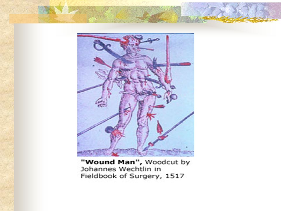
Wound Healing Vic V. Vernenkar, D.O. St. Barnabas Hospital Dept. of Surgery

2
Introduction Over the ages, many agents have been placed on wounds to improve healing. To date nothing has been identified that can accelerate healing in a normal individual. Many hinder the healing process. A surgeon’s goal in wound management is to create an environment where the healing process can proceed optimally.

3
Early Wound Healing Events

4
Stages of Wound Healing

5
Wounding Blood vessels are disrupted, resulting in bleeding. Hemostasis is the first goal achieved in the healing process. Cellular damage occurs, this initiates an inflammatory response. The inflammatory response triggers events that have implications for the entire healing process. Step one then is hemostasis, resulting in Fibrin.

7
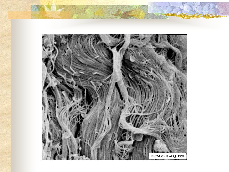
Early Events Fibrin and fibronectin form a lattice that provides scaffold for migration of inflammatory, endothelial, and mesenchymal cells. Fibronectin is produced by fibroblasts, has a dozen or so binding sites. Binds cytokines Its breakdown products stimulate angiogenesis.

8
Signs of Inflammation Erythema Edema Pain Heat

9
Signs of Inflammation Immediately after injury, intense vasoconstriction leads to blanching, a process mediated by epinephrine, NE, and prostaglandins released by injured cells. Vasoconstriction reversed after 10min, by vasodilatation. Now redness and warmth. Vasodilatation mediated by histamine, linins, prostaglandins.

10
Inflammation As microvenules dilate, gaps form between the endothelial cells,resulting in vascular permeability. Plasma leaks out into extravascular space. Leukocytes now migrate into the wound by diapedesis, adhere to endothelial cells, to wounded tissues. Alteration in pH from breakdown products of tissue and bacteria, along with swelling causes the pain.

11
Inflammation Neutrophils, macrophages and lymphocytes come into wound. Neutrophils first on scene, engulf and clean up. Macrophages then eat them or they die releasing O2 radicals and destructive enzymes into wound. Monocytes migrate into extravascular space and turn into macrophages. Macrophages very important in normal wound healing.

12
Inflammation Macrophages eat bacteria, dead tissue, secrete matrix metalloproteinases that break down damaged matrix. Macrophages source of cytokines that stimulate fibroblast proliferation, collagen production. Lymphocytes produce factors like FGF, EGF, IL- 2. At 48-72 hrs, macrophages outnumber neuts. By days 5-7 few remain.

13
Intermediate Events

14
Proliferation Mesenchymal cell chemotaxis Mesenchymal cell proliferation Angiogenesis Epithelialization

15
Proliferation Fibroblasts are the major mesenchymal cells involved in wound healing,, although smooth muscle cells are also involved. Normally reside in dermis, damaged by wounding. Macrophage products are chemotactic for fibroblasts. PDGF, EGF, TGF, IL-1, lymphocytes are as well.

16
Proliferation Angiogenesis reconstructs vasculature in areas damaged by wounding, stimulated by high lactate levels, acidic pH, decreased O2 tension in tissues. Cytokines directly stimulate the endothelial cell migration and proliferation required for angiogenesis. Many are produced by Macs. FGF-1 is most potent angiogenic stimulant identified. Heparin important as cofactor, TGF- alpha, beta, prostaglandins also stimulate.

17
Epithelialization The process of epithelial renewal after injury. Particularly important in partial thickness injuries, but plays a role in all healing. Partial thickness wounds have epidermis and dermis damaged, with some dermis preserved. Epithelial cells involved in healing come from wound edges and sweat glands, sebaceous glands in the more central portion of wound.

18
Skin Anatomy Epidermis is composed of multiple layers of epithelial cells superficial to the dermis. The first layer above the dermis is the basal layer, which is composed of basaloid cells. The cells become more elongated as you go to superficial stratum corneum. Stratum corneum is mostly keratin and dead tissue.

19
Layers of Skin

20
Epithelialization In contrast in an incisional wound, cellular migration occurs over a short distance. Incisional wounds are re-epithelialized in 24-48h. The sequence of events here are cellular detachment, migration, proliferation, differentiation.

21
Epithelialization First 24h, basal cell layer thickens, then elongate, detach from basement membrane and migrate to wound as a monolayer across denuded area. Generation of a provisional BM which includes fibronectin, collagens type 1 and 5. Basal cells at edge of wound divide 48-72 h after injury. Epithelial cells proliferation contributes new cells to the monolayer. Contact inhibition when edges come together.

22
Late Wound Healing Events

23
Collagen Synthesized by fibroblasts beginning 3-5 days after injury. Rate increases rapidly, and continues at a rapid rate for 2-4 weeks in most wounds. As more collagen is synthesized, it gradually replaces fibrin as the primary matrix in the wound. After 4 weeks, synthesis declines, balancing destruction by collagenase.

25
Collagen Age, tension, pressure and stress affect rate of collagen synthesis. TGF-b stimulates it, glucocorticoids inhibit it. 19 types identified. Type 1(80-90%) most common, found in all tissue. The primary collagen in a healed wound. Type 3(10-20%) seen in early phases of wound healing. Type V smooth muscle, Types 2,11 cartilage, Type 4 in BM.
 most common, found in all tissue. The primary collagen in a healed wound. Type 3(10-20%) seen in early phases of wound healing. Type V smooth muscle, Types 2,11 cartilage, Type 4 in BM..")
26
Collagen Three polypeptide chains, right handed helix. Most polypeptide chains used in collagen assembly are alpha chains.

28
Collagen Every third AA residue is Glycine. Another critical component is hydroxylation of lysine and proline within the chains. Hydroxyproline is necessary for this. Requires Vit C, ferrous iron, and alpha ketoglutarate as co- enzymes. Steroids suppress much of this, resulting in underhydroxylated collagen, which is incapable of making strong cross-links leading to easy breakdown.

29
Wound Contraction Begins approximately 4-5 days after wounding. Represents centripetal movement of the wound edge towards the center of the wound. Maximal contraction occurs for 12-15 days, although it will continue longer if wound remains open.

30
Wound Contraction The wound edges move toward each other at an average rate of 0.6 to.75 mm/day. Wound contraction depends on laxity of tissues, so a buttocks wound will contract faster than a wound on the scalp or pretibial area. Wound shape also a factor, square is faster than circular.

31
Wound Contraction Contraction of a wound across a joint can cause contracture. Can be limited by skin grafts, full better than split thickness. The earlier the graft the less contraction. Splints temporarily slow contraction.

32
Terminal Wound Healing Event

33
Remodeling After 21 days, net accumulation of collagen becomes stable. Bursting strength is only 15% of normal at this point. Remodeling dramatically increases this. 3-6 weeks after wounding greatest rate of increase, so at 6 weeks you are at 80% to 90% of eventual strength and at 6mos 90% of skin breaking strength.

34
Remodeling The number of intra and intermolecular cross- links between collagen fibers increases dramatically. A major contributor to the increase in wound breaking strength. Quantity of Type 3 collagen decreases replaced by Type 1 collagen Remodeling continues for 12 mos, so scar revision should not be done prematurely.

35
Disturbances in Wound Healing

36
Local Factors Infection versus contamination Infection is when number or virulence of bacteria exceed the ability of local defenses to control them. 100000 organisms per gram of tissue. Foreign bodies, hematomas promote infection, impaired circulation, radiation. Systemic: AIDS, diabetes, uremia, cancer.

37
Local Factors Smoking stimulates vasoconstriction. Increases platelet adhesiveness Limits O2 carrying capacity Endothelial changes Diminished amount of collagen deposition.

38
Local Factors Radiation damages the DNA of cells in exposed areas. Fibroblasts that migrate into radiated tissues are abnormal. Collagen is synthesized to an abnormal degree in irradiated tissue causing fibrosis. Blood vessels become occluded. Damage to hair and sweat glands Vitamin A has been used to counteract this.

39
Systemic Factors Malnutrition Cancer Old Age Diabetes- impaired neutrophil chemotaxis, phagocytosis. Steroids and immunosuppression suppresses macrophage migration, fibroblast proliferation, collagen accumulation, and angiogenesis. Reversed by Vitamin A 25,000u per day.

40
Hypertrophic Scars and Keloids Excessive healing results in a raised, thickened scar, with both functional and cosmetic complications. If it stays within margins of wound it is hypertrophic. Keloids extend beyond the confines of the original injury. Dark skinned, ages of 2-40. Wound in the presternal or deltoid area, wounds that cross langerhans lines.

41
Keloids and Hypertrophic Scars Keloids more familial Hypertrophic scars develop soon after injury, keloids up to a year later. Hypertrophic scars may subside in time, keloids rarely do. Hypertrophic scars more likely to cause contracture over joint surface.

42
Keloids and Hypertrophic Scars Both from an overall increase in the quantity of collagen synthesized. Recent evidence suggests that the fibroblasts within keloids are different from those within normal dermis in terms of their responsiveness. No modality of treatment is predictably effective for these lesions.

Similar presentations


 Immediate threat: –Dehydration and electrolyte.>")


