Download presentation
Presentation is loading. Please wait.

1
Review of clinical anatomy & physiology of the eyelids & common infective and inflammatory disorders of the eyelids Dr. Ayesha S Abdullah

2
Learning objectives By the end of this lecture the students would be able to:- Correlate the structure of the eyelids with their functions and clinical presentation in common infective and inflammatory disorders. Define stye, chalazion, trichiasis & blepharitis. Differentiate between stye & chalazion on the basis of clinical presentation and describe the treatment.

3
Important superficial anatomical landmarks
Lid margin Upper lid crease Palpebral fissure height (max) Palpebral fissure length (max) Movable folds of skin that close and cover the eye. Upper lid is much more mobile than the lower Palpebral fissure is an almond shaped opening bounded by both lids Average height is 10mm & width is 25-30mm Medial & lateral Canthi are the angles formed at the junction of the lids Medial canthus is generally lower than lateral canthus
 Palpebral fissure length (max) Movable folds of skin that close and cover the eye. Upper lid is much more mobile than the lower. Palpebral fissure is an almond shaped opening bounded by both lids. Average height is 10mm & width is 25-30mm. Medial & lateral Canthi are the angles formed at the junction of the lids. Medial canthus is generally lower than lateral canthus.")
4
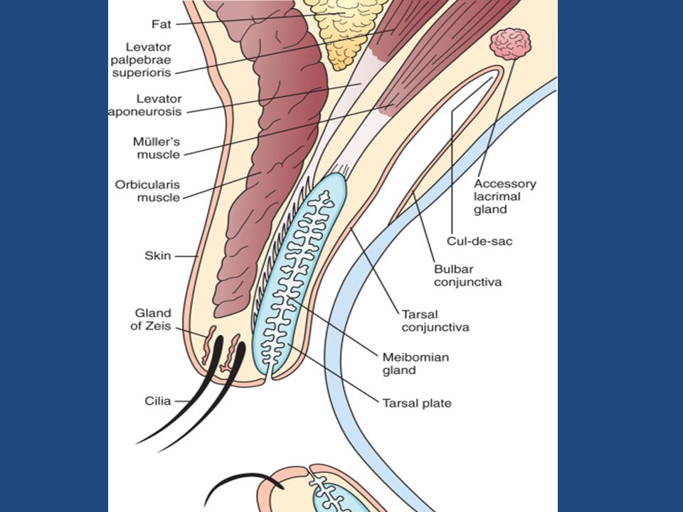
Structure of the lids The eyelid has five layers of different structures Skin Subcutaneous tissue Muscular layer Tarsal plate Conjunctiva

5
Surgical anatomy Anterior Lamina Skin Orbicularis muscle
Posterior Lamina Tarsal plate Conjunctiva Submuscular areolar tissue consists of variable, loose connective tissue below the orbicularis oculi muscle. The lid may be split into anterior and posterior portions through this potential plane, which is reached by division at the gray line of the lid margin. In the upper lid, this plane is traversed by fibers of the levator aponeurosis, some of which pass through the orbicularis to attach to the skin to form the lid crease

8
Anterior lamina Muscular layer
Orbital part Palpebral Part Preseptal part Pretarsal part The skin of the eyelids is the thinnest of the body (< 1 mm).The Orbicularis oculi muscle may be arbitrarily divided into the orbital and palpebral parts, with the latter being divided further into the preseptal and pretarsal portions. The palpebral portion is used in blinking and voluntary winking, while the orbital portion is used in forced closure. Facial nerve innervation is from the temporal branches and from zygomatic branches of the facial nerve. The nerves are orientated horizontally and innervate the muscle from the undersurface
.The Orbicularis oculi muscle may be arbitrarily divided into the orbital and palpebral parts, with the latter being divided further into the preseptal and pretarsal portions. The palpebral portion is used in blinking and voluntary winking, while the orbital portion is used in forced closure. Facial nerve innervation is from the temporal branches and from zygomatic branches of the facial nerve. The nerves are orientated horizontally and innervate the muscle from the undersurface.")
9
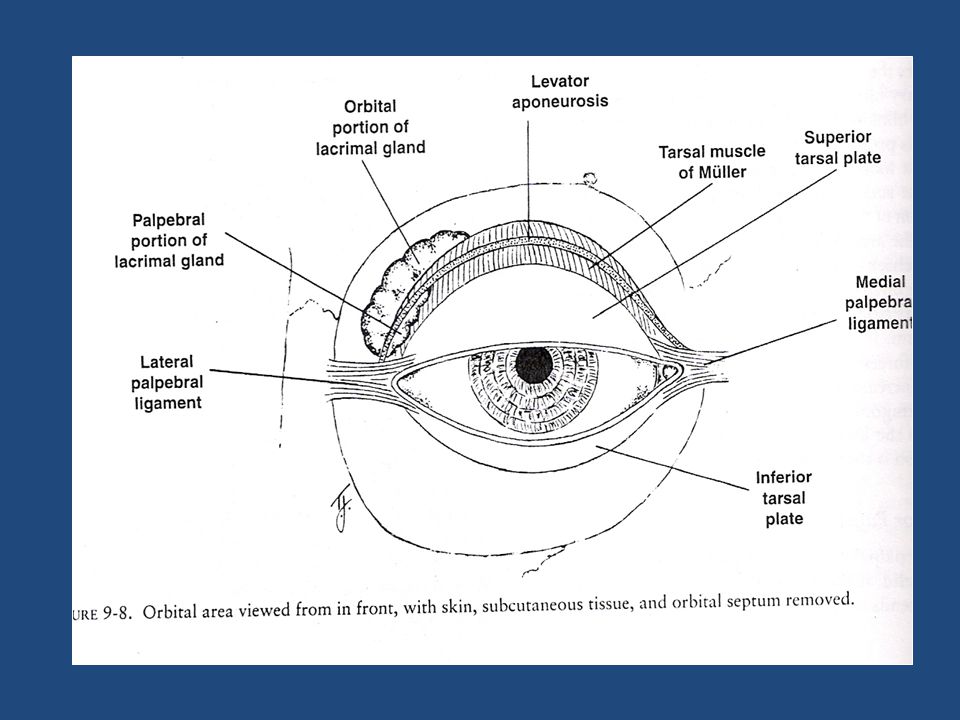
Posterior lamina The tarsal plates are composed of dense fibrous tissue and are responsible for the structural integrity of the lids. Each tarsus is approximately 29 mm long and 1 mm thick. The crescentic superior tarsus is 10 mm in vertical height centrally, narrowing medially and laterally. Each tarsus encloses about 25 sebaceous meibomian glands, which span the vertical height of the tarsus. Their ducts open at the lid margin posterior to the grey line and just anterior to the mucocutaneous junction. The medial and lateral ends of the tarsi are attached to the orbital rims by the medial and lateral palpebral ligaments.

10
Orbital Septum

13
Structures at the lid margin

16
HW msqheartline@hotmail.com
Nerve supply- sensory Blood supply Lymphatic drainage HW

17
Functions Protect the anterior surface of the globe
Aid in regulation of light reaching the eye Tear film maintenance; distribution & flow Lipid/ oily layer of the tear film

18
Disorders of eyelids Infective Inflammatory Neoplastic
Structural / disorders of malposition

19
Trichiasis In-turned eyelashes

20
Complications Corneal opacity & blindness

21
Treatment Epilation - but recurrences within few weeks
Electrolysis - but frequently repeated treatments Cryotherapy - for many lashes Laser ablation - for few scattered lashes Surgical correction - for resistant, localized crop Ocular lubricants

22
Blepharitis “ inflammation of the lid margin” Anterior Blepharitis
Affects base of eyelashes Associated with staphylococcal infection or Seborrhea Complicated by recurrent stye ,scarring of the lid margin and loss of eyelashes Posterior Blepharitis Affects meibomian gland openings Associated with meibomian gland dysfunction Complicated by recurrent chalazia, tear film instability and scarring of the lid margin

23
Anterior Blepharitis Posterior Blepharitis

24
Management Maintain lid hygiene
Apply warm compress Gently massage posterior lid to express meibomian gland contents Scrub lashes and lid margins with dilute baby shampoo Wipe lid margins with warm cloth after scrubbing Acute infectious flare-up (e.g. staphylococcal Blepharitis) Antibiotic ophthalmic ointment Meibomian gland dysfunction Ocular lubricants ( artificial tears/ tear substitutes) Tetracycline / Doxycycline orally Steriods for limited period
 Antibiotic ophthalmic ointment. Meibomian gland dysfunction. Ocular lubricants ( artificial tears/ tear substitutes) Tetracycline / Doxycycline orally. Steriods for limited period.")
25
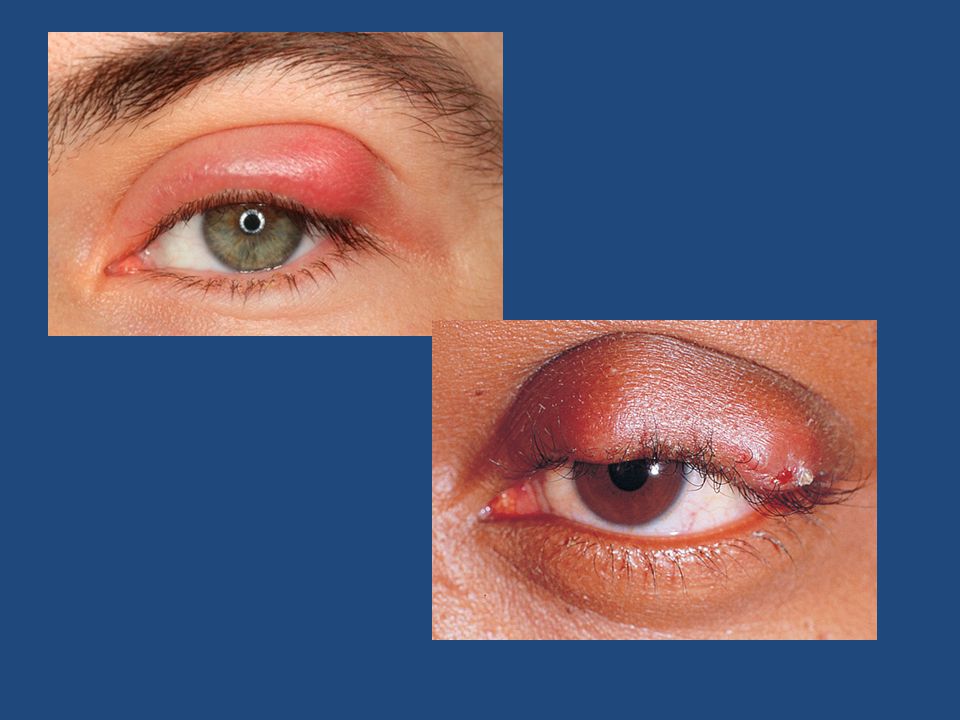
Stye “ Acute staphylococcal infection (abscess) of a lash follicle”
Common in children Tender hot swelling at the lid margin May spread to the entire lid causing prespetal or at time orbital cellulitis

28
Treatment Hot compresses
Self-limiting; epilation of the infected lash hastens resolution If spread of infection is likely with gross redness and swelling of the lid than topical and systemic antibiotics can be given along with analgesics/ NSAIDs

29
Chalazion “Chronic granulomatous inflammation of the Meibomian glands secondary to blockage of the gland orifice”. Common in patients with posterior blepharitis Non-tender swelling a little away from the lid margin

31
Treatment Small – no treatment
Incision and curretage of the affected gland.

32
Summary Important anatomical landmarks Trichiasis Blepharitis Stye
Chalazion

Similar presentations