Download presentation
Presentation is loading. Please wait.

1
Exercise 9: Cytoskeletal Structures

2
Announcements Post Lab 11 is due by your next lab. LNA Cytoskeletal Structure assigned today, and is due next lab (week of April 27). Next Lab Exam 2 Review. Exam 2: Week of May 4-6. Your exam time for Exam 2 is the same as it was for Exam 1 Final Exam: Friday, May 8from 8 – 11 AM If you have a conflict with the Final Exam, you must fill out the Conflict Final Exam Request Form found on the Course Website.
. Next Lab Exam 2 Review. Exam 2: Week of May 4-6. Your exam time for Exam 2 is the same as it was for Exam 1 Final Exam: Friday, May 8from 8 – 11 AM If you have a conflict with the Final Exam, you must fill out the Conflict Final Exam Request Form found on the Course Website..")
3
Goals Become familiar with the three different cytoskeletal systems Understand the role and structures of the mitotic spindle during cell division

4
Cytoskeleton Composed of three distinct systems (classified by size) –Microfilaments (actin) - thinnest –Intermediate filaments (lamin) –Microtubules (tubulin)- thickest
 –Microfilaments (actin) - thinnest –Intermediate filaments (lamin) –Microtubules (tubulin)- thickest")
5
Microfilaments (Actin) Microfilaments Made up of strands of the protein actin and often interact with strands of other proteins. They change cell shape and drive cellular motion, including contraction, cytoplasmic streaming, and the “pinched” shape changes that occur during cell division. Microfilaments and myosin strands together drive muscle action.

6
Intermediate Filaments (Lamins) Intermediate filaments Made up of fibrous proteins organized into tough, ropelike assemblages that stabilize a cell’s structure and help maintain its shape. Some intermediate filaments help to hold neighboring cells together (Cell junctions). Others make up the nuclear lamina.
. Others make up the nuclear lamina..")
7
Microtubules (Tubulin) Microtubules Long, hollow cylinders made up of many molecules of the protein tubulin. Tubulin consists of two subunits, -tubulin and - tubulin. Microtubules lengthen or shorten by adding or subtracting tubulin dimers. Microtubule shortening moves chromosomes. Interactions between microtubules drive the movement of cells. Microtubules serve as “tracks” for the movement of vesicles.

8
The Exercise Part I: Actin and Myosin Filaments –View prepared slides of smooth, skeletal, and cardiac muscle –Draw each muscle type and label nucleus, sarcomere, I-disk, A & I bands –You may find the Muscle Poster helpful Part II: Flagella and Cilia –Prepare slides –Observe the locomotion of each organism –Also available as prepared slides if needed Part III: Mitotic Spindle in plant and animal cells –View prepared slides of whitefish blastula and onion root tip –Compare/Contrast mitotic spindles found in plant and animal cells

9
Part I: Muscle Cells Skeletal: voluntary movement, breathing Smooth: involuntary, movement of internal organs Cardiac: beating of heart

10
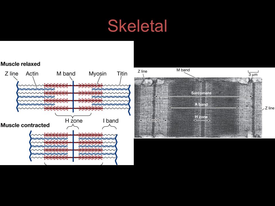
Skeletal

12
Cardiac Muscle Each muscle cell contains only one nucleus. Adjoining cells interdigitate forming a meshwork that is resistant to tearing (intercalated disk).
..")
13
Smooth Muscle Long and spindled shaped. Each cell has a single nucleus Actin and myosin filaments are not regularly arranged and therefore, do not produce the striated appearance

14
Summary of Muscle Types

15
The Exercise Part I: Actin and Myosin Filaments –View prepared slides of smooth, skeletal, and cardiac muscle –Draw each muscle type and label nucleus, sarcomere, I-disk, A & I bands Part II: Flagella and Cilia (microtubules) –Prepare slides with Protoslo –Observe the locomotion of each organism –Also available as prepared slides if needed Part III: Mitotic Spindle in plant and animal cells –View prepared slides of whitefish blastula and onion root tip –Compare/Contrast mitotic spindles found in plant and animal cells
 –Prepare slides with Protoslo –Observe the locomotion of each organism –Also available as prepared slides if needed Part III: Mitotic Spindle in plant and animal cells –View prepared slides of whitefish blastula and onion root tip –Compare/Contrast mitotic spindles found in plant and animal cells")
16
Protozoa Cultures Amoeba Ciliate –Stentor Flagellate –Euglena

17
Amoeba

18
Stentor Cilia

19
Euglena Flagella #3

20
The Exercise Part I: Actin and Myosin Filaments –View prepared slides of smooth, skeletal, and cardiac muscle –Draw each muscle type and label nucleus, sarcomere, I-disk, A & I bands Part II: Flagella and Cilia –Prepare slides –Observe the locomotion of each organism Part III: Mitotic Spindle in plant and animal cells –View prepared slides of whitefish blastula and onion root tip –Compare/Contrast mitotic spindles found in plant and animal cells

21
Mitotic Spindle Constructed to enable the separation of the chromatids formed during replication Consists of microtubules radiating out from the two centrosomes Centrosome consists of a pair of centrioles

22
Cell Division

23
The Mitotic Spindle Consists of Microtubules

Similar presentations


 is a cellular scaffolding or skeleton contained within the cytoplasm that is made out of protein. The cytoskeleton.>")


 of cell membrane 2. Structure (and function) of organelles 3. Interconnections between cells to maintain structural.>")


