Download presentation
Presentation is loading. Please wait.

1
Integrated Studies Applied Neuroscience Seminar Evan Fletcher, Mario Ortega Dr. Charles DeCarli, IDeA Lab Director

2
Overview of Course -OVERVIEW -Introductory Lecture -Lab Hours involving computer based image analysis (Mario, Evan) -LECTURE -Introductory Lecture (today) including: -Basics of Neuroanatomy, Software Overview of Laboratory -MRI Imaging Basics -Memory Decline with Aging -LABORATORY -Longitudinal Hippocampus Project -Testing a possibility programs under development -Trace a hippocampus at initial time and apply it to future scan using computer modeling and “warping”
 -LECTURE -Introductory Lecture (today) including: -Basics of Neuroanatomy, Software Overview of Laboratory -MRI Imaging Basics -Memory Decline with Aging -LABORATORY -Longitudinal Hippocampus Project -Testing a possibility programs under development -Trace a hippocampus at initial time and apply it to future scan using computer modeling and warping")
3
Studies of Brain Form and Function Postmortem brains (not us!) Used for precise anatomical measurements Living subjects (computer analysis) Use MRI imaging to track form and function in living individuals
 Used for precise anatomical measurements Living subjects (computer analysis) Use MRI imaging to track form and function in living individuals")
4
Basics of Brain Anatomy Next slides show postmortem images to illustrate large anatomical features Good for precise anatomical measurements Cannot study changes during life (obviously!). For that, need MRI

5
The Human Brain Cerebrum -Divided into four regions, Frontal, Parietal. Occipital, Temporal -Highly convoluted surface with 6 layers of cells in the cortex.

6
Meninges -the brain has several layers of protection, internal to the skull, these are the meninges. -On an MRI image the different layers are not discernable. -They provide protection, drain the Cerebrospinal Fluid, and blood input and output.

7
Four Cerebral Lobes (viewed from midline)
")
8
MRI Images Magnetic Resonance Imaging Intense magnetic and radio frequency fields Quantum mechanics Pinpoint small features in living brain Lauterbur and Mansfield – Nobel Prize 2003

9
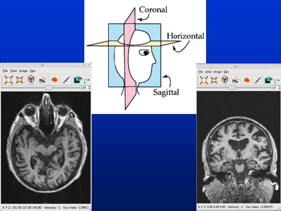
Sample MRI Images High resolution anatomical MRI 3D and 2D views Skull in place and stripped

11
These images were viewed using “Slice Viewer” a program you will see in this lab. We will show you images so you can learn the basic structures and how to separate brain from meninges.

12
Anatomical Basics from MRI The next slides show brain features relevant to longitudinal studies of living individuals Longitudinal studies -Track changes in form and function for same individual over time

13
More Anatomical Basics

14
Young and Old These images show comparable slices of young and old brain images. Compare features for changes: –Ventricles –Hippocampi –Sulci

15
Our focus: Hippocampus Next slides will explore the hippocampus which is the focus of your research project Points about the hippocampus: 1.Seat of long-term memory formation 2.Subject to early deterioration in Alzheimer’s Disease

16
The Hippocampus

17
Hippocampi and Ventricles Hippocampus (purple)
")
18
An elephant never forgets This shows the huge and convoluted hippocampus of an elephant (red) Greater hippocampus volume to brain ratio than in humans
 Greater hippocampus volume to brain ratio than in humans")
19
Our Research Project: Longitudinal Hippocampal Change Hippocampus is a main site of degeneration in dementia Acquire brain image at time 1 and time 2 Trace (manually) hippocampus at time 1 “Warp” time 1 onto time 2 Use the warp to automatically compute hippocampal volume at time 2
 hippocampus at time 1 Warp time 1 onto time 2 Use the warp to automatically compute hippocampal volume at time 2")
20
Warping No two brain images are alike Even for time 1 and time 2, local changes will occur Warping is a technique for computing these changes and morphing one image onto another It can highlight brain changes and compute differences

21
Uses of warping Slides show two views of longitudinal change Left panels: earlier time Middle: later time Right: color-coded views of shifts Changes computed by warps

22
Your Role in Research Overall goal: track hippocampal volume changes in 20 subjects Steps –Strip skulls from images –Align time 1 and time 2 brains –Trace hippocampus in each time –Warp time 1 to time 2 –Compute hippocampal volume changes

23
Things you will learn 1.Navigation in linux computers: Basic linux commands 2.Using software tools in linux environment 3.Basic brain anatomy focusing on hippocampus (Mario) 4.Tracing techniques (Mario) 5.Concepts of image warping and volumetric calculations
 4.Tracing techniques (Mario) 5.Concepts of image warping and volumetric calculations")
Similar presentations


>")


>")


 This study looks at the brains of London taxi drivers and examines the role of the hippocampus in helping them to navigate their way around.>")

 Temporal Lobe (& auditory cortex),>")