Download presentation
Presentation is loading. Please wait.

1
Wet Lab Radiation Damage Measured by Micronucleus (MN) Assay Background Background Equipment Equipment Supplies Supplies Procedures Procedures Lab Demonstrations Lab Demonstrations
 Assay Background Background Equipment Equipment Supplies Supplies Procedures Procedures Lab Demonstrations Lab Demonstrations")
2
Background Types of micronucleus assaysTypes of micronucleus assays Applications of micronucleus assaysApplications of micronucleus assays

3
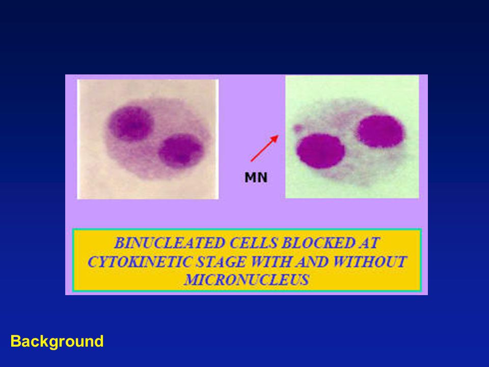
Background MN

4
Micronucleus Assay (MN) It can be used to detect the clastogenic and aneugenic effects of test agents both in vitro and in vivo.It can be used to detect the clastogenic and aneugenic effects of test agents both in vitro and in vivo. –Clastogenic: any substance or process causing chromosome breaks. –Aneugenic: Agents which affect cell division and the mitotic spindle apparatus resulting in the loss or gain of whole chromosomes, thereby inducing an aneuploidy. Background

5
Micronucleus Assay (MN) It is much more cost effective than the metaphase chromosome aberration assay.It is much more cost effective than the metaphase chromosome aberration assay. The in vitro micronucleus assay is considered as the replacement for conventional metaphase analysis as the screening test of choice for clastogenicity.The in vitro micronucleus assay is considered as the replacement for conventional metaphase analysis as the screening test of choice for clastogenicity. Background

6
Micronucleus Assay (MN) The in vivo assay, usually conducted in mice, is especially important since no in vitro alternative test has been validated to replace the MN test.The in vivo assay, usually conducted in mice, is especially important since no in vitro alternative test has been validated to replace the MN test. The cells evaluated in this assay are typically erythrocyte populations in either the peripheral blood or bone marrow compartment.The cells evaluated in this assay are typically erythrocyte populations in either the peripheral blood or bone marrow compartment. Background

7
Micronucleus Assays (MN) Cytokinesis-block micronucleus (CBMN) assayCytokinesis-block micronucleus (CBMN) assay lymph, cell lineslymph, cell lines Flow cytometric micronucleus (FCMN) assayFlow cytometric micronucleus (FCMN) assay red blood cells red blood cells Background
 Cytokinesis-block micronucleus (CBMN) assayCytokinesis-block micronucleus (CBMN) assay lymph, cell lineslymph, cell lines Flow cytometric micronucleus (FCMN) assayFlow cytometric micronucleus (FCMN) assay red blood cells red blood cells Background")
8
CBMN Assay (lymphocytes) Cytochalasin-B (actin inhibitor)Cytochalasin-B (actin inhibitor) Background
 Cytochalasin-B (actin inhibitor)Cytochalasin-B (actin inhibitor) Background")
10
CBMN + FISH Background

11
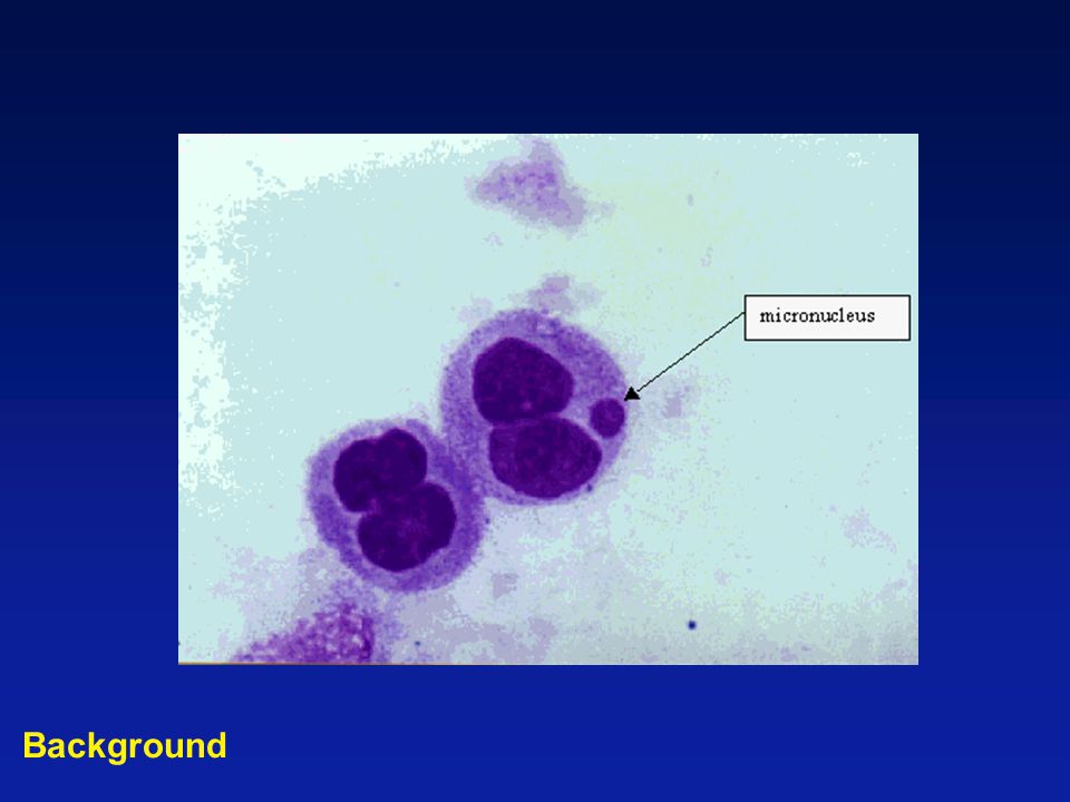
Criteria for CBMN The diameter of the MN should be less than one-third of the main nucleus.The diameter of the MN should be less than one-third of the main nucleus. MN should have similar staining as the main nucleus.MN should have similar staining as the main nucleus. MN should be separated from or marginally overlap with main nucleus.MN should be separated from or marginally overlap with main nucleus. Background

13
CBMN vs. Chromosome Spreading AdvantagesAdvantages –Rapidity –Cheap –Simplicity –Statistical power DisadvantagesDisadvantages –Not all chromosome aberrations (acentric fragments) –Toxicity of cyto-B Lymph. vs. cell linesLymph. vs. cell lines Background
 –Toxicity of cyto-B Lymph. vs. cell linesLymph. vs. cell lines Background.")
14
Applications of MN Assay Biological dosimetryBiological dosimetry Risk assessment of cancerRisk assessment of cancer –Lung: smoking –Oral mucosa and bladder cancer: arsenic Genotoxic effects of pesticidesGenotoxic effects of pesticides Cytotoxic effects of ROSCytotoxic effects of ROS Radiosensitivity indicator in head and neck tumor patientsRadiosensitivity indicator in head and neck tumor patients Background

15
Dose Response Dose (Gy) Dicentrics/Cell 1.0 2.0 0 Background Dose (Gy) % MN 2.0 4.0 0 ??? chromosome MN Post-irradiation time

16
FCMN Assay Rapid scoringRapid scoring Large number of cellsLarge number of cells Small changes in MN frequencySmall changes in MN frequency Reticulocytes from peripheral bloodReticulocytes from peripheral blood DNA contentsDNA contents

17
DNA content Where are micro-nucleated cells?

18
Whole blood White blood cells Red blood cells (size) Reticulocytes (CD71) Platelets (anti-platelet) CD61 Nucleated red cells (DNA contents) Ret-MN XX X Litron Laboratory Steve Dertinger, Ph.D.
 Reticulocytes (CD71) Platelets (anti-platelet) CD61 Nucleated red cells (DNA contents) Ret-MN XX X Litron Laboratory Steve Dertinger, Ph.D.")
19
Cell size (red cells)
")
20
Single cells total mature RBC Single cells platelets Single cells Reticulocytes for MN (anti-CD61-PE)
")
21
Single cells Reticulocytes No platelets MN MN: DNA content

22
Equipment Tissue culture equipmentTissue culture equipment –Sterile tissue culture hood, Incubator (CO 2, 37 o C, humidified), Microscopes( upright, inverted, dissecting), Pipettes, Hemacytometers (Coulter counter). Radiation sourceRadiation source Flow cytometerFlow cytometer

23
Supplies Tissue culture flasks (dishes)Tissue culture flasks (dishes) Tissue culture mediumTissue culture medium Glass slidesGlass slides Cytochalasin-BCytochalasin-B Giemsa stainGiemsa stain MN DNA staining kit (Litron)MN DNA staining kit (Litron)
Tissue culture flasks (dishes) Tissue culture mediumTissue culture medium Glass slidesGlass slides Cytochalasin-BCytochalasin-B Giemsa stainGiemsa stain MN DNA staining kit (Litron)MN DNA staining kit (Litron)")
24
Procedures Calibration and color compensation for flow cytometer Rat blood cellsRat blood cells –DNA (PI), CD71- (FITC), anti-platelets+ (PE) Parasite infected mouse blood cellsParasite infected mouse blood cells –DNA (PI), CD71+ (FITC) Parasite infected mouse blood cellsParasite infected mouse blood cells –DNA (PI), CD71+ (FITC), anti-platelets+ (PE)
, CD71- (FITC), anti-platelets+ (PE) Parasite infected mouse blood cellsParasite infected mouse blood cells –DNA (PI), CD71+ (FITC) Parasite infected mouse blood cellsParasite infected mouse blood cells –DNA (PI), CD71+ (FITC), anti-platelets+ (PE)")
25
Rat blood cells DNA (PI) CD71- anti-platelet (anti-CD61-PE) Single cells mature RBC Single cells platelets Single cells Reticulocytes for MN
 CD71- anti-platelet (anti-CD61-PE) Single cells mature RBC Single cells platelets Single cells Reticulocytes for MN")
26
Rat blood cells

27
Parasite infected mouse blood cells DNA (PI) CD71+ (anti-CD61-PE) DNA ratio 20:6000
 CD71+ (anti-CD61-PE) DNA ratio 20:6000")
28
Parasite infected mouse blood cells

29
Parasite infected mouse blood cells DNA (PI) CD71 anti-platelet (anti-CD61-PE)
 CD71 anti-platelet (anti-CD61-PE)")
30
Parasite infected mouse blood cells

31
MN Unirradiated (0 Gy) mouse blood cells DNA (PI) CD71+ CD61+
 mouse blood cells DNA (PI) CD71+ CD61+")
32
Parasite infected mouse blood cells by FACS Vantage sorter DNA (PI) CD71+ CD61+
 CD71+ CD61+")
33
Human blood cells from irradiated (therapy) patient DNA (PI) CD71+ CD61+
 patient DNA (PI) CD71+ CD61+")
34
Lab Demonstrations room 3-4151, 3-4157 FCMN assayFCMN assay room B-6624, B-6625 CBMN assayCBMN assay

Similar presentations


 Qingshan Qu, MD (PI) Roy Shore, PhD (Co-I) Dept. of Environmental Med. NYU School of Medicine.>")


 Analysis Lecture Module 6.>")


 and intercellular.>")
 on local tumor response and lung metastatic potential in boron neutron capture therapy (BNCT), referring to.>")