Download presentation
Presentation is loading. Please wait.

1
Rad Protection in Fluoro & INTRO RHB regs
RT Week 1,2 Wed

2
Regulatory Requirements
1. Regarding the operation of fluoroscopy units 2. Regarding personnel protection 3. Regarding patient protection shielding for image intensifier cumulative timer dead-man switch

3
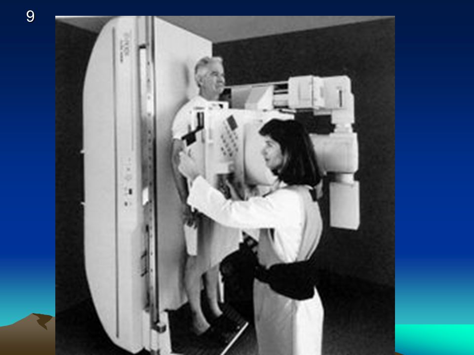
Fluoroscopic Positioning Previewing
Radiographers are trained in positioning Unnecessary radiation exposure to patient is unethical Fluoroscopic equipment should not be used to preview patient’s position

4
Patient Protection Tabletop exposure rate Maximum 10 R/min
Typically 1 – 3 R/min Some books ave is 4 R/min **

5
Patient Protection Minimum source-to-skin distance
12” for mobile equipment 15” for stationary systems Audible alarm at 5 mins. Same rules for collimation

6
Patient Protection Typical exposure rates Cinefluorography Cassettes
7.2 R/min Cassettes 30 mR/exposure 105 mm film 10 mR/exposure

7
Protection of Radiographer and Radiologist
Lead apron 0.25 mm Pb/eq Highest energy scatter 90o angle to the incident beam Same level as radiographer /radiologist’s gonads

8
Protection of Radiographer and Radiologist
Single step away from the table decreases exposure exponentially Bucky slot cover Lead rubber drape Radiologist as shielding

11
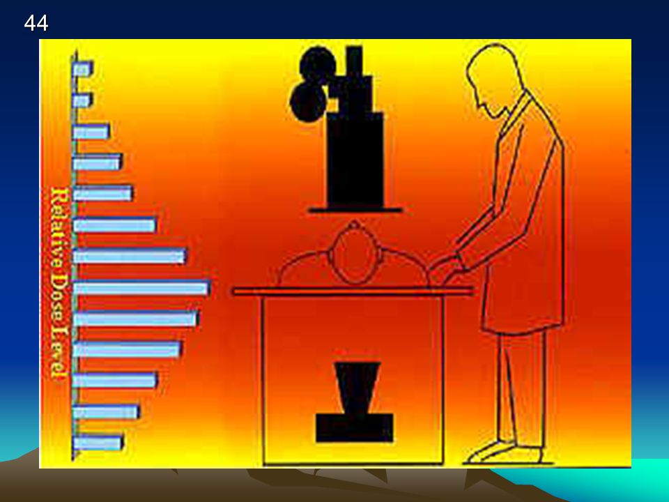
ISOEXPOSURE CURVES

12
Protection of Others Radiographer’s responsibility to inform others in the room to wear lead apron Do not initiate fluoroscopy until all persons have complied

13
PUBLIC EXPOSURE 10 % OF OCCUPATIONAL NON MEDICAL EXPOSURE
.5 RAD OR 500 MRAD UNDER AGE 18 AND STUDENT .1 rem mSv

14
COLLIMATION (for X-ray TUBE Collimator)
The PATIENT’S SKIN SURFACE SHOULD NOT BE CLOSER THAN ___________ CM BELOW THE COLLIMATOR? ____________ INCHES? 15 cm / 6.5 inches

15
The Patient & Scatter

16
Radiation Protection (recap)
Tube in never closer to the patient than 15” in stationary tubes and 12” with a C arm As II moves away from the patient the tube is being brought closer Bucky tray is connected to a lead shield called the Bucky slot cover. It must be 0.25 mm Pb There should be a protective apron of at least 0.25 mm Pb that hangs down from the II Every machine is required to have an audible timer that signals 5 minutes of fluoroscopy time Exposure switch must be a “dead man” type 16

17
Regulations about the operation
Fluoroscopic tubes operate at currents that range from0.5 to 5 mA with 3 the most common AEC rate controls: equipment built after 1974 with AEC shall not expose in excess of 10 R/min; equipment after 1974 without AEC shall not expose in excess of 5 R/min

18
Other regulations Must have a dead man switch
Must have audible 5 min. exposure timer Must have an interlock to prevent exposure without II in place Tube potential must be tested (monitored)weekly Brightness/contrast must be tested annually Beam alignment and resolution must be tested monthly Leakage cannot exceed 100mR/hr/meter
weekly. Brightness/contrast must be tested annually. Beam alignment and resolution must be tested monthly. Leakage cannot exceed 100mR/hr/meter.")
19
Fluoroscopy exposure rate
For radiation protection purposes the fluroscopic table top exposure rate must not exceed 10 mR/min. The table top intensity should not exceed 2.2 R/min for each mA of current at 80 kVp

20
Patient Protection A 2 minute UGI results in an exposure of approximately 5 R!! After 5 minutes of fluoro time the exposure is R Use of pulsed fluoro is best (means no matter how long you are on pedal there is only a short burst of radiation) ESE must not be more than 5 rads/min
 ESE must not be more than 5 rads/min.")
21
Rad Protection Always keep the II as close to the patient as possible to decrease dose Highest patient exposure happens from the photoelectric effect (absorption) Boost control increases tube current and tube potential above normal limits Must have continuous audible warning Must have continuous manual activation
 Boost control increases tube current and tube potential above normal limits. Must have continuous audible warning. Must have continuous manual activation.")
22
ESE FOR FLUORO TLD PLACED AT SKIN ENTRACE POINT
1 – 5 R/MINUTE AVE IS 4 R/MIN INTERGRAL DOSE – 100 ERGS OF TISSUE = 1 RAD EXPOSURE OR 1 GM RAD = 100 ERGS

23
SSD – TUBE TO SKIN DISTANCE
FIXED UNITS 18” PREFERRED 15 “ MINIMUM MOBILE UNITS ( C-ARMS) 12’ MINIMUM
 12’ MINIMUM.")
24
PATIENT PROTECTION LIMIT SIZE OF BEAM BEAM ON TIME
DISTANCE OF SOURCE TO SKIN PBL FILTRATION (2.5 mm Al 70 SHEILDING SCREEN/FILM COMBO

26
GONAD SHIELDING MUST BE . 5 MM OF LEAD
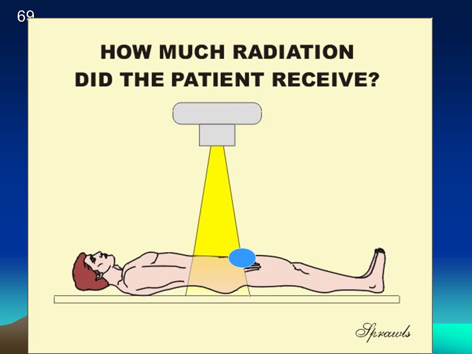
MUST BE USED WHEN GONADS WILL LIE WITHING 5 CM OF THE COLLIMATED AREA (RHB) KUB. Lumbar Spine Pelvis male vs female shielding
 KUB. Lumbar Spine Pelvis. male vs female shielding.")
27
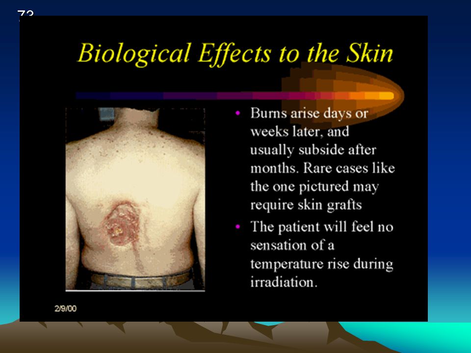
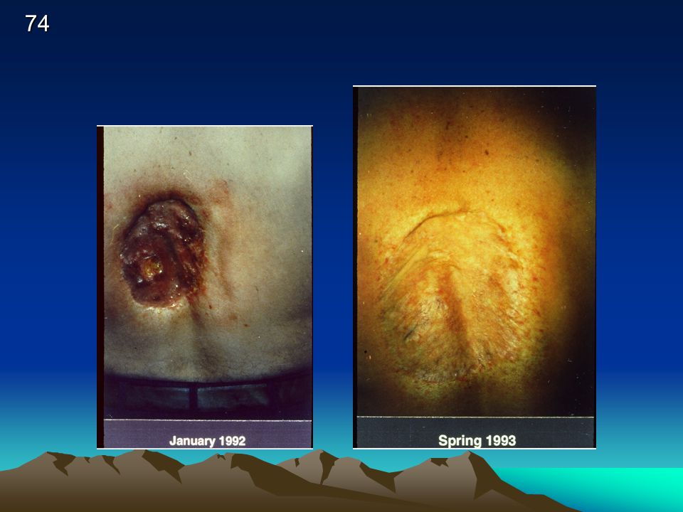
♀ receive 3x more dose than ♂ for pelvic x-rays
Gonad shielding & dose ♀ receive 3x more dose than ♂ for pelvic x-rays 1 mm lead will reduce exposure (primary) by about 50% ♀ by about 90 – 95 % ♂
 by about 50% ♀ by about 90 – 95 % ♂")
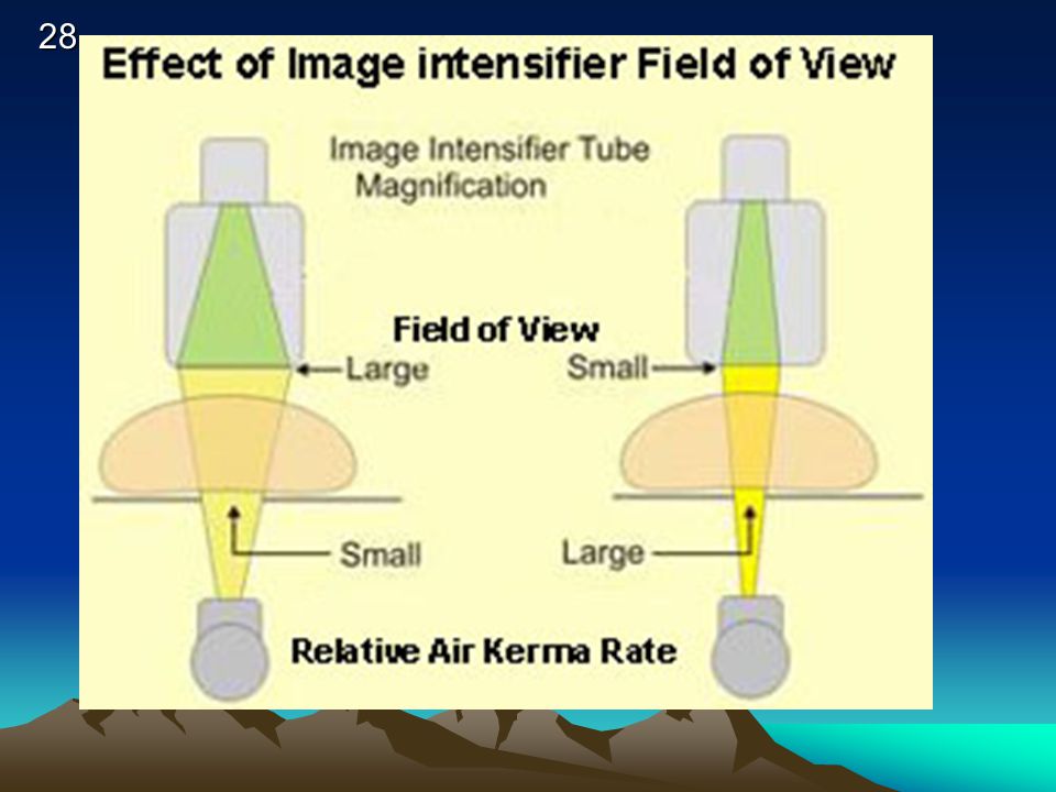
29
KEEP I.I. CLOSE TO PATIENT

30
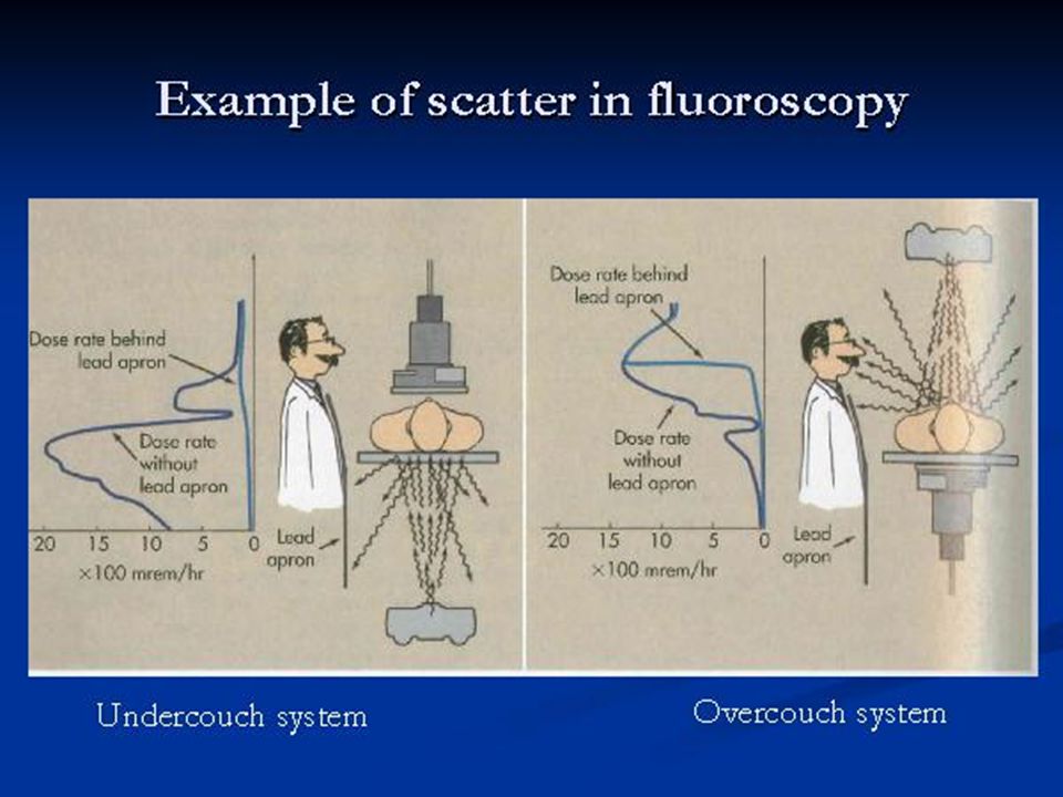
Over vs under the table fluoro tubes

31
REDUCE DISTANCE OF IMAGE INTESIFIER
PATIENT EXPOSURE REDUCE DISTANCE OF IMAGE INTESIFIER INCREASE DISTANCE FROM THE TUBE

32
Patient entrance skin exposure (ESE) is higher when the fluoroscopic x-ray tube is too close to the tabletop.
 is higher when the fluoroscopic x-ray tube is too close to the tabletop.")
33
PATIENT EXPOSURE REDUCE SIZE OF COLLIMATED BEAM WHEN POSSIBLE

34
Framing and patient dose syll = Pg 31
The use of the available film area to control the image as seen from the output phosphor. Underframing Exact Framing, (58 % lost film surface) Overframing,(part of image is lost) Total overframing
 Overframing,(part of image is lost) Total overframing.")
35
EXPOSURE RATES FLUORO MA IS 0.5 MA TO 5 MA PER MIN
AVE DOSE IS 4 R / MIN IF MACHINE OUTPUT IS 2 R/MA/MIN = WHAT IS PT DOSE AT 1.5 MA FOR 5 MIN STUDY? 15R

36
EXPOSURE RATES FOR FLUORO
CURRENT STANDARD 10 R/MIN (INTENSIFIED UNITS) HLC: BOOST MODE 20 R/MIN OLD (1974) NO ABC NON IMAGE INTES 5 R/MIN
 HLC: BOOST MODE 20 R/MIN. OLD (1974) NO ABC NON IMAGE INTES. 5 R/MIN.")
37
DOSE REGULATIONS BEFORE 1974 - AT TABLETOP 5R/MIN (WITHOUT AEC)
5R/MIN (WITHOUT AEC) – BOOST MODE After with AEC 10 R/MIN R/MIN BOOST
 – BOOST MODE. After 1974 with AEC. 10 R/MIN 20R/MIN BOOST.")
38
RADIATION PROTECTION The Patient is the largest scattering object
Lower at a 90 DEGREE ANGLE from the patient + PRIMARY BEAM AT 1 METER DISTANCE - 1/1000 OF INTENSITY PRIMARY XRAY or 0.1%

39
BUCKY SLOT COVER .25 MM LEAD

40
Bucky Slot Cover

41
ISOEXPOSURE CURVES

43
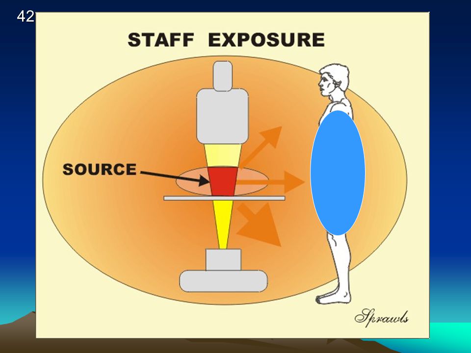
PERSONNEL PROTECTION SCATTER FROM THE PATIENT
TABLE TOP, COLLIMATOR, TUBE HOUSING, BUCKY STRAY RADIATION – LEAKAGE OR SCATTER RADIATION

45
TOWER CURTAIN .25 MM LEAD EQ

46
Lead curtain & dose reduction

47
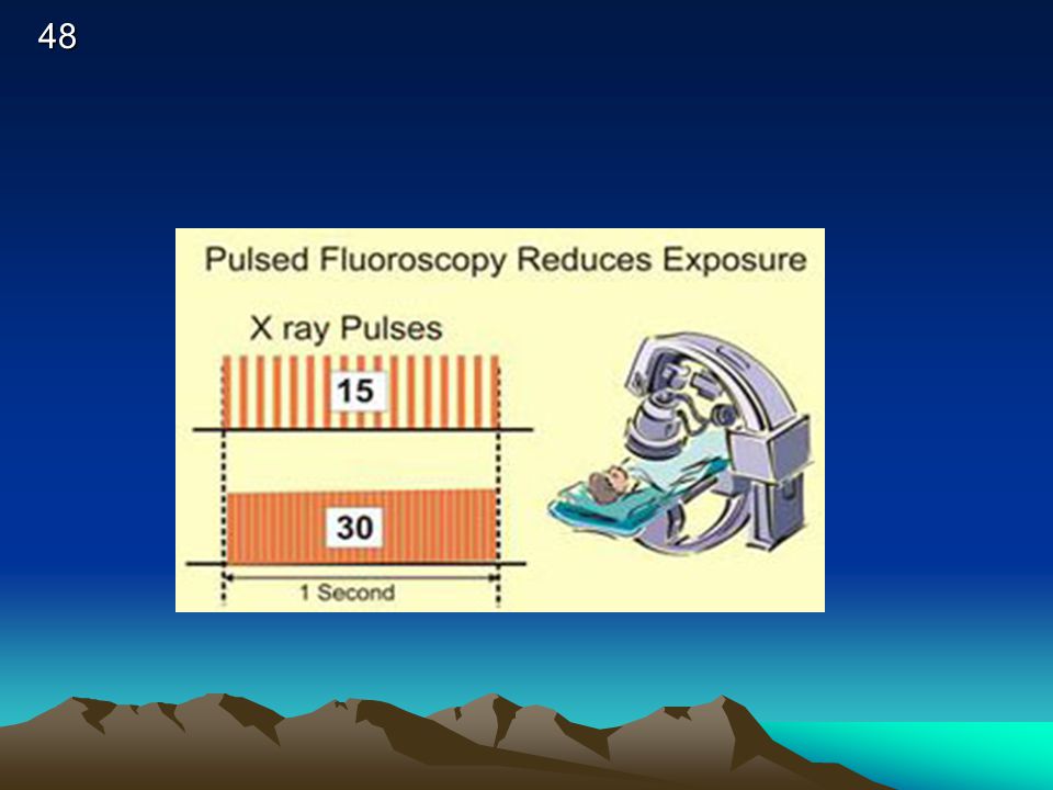
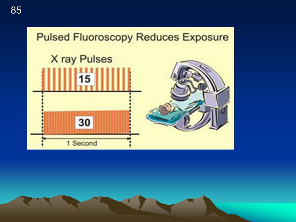
Pulsed Fluoro Some fluoroscopic equipment is designed for pulsed-mode operation. With the pulsed mode, it can be set to produce less than the conventional 25 or 30 images per second. This reduces the exposure rate. Collimation of the X ray beam to the smallest practical size and keeping the distance between the patient and image receptor as short as possible contribute to good exposure management.

49
PERSONNEL PROTECTION STANDING BEHIND A PROTECTIVE PRIMARY (1/16TH pb) BARRIER: PRIMARY RADIATION EXPOSURE – 99.87% REDUCED PORTABLE BARRIER = 99 % REDUCTION

50
PERSONNEL PROTECTION PROTECTIVE APRONS – 0.25 PB = 97% ↓ TO SCATTER
THYROID SHEILDS (0.25 & 0.5) GLOVES (0.25 & 0.5)
 GLOVES (0.25 & 0.5)")
51
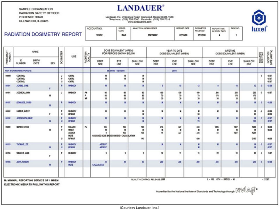
PERSONNEL PROTECTION MONITORING
FILM BADGE TLD POSL POCKET DOSIMETER RING BADGE

52
PERSONNEL PROTECTION MONITORING
DOSE LIMITS WHOLE BODY EYES EXTREMITIES (BELOW ELBOW/KNEES)
")
54
Report at least every quarter Preserved for a minimum of 3 years

55
RHB NOTIFICATION (EXP IN 24 HOURS) (RP Syllabus – pg 68)
IMMEDIATE reporting – WITHIN 24 HOURS TOTAL DOSE OF 25 rems Eye dose – 75 rem Extremity – 250 RADS OVEREXPOSURE – received w/in 24 hrs Must be ReportedWITHIN 30 DAYS TOTAL DOSE OF 5 rems Eye dose – 15 rem Extremity REMS

56
LICENSE RENEWAL WITHIN 30 DAYS OF EXPRIATION
NOTIFICATION OF CHANGE OF ADDRESS

57
HIGH RADIAITON AREA – RADIAITON AREA – 100 mRem ( 0.1 rem / (1 msV)
@ 30 cm from the source of radiaton RADIAITON AREA – RHB: 5 mRem ( rem / (.05 msV) @ 30 cm from the source of radiation PUBLIC 2 mrem per week* (STAT)
")
58
A “controlled area” is defined as one
that is occupied by people trained in radiologic safety that is occupied by people who wear radiation monitors whose occupancy factor is 1

59
RHB “RULES” RHB RP PG61 LICENTIATES OF THE HEALING ARTS
(MD, DO, DC, DPM) MUST HAVE A RADIOLOGY SUPERVISOR & OPERATORS PERMIT & CERTIFICATE TO OPERATE OR SUPERVISE THE USE OF X-RAYS ON HUMANS SUPEVISORS MUST POST THEIR LICENSES
 MUST HAVE A. RADIOLOGY SUPERVISOR & OPERATORS PERMIT & CERTIFICATE. TO OPERATE OR SUPERVISE THE USE OF X-RAYS ON HUMANS. SUPEVISORS MUST POST THEIR LICENSES.")
60
RHB “RULES” RHB RP PG62 ALL XRAYS MUST BE ORDERED BY A PHYSICIAN
VERBAL OR WRITTEN PRESCRIPTION See Section C – “Technologist Restrictions”

61
DOSE CINE - 2mR per frame (60f/sec) 400 mr per “look”
 400 mr per look")
62
WHATS WRONG?

63
Declared Pregnant Worker
Must declare pregnancy – 2 badges provided 1 worn at collar (Mother’s exposure) 1 worn inside apron at waist level Under 5 rad – negligible risk Risk increases above 15 rad Recommend abortion (spontaneous) 25 rad (“Baby exposure” approx 1/1000 of ESE)
 1 worn inside apron at waist level. Under 5 rad – negligible risk. Risk increases above 15 rad. Recommend abortion (spontaneous) 25 rad. ( Baby exposure approx 1/1000 of ESE)")
64
Remote – over the table tube

65
Under table tube vs Over table tube (remote units) Need moving shield for Remote rooms
 Need moving shield for Remote rooms")
67
IsoExposure Curves Where is it SAFE??

68
Back to the Patient & Protection

72
RADIATION DOSE TO PATIENTS
ESE - ENTRACE SKIN EXPOSURE (MEASURED BY A TLD) SKIN DOSE GONADAL DOSE BONE MARROW DOSE (MEAN GLADULAR DOSE- MAMMO) ** SEE TABLE 1-5 PG 20 & 1-8 PG 21 (STAT)
 SKIN DOSE. GONADAL DOSE. BONE MARROW DOSE. (MEAN GLADULAR DOSE- MAMMO) ** SEE TABLE 1-5 PG 20 & 1-8 PG 21 (STAT)")
75
ESE FOR FLUORO TLD PLACED AT SKIN ENTRACE POINT
1 – 5 R/MINUTE AVE IS 4 R/MIN INTERGRAL DOSE – 100 ERGS OF TISSUE = 1 RAD EXPOSURE OR 1 GM RAD = 100 ERGS

76
1 rad (of ionizing radiation) deposits 100 ergs of energy per gram
1 RAD -100 ERGS – 1 GRAM 1 rad (of ionizing radiation) deposits 100 ergs of energy per gram
 deposits 100 ergs of energy per gram.")
77
DOSE REGULATIONS BEFORE 1974 - AT TABLETOP 5R/MIN (WITHOUT AEC)
5R/MIN (WITHOUT AEC) – BOOST MODE After with AEC 10 R/MIN R/MIN BOOST
 – BOOST MODE. After 1974 with AEC. 10 R/MIN 20R/MIN BOOST.")
79
SSD – TUBE TO SKIN DISTANCE
FIXED UNITS 18” PREFERRED 15 “ MINIMUM MOBILE UNITS ( C-ARMS) 12’ MINIMUM
 12’ MINIMUM.")
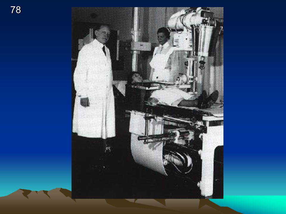
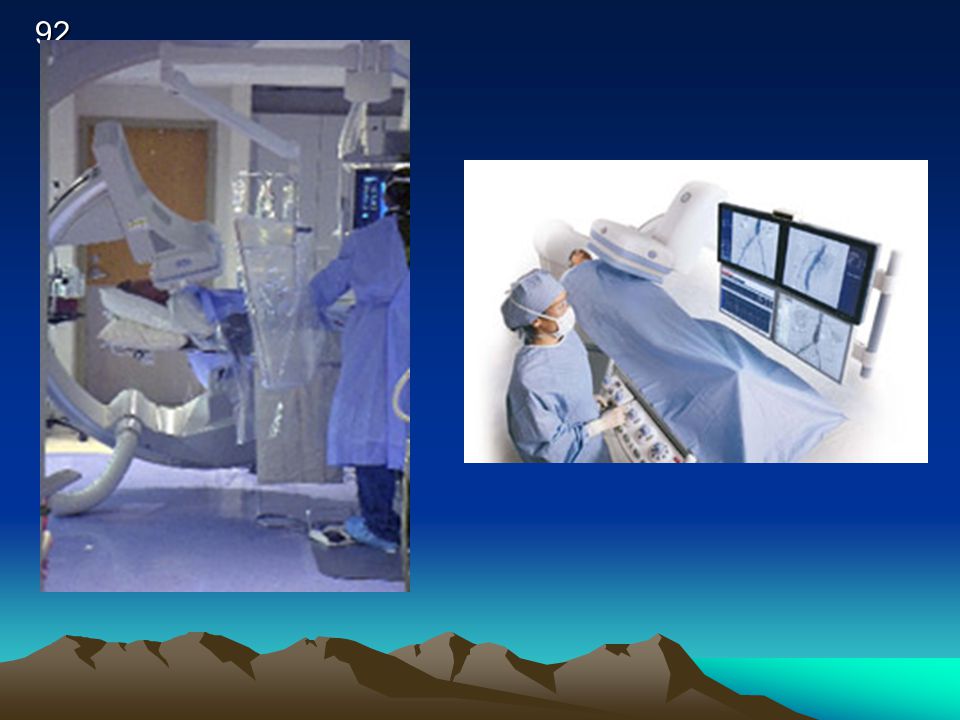
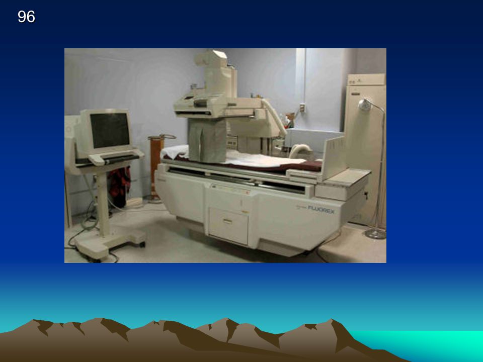
80
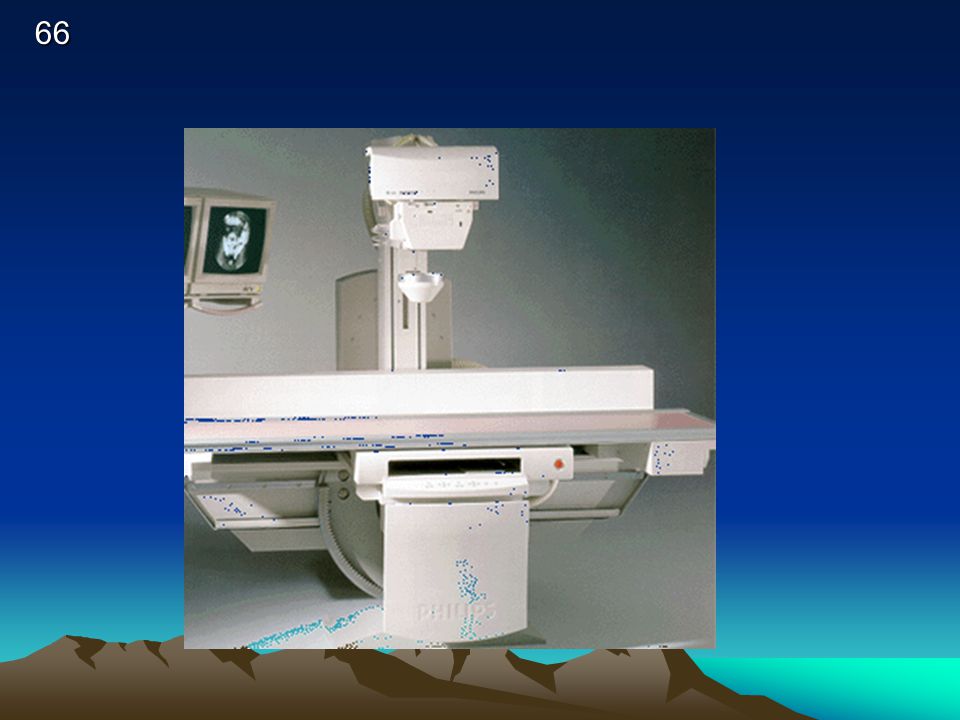
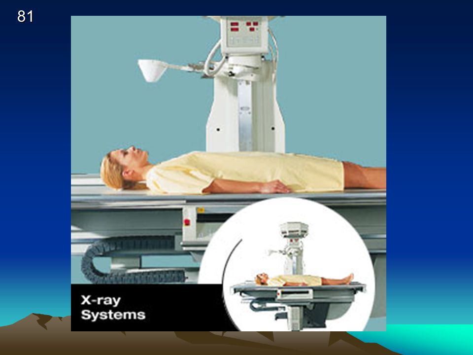
Conventional “FIXED” Fluoro

82
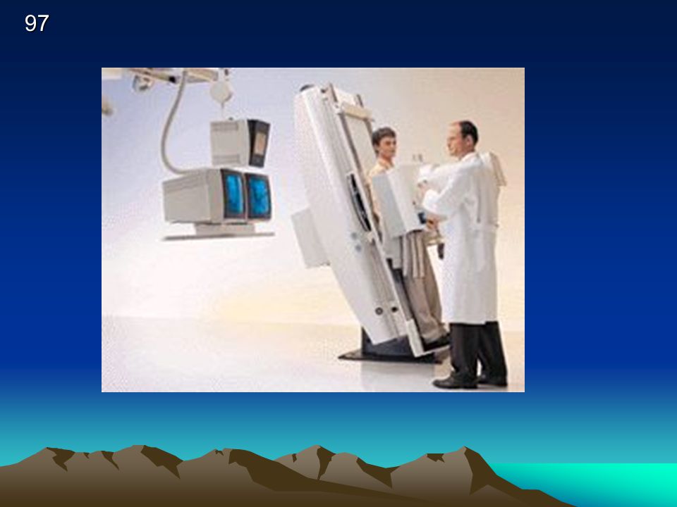
Mobile – C- arm fluoro

84
Pulsed Fluoro Some fluoroscopic equipment is designed for pulsed-mode operation. With the pulsed mode, it can be set to produce less than the conventional 25 or 30 images per second. This reduces the exposure rate. Collimation of the X ray beam to the smallest practical size and keeping the distance between the patient and image receptor as short as possible contribute to good exposure management.

86
Dose rate to patients

88
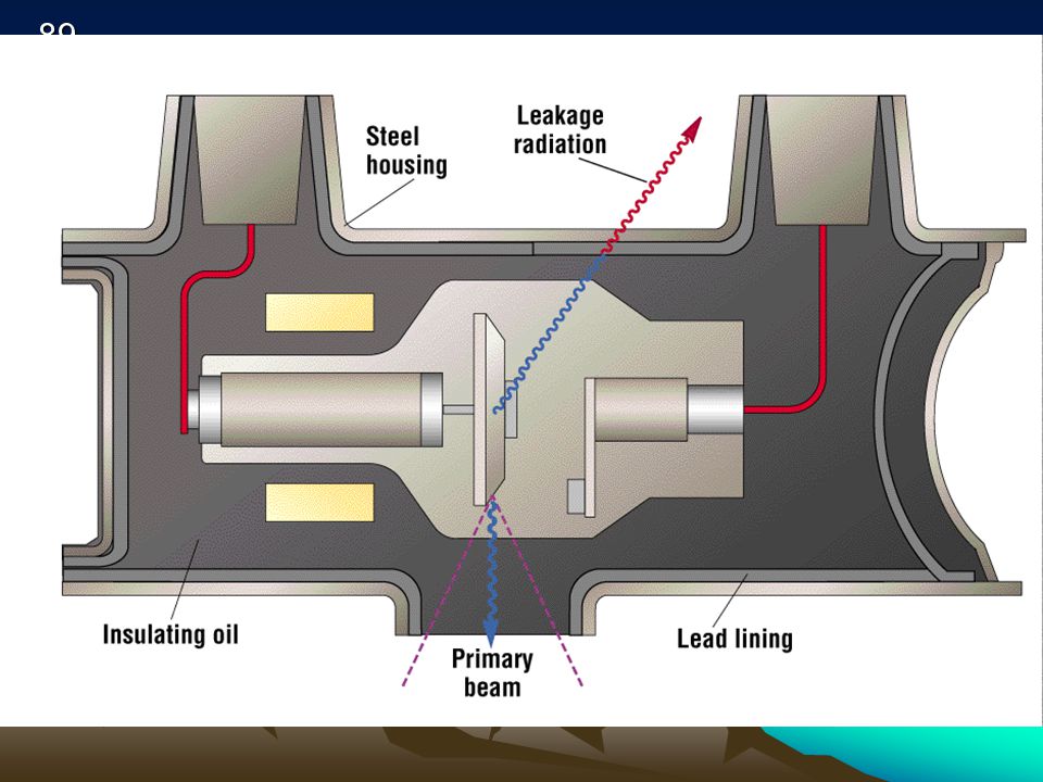
TUBE HOUSING 100MR / HR @ 1 METER
LEAKAGE RADIATION TUBE HOUSING 100MR / 1 METER

90
STAT 8 & 9 Protecting Patients & Personnel
COMMUNICATE COLLIMATE SHIELD

91
IONIZATION CHAMBER- CUTIE PIE Q = t x תּ
Quantity of Radiation = time x pie (diameter of cylinder)
")
93
Mini c-arm

98
Some References Physics of diagnostic radiology, Curry et al, Lea & Febiger, 1990 Imaging systems in medical diagnostics, Krestel ed., Siemens, 1990 The physics of diagnostic imaging, Dowsett et al, Chapman&Hall, 1998

Similar presentations