Download presentation
Presentation is loading. Please wait.

1
Section B Amino acid and protein
General introduction Amino acid structure (B1) Acid and bases (B2) Protein structure (B3) Myoglobin and Hemoglobin (B4) Collagen (B5) Protein purification (B6, B7,B8) Protein sequencing (B9)
 Acid and bases (B2) Protein structure (B3) Myoglobin and Hemoglobin (B4) Collagen (B5) Protein purification (B6, B7,B8) Protein sequencing (B9)")
2
B0. General Introduction
Proteins are polymers(多聚体) of amino acids. -20 amino acids (氨基酸) millions of proteins with different properties and activities. Protein structures are studied at primary, secondary, tertiary and quaternary levels.
 of amino acids. -20 amino acids (氨基酸) millions of proteins with different properties and activities. Protein structures are studied at primary, secondary, tertiary and quaternary levels.")
5
Proteins have widely diverse forms and functions.
enzymes(酶), hormones(激素,荷尔蒙), antibodies(抗体), transporters(转运蛋白), muscle(肌肉), lens protein of eyes(眼睛晶状体), spider webs(蜘蛛网), rhinoceros horn(犀牛角), antibiotics(抗生素), mushroom poisons(蘑菇毒素).
, hormones(激素,荷尔蒙), antibodies(抗体), transporters(转运蛋白), muscle(肌肉), lens protein of eyes(眼睛晶状体), spider webs(蜘蛛网), rhinoceros horn(犀牛角), antibiotics(抗生素), mushroom poisons(蘑菇毒素).")
6
A few examples on protein diversity…
Fireflies emit light catalyzed by luciferase(荧光素酶) with ATP
 with ATP.")
7
Erythrocytes(红细胞) contain a large amount of
hemoglobins(血红蛋白), the oxygen-transporting protein.
, the oxygen-transporting protein.")
8
The protein keratin (角蛋白) is the chief structural components of hair, horn, wool, nails and feathers.
 is the chief structural components of hair, horn, wool, nails and feathers.")
9
Section B1 Amino acid structure
1. All natural proteins were found to be built from the same set of 20 standard -amino acids. 2. The 20 amino acids are usually grouped according to the properties (mainly polarity) of their R groups. 3. Nonstandard amino acids are found in certain proteins, generally as a result of post-translational modifications. 4. The amino acids ionize in aqueous solutions.
 of their R groups. 3. Nonstandard amino acids are found in certain proteins, generally as a result of post-translational modifications. 4. The amino acids ionize in aqueous solutions.")
10
1. All natural proteins were found to be built from the same set of 20 standard -amino acids
The earliest studies of proteins focused on the free amino acids derived from these proteins. The 1st amino acid (asparagine) was discovered in 1806 from asparagus (a green vegetable). The last (threonine) was not identified until 1938! Glutamate from wheat gluten (sticky). Tyrosine from cheese .
 was discovered in 1806 from asparagus (a green vegetable). The last (threonine) was not identified until 1938! Glutamate from wheat gluten (sticky). Tyrosine from cheese .")
11
The names of the amino acids are often abbreviated, either to three letters of to a single letter.
Glycine (Gly, G) 甘氨酸 Alanine (Ala, A) 丙氨酸 Valine (Val, V) 缬氨酸 Leucine (Leu, L) 亮氨酸 Isoleucine (Ile, I) 异亮氨酸 Methionine(Met, M) 蛋氨酸 Proline (Pro, P) 脯氨酸 Cysteine (Cys, C) 半胱氨酸 Hydrophobic, aliphatic amino acids
 甘氨酸. Alanine (Ala, A) 丙氨酸. Valine (Val, V) 缬氨酸. Leucine (Leu, L) 亮氨酸. Isoleucine (Ile, I) 异亮氨酸. Methionine(Met, M) 蛋氨酸. Proline (Pro, P) 脯氨酸. Cysteine (Cys, C) 半胱氨酸. Hydrophobic, aliphatic amino acids.")
12
Phenylalanine (Phe,F) 苯丙氨酸 Tyrosine (Tyr) 酪氨酸 Tryptophan (Trp, W) 色氨酸
Hydrophobic, aromatic amino acids Phenylalanine (Phe,F) 苯丙氨酸 Tyrosine (Tyr) 酪氨酸 Tryptophan (Trp, W) 色氨酸
 苯丙氨酸. Tyrosine (Tyr) 酪氨酸. Tryptophan (Trp, W) 色氨酸.")
13
Arginine (Arg, R) 精氨酸 Lysine (Lys, K) 赖氨酸 Histidine (His, H) 组氨酸
Polar, charged amino acid Arginine (Arg, R) 精氨酸 Lysine (Lys, K) 赖氨酸 Histidine (His, H) 组氨酸 Aspartate (Asp, D) 天冬氨酸 Glutamate (Glu, E) 谷氨酸
 精氨酸. Lysine (Lys, K) 赖氨酸. Histidine (His, H) 组氨酸. Aspartate (Asp, D) 天冬氨酸. Glutamate (Glu, E) 谷氨酸.")
14
Asparagine (Asn, N) 天冬酰胺 Glutamine (Gln, Q) 谷氨酰氨
Polar, uncharged amino acid Serine (Ser, S) 丝氨酸 Threonine (Thr, T) 苏氨酸 Asparagine (Asn, N) 天冬酰胺 Glutamine (Gln, Q) 谷氨酰氨
 丝氨酸. Threonine (Thr, T) 苏氨酸. Asparagine (Asn, N) 天冬酰胺. Glutamine (Gln, Q) 谷氨酰氨.")
15
1.1 The 20 a-amino acids share common
structural features. Each has a carboxyl group and an amino group (but one has an imino group in proline) bonded to the same carbon atom, designated as the a-carbon.
 bonded to the same carbon atom, designated as the a-carbon.")
16
Each has a different side chain (or R group, R=“Remainder of the molecule”).
.")
17
The a-carbons for 19 of them are asymmetric (or chiral), thus being able to have two stereoisomers. Glycine has no chirality.

18
Enantiomers (The C atom is an asymmetric center or chiral center)
")
19
某些光学活性介质能使平面偏振光的偏 振面发生旋转
旋光性(optical rotation) 光学活性(optical activity) “手性” (chiral) 入射光的偏振面 出射光的偏振面 光学活性介质 某些光学活性介质能使平面偏振光的偏 振面发生旋转
 光学活性(optical activity) 手性 (chiral) 入射光的偏振面. 出射光的偏振面. 光学活性介质. 某些光学活性介质能使平面偏振光的偏 振面发生旋转.")
20
About AA stereoisomers:
Only the L-amino acids have been found in proteins (D-isomers have been found only in small peptides of bacteria cell walls and in some peptide antibiotics). The correlation of structure (or configuration) with optical rotation is very complex and has not been successful to date! (i.e., the D- and L-signs do not tell anything about their optical rotation!)
. The correlation of structure (or configuration) with optical rotation is very complex and has not been successful to date! (i.e., the D- and L-signs do not tell anything about their optical rotation!)")
21
1.2 Each amino acid is given a three-letter abbreviation and a one-letter symbol. They often use the first three letter and the first letter. When there is confusion, an alternative is used. These must be remembered.

22
1.3 All proteins in all species (from bacteria to human) are constructed from the same set of 20 amino acids. All proteins, no matter how different they are in structure and function, are made from the 20 standard amino acids. This fundamental alphabet of the protein language is at least two billion years old.

23
2. The 20 amino acids are usually grouped according to the properties (mainly polarity) of their R groups 2.1 Six amino acids have nonpolar, aliphatic (hydrophobic) R groups. They are Gly, Ala, Val, Leu, Ile, and Met. Gly has a hydrogen as its R group, having minimal steric hindrance. In protein structure Gly offers the most flexibility! Ala, Val, Leu, Ile and Met have hydrocarbon R groups, often involved in hydrophobic interactions.
 R groups. They are Gly, Ala, Val, Leu, Ile, and Met. Gly has a hydrogen as its R group, having minimal steric hindrance. In protein structure Gly offers the most flexibility! Ala, Val, Leu, Ile and Met have hydrocarbon R groups, often involved in hydrophobic interactions.")
24
Gly, G, 甘氨酸 Ala, A, 丙氨酸 Val, V, 缬氨酸 Leu, L, 亮氨酸 Ile, I, 异亮氨酸 Met, M, 蛋氨酸, 甲硫氨酸

25
2.2 Ser, Thr, Asn, Gln, Cys, and Pro have polar, uncharged R groups.
The R groups are more hydrophilic, due to the presence of hydroxyl groups, sulfur atoms, or amide groups. -SH group of two Cys in proteins can be oxidized to form a covalent disulfide bond. Cys often participates in hydrophobic interactions. Pro has an imino group, instead of an amino group, forming a five-membered ring structure, being rigid in conformation. Pro is often found in the bends of folded protein chains and often present on the surface of proteins. It offers the least flexibility.

26
Ser, S, 丝氨酸 Thr, T, 苏氨酸 Cys, C, 半胱氨酸 Pro, P, 脯氨酸 Asn, N, 天冬酰胺 Gln, Q, 谷氨酰胺

27
Cystine (胱氨酸) is a dimer of cysteine (半胱氨酸)
 is a dimer of cysteine (半胱氨酸)")
28
2.3 Phe, Tyr, and Trp have aromatic (芳香族) R groups
Phe and Tyr both have benzene rings. Tryptophan has an indole ring. All three participate in hydrophobic interactions. The -OH group in Tyr is an important functional group in proteins. (phosphorylation, hydrogen bond, etc) They are jointly responsible for the light absorption of proteins at 280 nm, a property used as a measure of the concentration of proteins.
 They are jointly responsible for the light absorption of proteins at 280 nm, a property used as a measure of the concentration of proteins.")
29
Phe, F, 苯丙氨酸; Tyr, Y, 酪氨酸; Trp, W, 色氨酸

30
Lambert-Beer’s law: A=Log Io/I = ecl e, extinction coefficient; c, concentration; l, optical length

31
2.4 Positively charged (basic) and negatively
charged (acidic) R groups The most hydrophilic R groups are those either positively or negatively charged. Asp and Glu have carboxyl in their R groups. They have net negative charge at pH 7.0, thus usually named as aspartate and glutamate (conjugate base names, instead of aspartic acid and glutamic acid, un-ionized form).
 R groups. The most hydrophilic R groups are those either positively or negatively charged. Asp and Glu have carboxyl in their R groups. They have net negative charge at pH 7.0, thus usually named as aspartate and glutamate (conjugate base names, instead of aspartic acid and glutamic acid, un-ionized form).")
32
Arg, Lys, and His have positively charged R groups at pH 7.0.
Their R groups contain guanidino(胍基), amino, imidazole(咪唑基) groups respectively. The side chain of His can be positively or uncharged depending on the local environment near pH 7.0
, amino, imidazole(咪唑基) groups respectively. The side chain of His can be positively or uncharged depending on the local environment near pH 7.0.")
33
Lys, K, 赖氨酸; Arg, R, 精氨酸; His, H, 组氨酸

34
Asp, D,天冬氨酸; Glu, E,谷氨酸

36
3. Nonstandard amino acids are found in certain proteins, generally as a result of post-translational modifications. 4-Hydroxyglutamate and 5-hydroxylysine in collagen(胶原). g-carboxyglutamate is found in the blood-clotting prothrombin (凝血酶原,an enzyme). Many additional nonstandard amino acids are found in cells, but not in proteins (e.g., ornithine鸟氨酸and citrulline瓜氨酸, intermediates in amino acid metabolism).
. g-carboxyglutamate is found in the blood-clotting prothrombin (凝血酶原,an enzyme). Many additional nonstandard amino acids are found in cells, but not in proteins (e.g., ornithine鸟氨酸and citrulline瓜氨酸, intermediates in amino acid metabolism).")
37
Two additional amino acids have been identified
Selenocysteine: encoded by the RNA nucleotide triplet UGA found in Archaea, eubacteria and animals including mammals. Pyrrolysine: encoded by the RNA nucleotide triplet UAG, found in Archaea, and eubacteria. Science (2002) vol. 296, 1409 Nature may yet surprise us with more directly encoded amino acids.
 vol. 296, Nature may yet surprise us with more directly encoded amino acids.")
40
B2 Acids and Bases Acids, bases and pH Buffers
Ionization of amino acids

41
1 Weak acids (proton donors) and weak bases (proton acceptors) do not ionize completely when dissolved in water. 1.1 strong acids (e.g., hydrochloric acids, sulfuric and nitric acids) and strong bases (e.g., NaOH and KOH) ionizes completely when dissolved in water. 1.2 A proton donor and its corresponding proton acceptor make up a conjugate acid-base pair.
 and strong bases (e.g., NaOH and KOH) ionizes completely when dissolved in water. 1.2 A proton donor and its corresponding proton acceptor make up a conjugate acid-base pair.")
42
1.4 For convenience, Ka is converted to pKa (the negative logarithm).
1.3 The characteristic tendency of each weak acid for losing its proton is reflected by its dissociation constant Ka (stronger acids have larger Ka value). 1.4 For convenience, Ka is converted to pKa (the negative logarithm).
. 1.4 For convenience, Ka is converted to pKa (the negative logarithm).")
43
2 The titration curves of weak acids can be fitted by the Henderson-Hasselbach equation
2.1 The amount of weak acids in a solution can be determined by titrating with a strong base of known concentration . 2.2 Titration curves are made from plotting the pH of the solution against the amount of strong base added to neutralize the weak acid (until [proton donor]~0, [proton acceptor]=initial weak acid concentration, that is, all weak acid molecules are ionized, no more buffering effect, pH increases rapidly).
.")
45
pH = pKa + log[proton acceptor]/[proton donor]
2.3 Titration curves of weak acids have nearly identical shapes (reflecting the same law behind the phenomenon) 2.4 The Henderson-Hasselbalch equation fits the titration curves of all weak acids! pH = pKa + log[proton acceptor]/[proton donor] 2.5 The pH value at which the conjugate acid-base pair is at equimolar concentration equals to the pKa value of the weak acid. The plateau of the curve gives the pKa.
![pH = pKa + log[proton acceptor]/[proton donor]](http://slideplayer.com/slide/4463379/14/images/45/pH+%3D+pKa+%2B+log%5Bproton+acceptor%5D%2F%5Bproton+donor%5D.jpg "2.3 Titration curves of weak acids have nearly identical shapes (reflecting the same law behind the phenomenon) 2.4 The Henderson-Hasselbalch equation fits the titration curves of all weak acids! pH = pKa + log[proton acceptor]/[proton donor] 2.5 The pH value at which the conjugate acid-base pair is at equimolar concentration equals to the pKa value of the weak acid. The plateau of the curve gives the pKa.")
46
3 The titration curves of weak acids indicate that a conjugate acid-base pair can act as a buffer (resisting to pH changes of the system). 3.1 There is a relatively flat zone on all titration curves of weak acids extending about 0.5 pH units on either side of the pKa values. 3.2 pH changes little in the flat zone when H+ or OH- are added to the system .

47
(e.g., the phosphate buffer H2PO4-/HPO42- in cytoplasm
3.3 pH values in biological systems are usually strictly kept constant (near pH 7.0) by buffering pairs of conjugate acid-bases. (e.g., the phosphate buffer H2PO4-/HPO42- in cytoplasm and the bicarbonate buffer H2CO3/HCO3- in blood, better with CO2 gas to dissolved H2CO3 conversion).
 by buffering pairs of conjugate acid-bases. (e.g., the phosphate buffer H2PO4-/HPO42- in cytoplasm. and the bicarbonate buffer H2CO3/HCO3- in blood, better with CO2 gas to dissolved H2CO3 conversion).")
54
4. The amino acids ionize in aqueous solutions.
4.1 Crystalline amino acids (in neutral aqueous solutions) have melting points much higher than those of other organic molecules of similar size. 4.2 The amino acids ionize to various states depending on pH values.
 have melting points much higher than those of other organic molecules of similar size. 4.2 The amino acids ionize to various states depending on pH values.")
55
4.3 The amino acids (of neutral side chains) exist predominantly as dipolar ions, known as zwitterions (German for “hybrid ions”). 4.4 Amino acids can act both as acids and bases. The zwitterion form of amino acids are ampholytes. Amino acids can be diprotic and triprotic acids.

57
4.5 Monoamino monocarboxylic a-amino acids
(e.g., Gly, Ser, Phe with no ionizable groups) all have similar two-stage titration curves. The first stage reflects the deprotonation of the a-COOH group (pK1). The second stage reflects the deprotonation of the a-NH 3 + group (pK2).
 all have similar two-stage titration curves. The first stage reflects the deprotonation of the a-COOH group (pK1). The second stage reflects the deprotonation of the a-NH 3 + group (pK2).")
58
Amino acids, being both weak acids and bases, have characteristic titration curves and pKa values

59
The pKa value of the a-COOH is more than 2
The pKa value of the a-COOH is more than 2.0 units smaller than that of acetic acid (pKa of 4.76), that is, a stronger (weak) acid. These amino acids have two buffering power regions.
, that is, a stronger (weak) acid. These amino acids have two buffering power regions.")
60
pI = (pK1+pK2)/2
/2")
61
There is a specific pH (designated pI) at which
an amino acid has equal positive and negative charge. An amino acid does not move in an electric field at its pI, called isoelectric point. The pI of monoamino monocarboxylic amino acids reflects a status at which the a-COOH group is fully deprotonated, but the a-NH3+ group has not yet started deprotonating pI = (pK1+pK2)/2
/2.")
62
4. 6 Acidic and basic amino acids have three-stage titration curves
4.6 Acidic and basic amino acids have three-stage titration curves. The additional stage is for the ionizable group on the side chains (pKR).
.")
63
The pI point of an acidic amino acid reflects a status at which the a-COOH is fully deprotonated, but the side chain -COOH and the a-NH3+ group have not yet started deprotonating pI = (pK1+pKR)/2 The pI point of a basic amino acid reflects a status at which the a-COOH and the side chain NH3+ have fully deprotonated, but the a- NH3+ group not yet deprotonated. pI = (pKR+pK2)/2
/2. The pI point of a basic amino acid reflects a status at which the a-COOH and the side chain NH3+ have fully deprotonated, but the a- NH3+ group not yet deprotonated. pI = (pKR+pK2)/2.")
64
An acidic amino acid pI=(pK1+pKR)/2
/2")
65
A basic amino acid pI=(pKR+pK2)/2
/2")
66
氨基酸的理化性质: 1.光学性质 2.两性解离与等电点 3.氨基酸的重要反应 与茚三酮反应 Sanger反应 Edman反应

67
B3 Protein structure Peptide The Three-dimensional Structure
of Proteins

68
1. Amino acids covalently join one another to form peptides
1.1. The a-carboxyl group of one amino acid joins with the a-amino group of another amino acid by a peptide bond (actually an amide bond) 1.1.1 This is a condensation reaction where a water molecule is liberated or eliminated. 1.1.2 The condensation reaction can occur repeatedly to form oligopeptides (with less than 50 aa), polypeptides (btw aa), and proteins (longer).
 This is a condensation reaction where a water molecule is liberated or eliminated The condensation reaction can occur repeatedly to form oligopeptides (with less than 50 aa), polypeptides (btw aa), and proteins (longer).")
69
1. Amino acids covalently join one another to form peptides
1.1. The a-carboxyl group of one amino acid joins with the a-amino group of another amino acid by a peptide bond (actually an amide bond) 1.1.1 This is a condensation reaction where a water molecule is liberated or eliminated. 1.1.2 The condensation reaction can occur repeatedly to form oligopeptides (with less than 50 aa), polypeptides (btw aa), and proteins (longer).
 This is a condensation reaction where a water molecule is liberated or eliminated The condensation reaction can occur repeatedly to form oligopeptides (with less than 50 aa), polypeptides (btw aa), and proteins (longer).")
71
1.2. The peptide chain is directional.
1.2.1 An amino acid unit in a peptide chain is called a residue. 1.2.2 The end having a free a-amino group is called amino-terminal or N-terminal. 1.2.3 The end having a free a-carboxyl group is called carboxyl-terminal or C-terminal. 1.2.4 By convention, the N-terminal is taken as the beginning of the peptide chain, and put at the left (C-terminal at the right). Biosynthesis starts from the N-terminal.
. Biosynthesis starts from the N-terminal.")
73
1.2.6 Amino acid residues have an order or sequence on a peptide.
1.2.5 The peptide chain consist of a regularly repeating main chain (or backbone) and the variable side chains of the residues. 1.2.6 Amino acid residues have an order or sequence on a peptide. 1.2.7 The 20 amino acids are analogous to the 26 letters in English; the number of different peptides made of them is unlimited.
 and the variable side chains of the residues Amino acid residues have an order or sequence on a peptide The 20 amino acids are analogous to the 26 letters in English; the number of different peptides made of them is unlimited.")
74
1.3. The size of a peptide can be described by its total number of residues (e.g., a pentapeptide, an octapeptide) or relative molecular mass (molecular weight). 1.3.1 The mean molecular weight of an amino acid residue in a peptide is ~110 dalton. 1.3.2 Most natural polypeptide chains contain between 50 and 2000 amino acid residues, thus having relative molecular mass between 5500 daltons and 220,000 daltons, or 5.5 kDa and 220 kDa, respectively.

75
1.4. Each peptide has a characteristic titration curve and an isoelectric point (pI).
.")
76
This tetrapeptide has one free a-amino group, one free a-carboxyl group, plus two ionizable R groups.

77
2. Many short peptides have important biological activities
2.1 Some short peptides, such as neuropeptides (神经肽), act as neurotransmitters(神经传递素), neurohormones(神经激素), and neuromodulators.
, act as neurotransmitters(神经传递素), neurohormones(神经激素), and neuromodulators.")
78
2.2 Some short peptides act as antibiotics
Gramicidin(短杆菌肽) A (15-residue) is a well studied peptide antibiotic Its structure has been determined. It contains alternating L- and D-amino acid residues. It is not synthesized on ribosomes!
 A (15-residue) is a well studied peptide antibiotic Its structure has been determined. It contains alternating L- and D-amino acid residues. It is not synthesized on ribosomes!")
79
2.3 Many short peptides are used as
defensive poisons. 2.3.1 a-Amanitin (鹅膏蕈碱) (8-residue, circular) in mushroom is an extremely toxic peptide (inhibiting RNA polymerases II and III at picomolar levels!) 2.3.2 Very toxic short peptides are also found in snake venom, spider.
 (8-residue, circular) in mushroom is an extremely toxic peptide (inhibiting RNA polymerases II and III at picomolar levels!) Very toxic short peptides are also found in snake venom, spider.")
80
2.4 Many vertebrate hormones are small polypeptides.
Insulin (胰岛素)(51-residue) is produced by the pancreas and acts to lower blood glucose level. Glucagon (胰高血糖素) (29-residue) is also produced by the pancreas and acts to increase the blood glucose level.
(51-residue) is produced by the pancreas and acts to lower blood glucose level. Glucagon (胰高血糖素) (29-residue) is also produced by the pancreas and acts to increase the blood glucose level.")
81
3. Proteins in general 3.1 While proteins and polypeptides are normally used interchangeably, polypeptides generally have MW of less than 10,000; and also proteins may have multiple polypeptide subunits.

83
3.2 Proteins have characteristic AA compositions

84
3.3 Some proteins contain chemical groups other than AA, which are called the prosthetic groups, and these proteins are referred as conjugated proteins while the amino acid part alone is called apoprotein.

86
3.4 Protein structure is studied at different levels.

87
Protein structure General studies of the peptide bond
Primary structure Secondary structure Tertiary structure Quaternary structure Protein stability

88
1. General studies of the peptide bond
1.1 The peptide (O=C-N-H) bond was found to be shorter than the C-N bond in a simple amine and atoms attached are coplanar. 1.1.1 This was revealed by X-ray diffraction studies of amino acids and of simple dipeptides and tripeptides. 1.1.2 The peptide (amide) bond was found to be about 1.32 Å (C-N single bond, 1.49; C=N double bond, 1.27), thus having partial double bond feature (should be rigid and unable to rotate freely).
 bond was found to be shorter than the C-N bond in a simple amine and atoms attached are coplanar This was revealed by X-ray diffraction studies of amino acids and of simple dipeptides and tripeptides The peptide (amide) bond was found to be about 1.32 Å (C-N single bond, 1.49; C=N double bond, 1.27), thus having partial double bond feature (should be rigid and unable to rotate freely).")
89
1.1.3 The partial double bond feature is a result of partial sharing (resonance) of electrons between the carbonyl oxygen and amide nitrogen. 1.1.4 The atoms attached to the peptide bond are coplanar with the oxygen and hydrogen atom in trans positions. 1.2 X-ray studies of a-keratin (the fibrous protein making up hair and wool) revealed a repeating unit of 5.4 Å (Astury in the 1930s).
 revealed a repeating unit of 5.4 Å (Astury in the 1930s).")
92
1.2 The peptide bond is rigid and planar

94
1.3 Protein structures have conventionally been understood at four different levels.
1.3.1 The primary structure is the amino acid sequence (including the locations of disulfide bonds).
.")
96
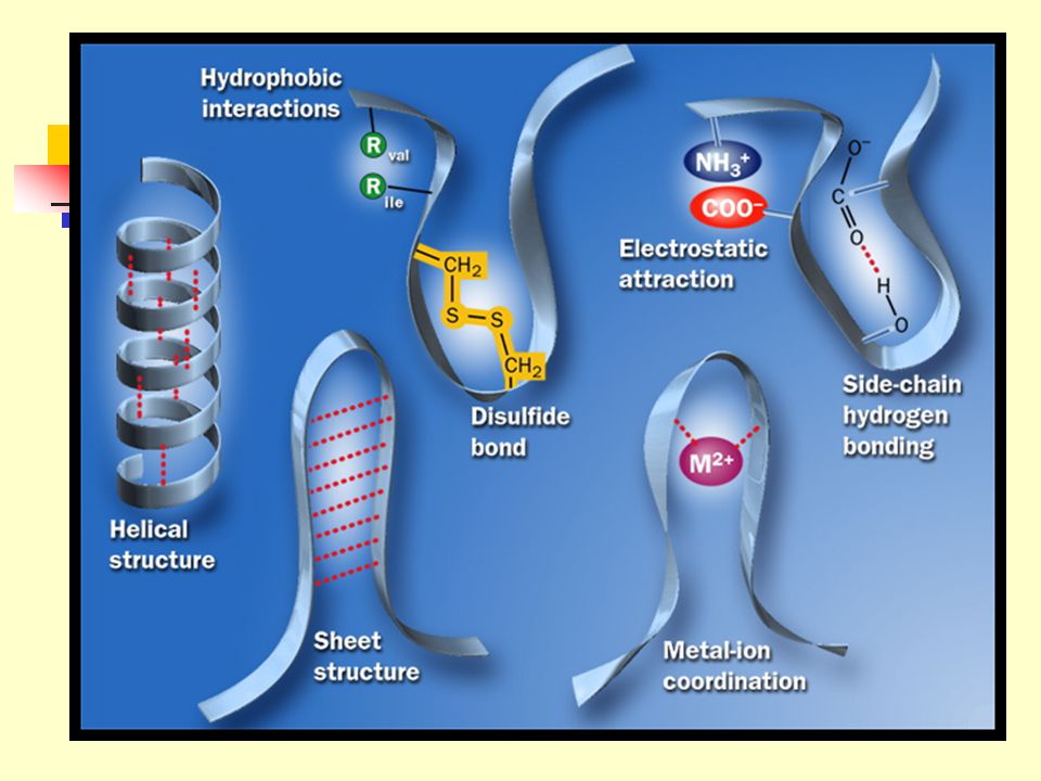
1.3.2 The secondary structure refers to the regular, recurring arrangements of adjacent residues resulting mainly from hydrogen bonding between backbone groups, with -helices and -pleated sheets as the two most common ones. 1.3.3 The tertiary structure refers to the spatial relationship among all amino acid residues in a polypeptide chain, that is, the complete three-dimensional structure. 1.3.4 The quaternary structure refers to the spatial arrangements of each subunit in a multisubunit protein, including nature of their contact.

98
2. Protein Secondary Structure
Secondary structure refers to the regular folding of regions of the polypeptide chain or local conformation of some part of the polypeptide. 2.1 Single bonds other than the peptide bond in the backbone chain are free to rotate. -helix -pleated sheet -turn

100
2.2 The simplest arrangement of the polypeptide chain was proposed to be a helical structure called a-helix (Pauling and Corey, 1951) 2.2.1 The polypeptide backbone is tightly wound around the long axis (rodlike). 2.2.2 R groups protrude outward from the helical backbone. 2.2.3 A single turn of the helix (corresponding to the repeating unit in -keratin) extends about 5.4 Angstroms, including 3.6 residues (each residue arises 1.5 Å and rotate 100 degrees about the helix axis).
 R groups protrude outward from the helical backbone A single turn of the helix (corresponding to the repeating unit in -keratin) extends about 5.4 Angstroms, including 3.6 residues (each residue arises 1.5 Å and rotate 100 degrees about the helix axis).")
101
2.2.4 The model made optimal use of internal hydrogen bonding for structure stabilization.
2.2.5 Each carbonyl oxygen of the residue is hydrogen bonded to the hydrogen on the amino group of the fourth amino acid away.

106
2.3 b-pleated sheet (or b conformation) was proposed to be the more extended conformation of the polypeptide chain. 2.3.1 The conformation is formed when two or more almost fully extended polypeptide chains are brought together side by side. 2.3.2 Regular hydrogen bonds are formed between the carbonyl oxygen and amide hydrogen between adjacent chains (look like a zipper). 2.3.3 The axial distance between the adjacent amino acid residues is ~3.5 Angstroms.
 The axial distance between the adjacent amino acid residues is ~3.5 Angstroms.")
107
2.3.4 The planes of the peptide bonds arrange as pleated sheets.
2.3.5 The R groups of adjacent residues protrude in opposite directions. 2.3.6 The adjacent polypeptide chains can be either parallel (the same direction) or antiparallel (the opposite direction).
 or antiparallel (the opposite direction).")
110
2.4 b turn (b -bend or hairpin发卡 turn) is also a common secondary structure found where a polypeptide chain abruptly reverses its direction.
 is also a common secondary structure found where a polypeptide chain abruptly reverses its direction.")
111
2.4.1 It often connects the ends of two adjacent segments of an antiparallel b-pleated sheet.
2.4.2 It is a tight turn of ~180 degrees involving four amino acid residues. 2.4.3 The essence of the structure is the hydrogen bonding between the C=O group of residue n and the NH group of the residue n+3. 2.4.4 Gly and Pro are often found in b turns. 2.4.5 b turns are often found near the surface of a protein.

112
2.5 Some amino acid residues are accommodated in the different types of secondary structure better than others. 2.6 Regions of polypeptide chain that are not in a regular secondary structure are said to have a coil or loop conformation.

113
Relative probabilities that a given amino acid will occur in the three common types of secondary structure.

115
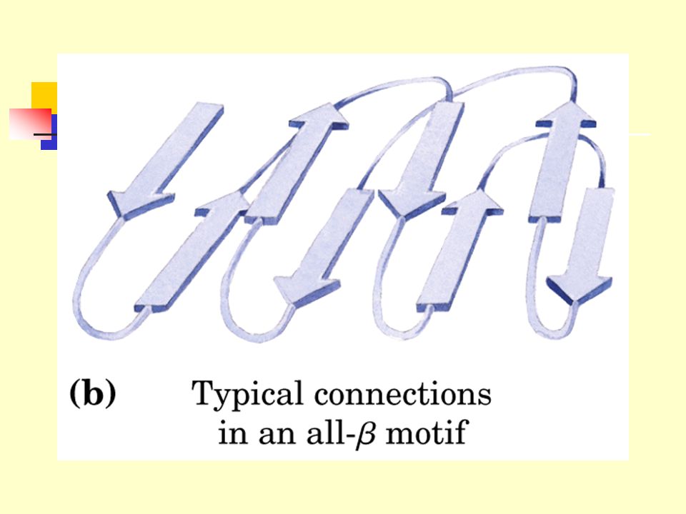
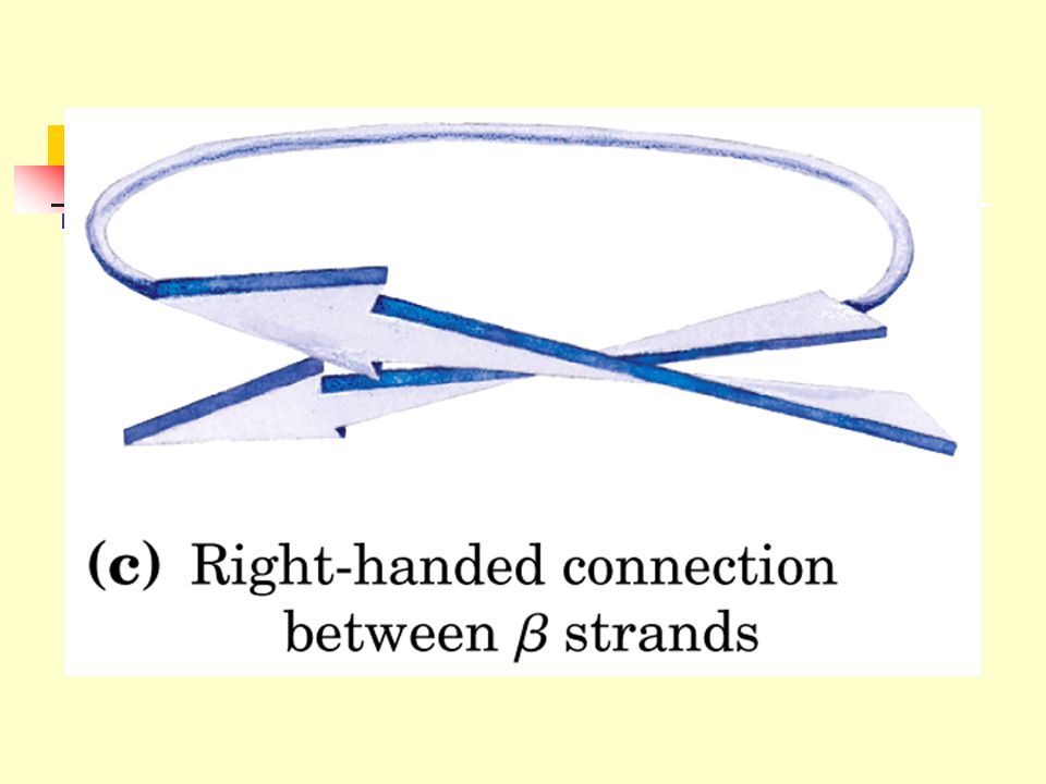
2.6 Supersecondary structures and domains
2.6.1 The supersecondary structures, also called motifs or simply folds, refers to clusters of secondary structures that repeatedly appear. 2.6.2 A compact region (usually including less than 200~400 residues) that is a distinct structural unit within a larger polypeptide chain is called a domain.
 that is a distinct structural unit within a larger polypeptide chain is called a domain.")
116
Structural domains in the polypeptide troponin (肌钙蛋白) C, two separate calcium-binding domains
 C, two separate calcium-binding domains")
125
Structural domains in the polypeptide troponin (肌钙蛋白) C, two separate calcium-binding domains
 C, two separate calcium-binding domains")
130
3. Protein Tertiary Structure---
the three-dimentional arrangement of the amino acid in the polypeptide chain.

133
The polypeptide chain folds spontaneously so that the majority of its hydrophobic side-chains are buried in the interior, and the majority of its polar, charged side-chain are on the surface. The native conformation is maintained not only by hydrophobic interactions, but also by salt bridges, multiple weak van der Waals interactions, hydrogen bonding and the covalent disulfide bonds.

134
3.1 Under physiological conditions of solvent and temperature, each protein folds spontaneously into one three-dimensional conformation, called the native conformation. 3.2 This conformation is usually the most stable, and predominates among the innumerable theoretically possible ones. 3.3 Usually only the native conformation is functional.

135
Globular protein structures are compact and varied

136
4. Quaternary structure: macromolecular assembly
deoxyhemoglobin

138
If a protein is made up of more than one polypeptide chain it is said to have quaternary structure. This refers to the spatial arrangement of the polypeptide subunits and the nature of the ineractions between them.

139
5. Proteins denature under stress
5.1 Loss of tertiary structure (native conformation) is accompanied by loss of function. 5.1.1 Proteins are relatively easy to lose their tertiary structures due to their marginal stability maintained by noncovalent interactions. 5.1.2 The process of total loss or randomization of three-dimensional structure of proteins is called denaturation. 5.1.3 Protein denaturation results from a change in the solvent environment that is sufficiently large to upset the forces that keep the protein structure intact.
 is accompanied by loss of function Proteins are relatively easy to lose their tertiary structures due to their marginal stability maintained by noncovalent interactions The process of total loss or randomization of three-dimensional structure of proteins is called denaturation Protein denaturation results from a change in the solvent environment that is sufficiently large to upset the forces that keep the protein structure intact.")
140
Extreme pH: upset the balance of a protein’s charge interactions.
5.1.4 Many means can cause protein to denature: Heating: unbalance the compensating enthalpic and entropic contributions, thermal motion causes melting. Extreme pH: upset the balance of a protein’s charge interactions. Miscible organic solvents (alcohol and acetone): solvent polarity and hydrogen bonding. Solutes (urea, guanidine): provide alternative hydrogen bonding. Detergents (SDS): introducing their hydrophobic tails into the protein’s interior. 5.1.5 The mechanism of many of these denaturing processes are fully understood.
: solvent polarity and hydrogen bonding. Solutes (urea, guanidine): provide alternative hydrogen bonding. Detergents (SDS): introducing their hydrophobic tails into the protein’s interior The mechanism of many of these denaturing processes are fully understood.")
141
5.2 Denaturation of some proteins is reversible.
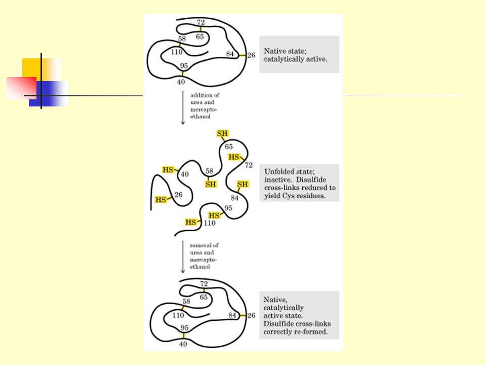
5.2.1 Some denatured globular proteins will regain their native structure and their biological activity once returned to conditions in which the native conformation is stable. This process is called renaturation. 5.2.2 The denaturation and renaturation phenomena were originally observed on ribonuclease A by chance by Christian Anfinsen (1960s). 5.2.3 Ribonuclease A became reduced and randomly coiled (denatured) in 8 M urea plus -mercaptoethanol, with a loss of the enzymatic activity.
 Ribonuclease A became reduced and randomly coiled (denatured) in 8 M urea plus -mercaptoethanol, with a loss of the enzymatic activity.")
143
Nobel Prize in Chemistry in 1972 to Anfinsen.
5.2.4 When urea and b-mercaptoethanol were removed, the enzymatic activity was slowly regained until full recovery under stable conditions. 5.2.5 All the physical and chemical properties of the refolded enzyme were virtually identical with those of the native enzyme. 5.2.6 Conclusion: the information needed to specify the complex tertiary structure of ribonuclease A is all contained in its amino acid sequence. 5.2.7 Subsequent studies have established the generality of this central principle of molecular biology: sequence specifies conformation. Nobel Prize in Chemistry in 1972 to Anfinsen.

144
5.3 The tertiary structures of proteins are not rigid.
5.3.1 Many studies have found that globular proteins have certain amount of flexibility in their backbones and undergo short-range internal fluctuations. 5.3.2 Many proteins undergo small conformational changes in the course of their biological function (e.g., O2-bound hemoglobin differs from O2-free hemoglobin, substrate binding to enzymes often causes conformational changes).
.")
145
6. Three-dimensional structures of proteins can be determined by several methods
6.1 X-Ray Diffraction (X-Ray crystallography) 6.2 Nuclear Magnetic Resonance (NMR) 6.3 Many proteins have since been determined, such as cytochrome c, lysozyme and ribonuclease A. 6.4 Some common characteristics of proteins All the water soluble globular proteins have a hydrophobic core and a mainly hydrophilic outer surface. Proteins are stabilized mainly by noncovalent interactions (sometimes by disulfide bonds). Each protein has a unique structure to perform its unique function.
 6.2 Nuclear Magnetic Resonance (NMR) 6.3 Many proteins have since been determined, such as cytochrome c, lysozyme and ribonuclease A. 6.4 Some common characteristics of proteins. All the water soluble globular proteins have a hydrophobic core and a mainly hydrophilic outer surface. Proteins are stabilized mainly by noncovalent interactions (sometimes by disulfide bonds). Each protein has a unique structure to perform its unique function.")
146
X-Ray Diffraction

147
One-dimensional NMR

148
Two-dimensional NMR

149
cytochrome c contains only about 40% a-helices, but many irregularly coiled and extended segments

150
Lysozyme, an enzyme that catalyzes the hydrolytic cleavage of polysaccharides in some bacterial cell walls, contains about 40% a-helices and about 12% b-sheets.

151
Ribonuclease A contains more b-sheet structures.

152
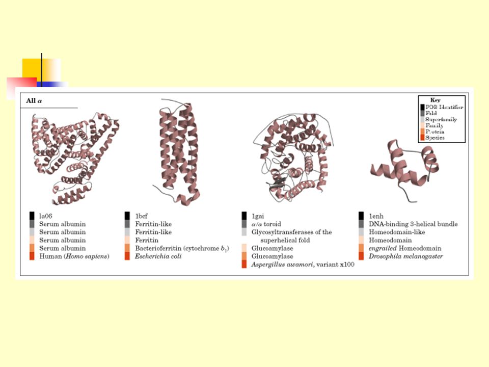
Five schemes of protein three- dimensional structures
Summary: Five schemes of protein three- dimensional structures 1) The three-dimensional structure of a protein is determined by its amino acid sequence. 2) The function of protein depends on its structure. 3) An isolated protein has a unique, or nearly unique, structure. 4) The most important forces stabilizing the specific structure of a protein are non-covalent interactions. 5) Amid the the huge number of unique protein structures, we can recognize some common structural patterns to improve our understanding of protein architecture.
 The three-dimensional structure of a protein is determined by its amino acid sequence. 2) The function of protein depends on its structure. 3) An isolated protein has a unique, or nearly unique, structure. 4) The most important forces stabilizing the specific structure of a protein are non-covalent interactions. 5) Amid the the huge number of unique protein structures, we can recognize some common structural patterns to improve our understanding of protein architecture.")
153
B4 Myoglobin and Hemoglobin

154
1. Oxygen-binding proteins
Hemoglobin and myoglobin are the two oxygen-binding proteins present in large multicellular organisms.

155
2. Sperm whale (抹香鲸, 巨头鲸) myoglobin (肌红蛋白), the oxygen carrier in muscle, was the first protein to be seen in atomic detail by X-ray analysis (John Kendrew, 1950s)
 myoglobin (肌红蛋白), the oxygen carrier in muscle, was the first protein to be seen in atomic detail by X-ray analysis (John Kendrew, 1950s).")
156
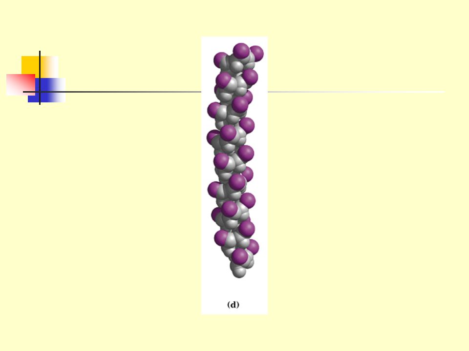
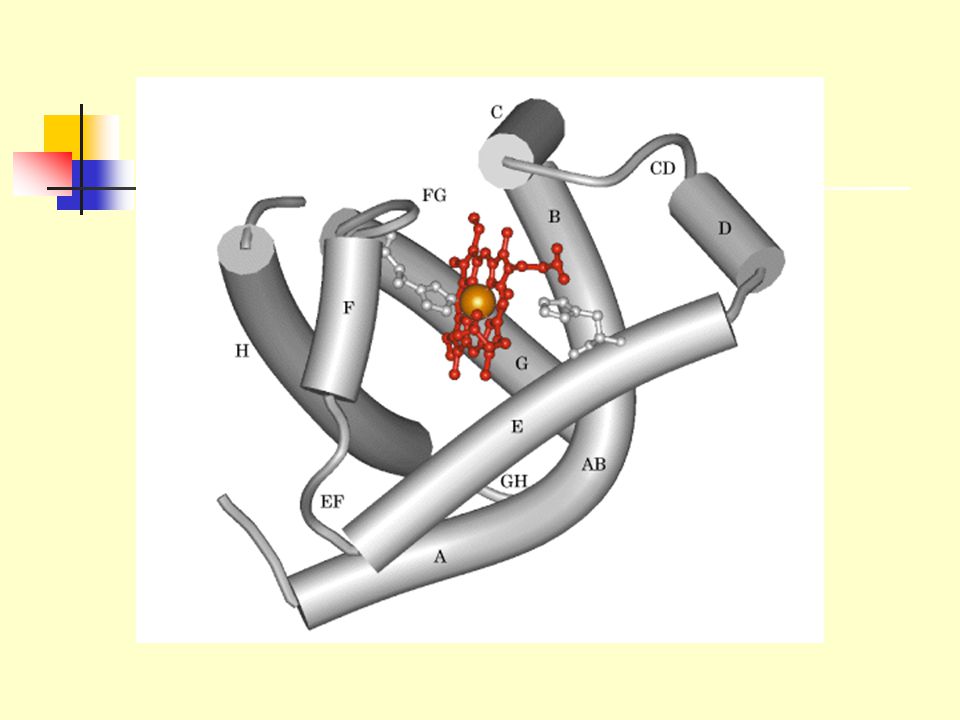
Tertiary structure of sperm whale myoglobin.
a) backbone; b) “mesh” image; c) surface contour image; d) ribbon; e) space-filling model.
 backbone; b) mesh image; c) surface contour image; d) ribbon; e) space-filling model.")
157
2.1 The existence of -helices were for the first time directly observed in a protein.
The myoglobin molecule contains eight -helices. All the -helices are right-handed. All the peptide bonds are in the planar trans configuration. There is no -pleated sheets observed in the molecule.

158
2.2 The myoglobin molecule has a dense hydrophobic core.
Many hydrophobic R groups (e.g., Leu, Val, Met, Phe) are found to be in the interior of the myoglobin molecule. Hydrophobic interaction is important for the stability of the protein structure. Only two hydrophilic histidine residues were found in the interior of the protein.
 are found to be in the interior of the myoglobin molecule. Hydrophobic interaction is important for the stability of the protein structure. Only two hydrophilic histidine residues were found in the interior of the protein.")
159
2.3 The polar R groups are located on the outer surface and hydrated.
Nonpolar residues are also present on the outer surface! 2.4 All Pro residues are found at bends. 2.5 The flat heme group (the prosthetic group) was revealed to rest in a crevice (pocket). The heme group consists of a complex organic ring structure, protoporphyrin, which is bound to an iron atom in its ferrous (Fe2+) state. The iron atom has six coordination bonds, with four in the plane of and bonded to the flat porphyrin molecule and two perpendicular to it.
 was revealed to rest in a crevice (pocket). The heme group consists of a complex organic ring structure, protoporphyrin, which is bound to an iron atom in its ferrous (Fe2+) state. The iron atom has six coordination bonds, with four in the plane of and bonded to the flat porphyrin molecule and two perpendicular to it.")
160
The heme group.

161
One of the perpendicular coordination bonds is bound to a nitrogen atom of an interior His residue.
The sixth coordination serves as the binding site for O2. The accessibility of the heme group to solvent is highly restricted, thus preventing the oxidation of the Fe2+ to the ferric ion (Fe3+), which is unable to bind O2.
, which is unable to bind O2.")
162
Sir John Kendrew and Max Perutz won the Nobel Prize in Chemistry in 1962 for determining the complete atomic structure of myoglobin and hemeglobin.

163
3. Hemoglobin is a multisubunit allosteric (变构) protein that carries O2 in erythrocyte.
3.1 Hemoglobin is a well-studied and well-understood protein. 3.1.1 It was one of the first proteins to have its molecular mass accurately determined. 3.1.2 The first protein to be characterized by ultracentrifuge. 3.1.3 The first protein to be associated with a specific physiological function.

164
3.1.6 The best understood allosteric protein.
3.1.4 The first protein with a single amino acid substitution being related to a genetic disease. 3.1.5 The first multisubunit protein with its detailed atomic structure determined by X-ray crystallography. 3.1.6 The best understood allosteric protein.

165
3.2 Determination of the atomic structure of hemoglobin A (from normal adult) is very revealing.
3.2.1 The protein molecule exists as a a2b2 tetramer. 3.2.2 Each subunit has a structure strikingly and unexpectedly similar to each other and to that of myoglobin, indicating quite different amino acid sequences can specify very similar 3-D structures.

167
3.2.3 Extensive interactions exist between the unlike subunits through noncovalent interactions.
3.2.4 Quaternary structure changes markedly when O2 binds. Crystals of deoxyhemoglobin shatter (break) when exposed to O2. 3.2.5 O2 binds to the sixth coordination position of the ferrous iron (as in myoglobin). 3.2.6 Oxygen binding of hemoglobin shows positive cooperativity. The binding of O2 (the ligand) at one heme facilitates the binding of O2 at the other hemes on the same tetramer.
 when exposed to O O2 binds to the sixth coordination position of the ferrous iron (as in myoglobin) Oxygen binding of hemoglobin shows positive cooperativity. The binding of O2 (the ligand) at one heme facilitates the binding of O2 at the other hemes on the same tetramer.")
171
3.3 The Bohr effect Increasing concentrations of H+ (with a decrease of pH) or CO2 lower the O2 affinity of hemoglobin (H+ and CO2 has no effect on O2 affinity of myoglobin). One molecule of 2,3-diphosphoglycerate (BPG) binds to the central cavity of one tetramer of hemoglobin, which also lowers its O2 affinity. This is called Bohr effect, which helps the release of O2 in the capillaries of actively metabolizing tissues.
 or CO2 lower the O2 affinity of hemoglobin (H+ and CO2 has no effect on O2 affinity of myoglobin). One molecule of 2,3-diphosphoglycerate (BPG) binds to the central cavity of one tetramer of hemoglobin, which also lowers its O2 affinity. This is called Bohr effect, which helps the release of O2 in the capillaries of actively metabolizing tissues.")
173
Bohr’s effect and its molecular mechanism
Binding of O2 in lungs with release of H+ and CO2. Release of O2 in peripheral tissues

174
3.4 Cooperativity is a particular case of an allosteric effect
3.4.1 Allosteric effect refers to the phenomenon in which a molecule (allosteric effector) bound to one site on a protein causes a conformational change in the protein such that the activity of another site on the protein is altered (increased or decreased). 3.4.2 H+, CO2, and BPG all show an allosteric effect for the O2 binding process of hemoglobin.
 bound to one site on a protein causes a conformational change in the protein such that the activity of another site on the protein is altered (increased or decreased) H+, CO2, and BPG all show an allosteric effect for the O2 binding process of hemoglobin.")
175
3.5 The functional characteristics of an allosteric protein are regulated by specific molecules in its environment. In other words, in the evolutionary transition from myoglobin to hemoglobin, a macromolecule capable of perceiving information from its environment has emerged.

176
3.6 Sickle-cell anemia was found to be caused by a single amino acid change in the b chain of hemoglobin molecules. 3.6.1 The hemoglobin molecule from sickle-cell anemia patients (HbS) was found to have a higher pI value (having more net positive charges). 3.6.2 Amino acid sequencing revealed that HbS contains Val instead of Glu at position 6 of the b chain! 3.6.3 DNA sequencing of the genes further revealed that the codon of b6 changed from GAA(Glu) to GTA(Val).
 was found to have a higher pI value (having more net positive charges) Amino acid sequencing revealed that HbS contains Val instead of Glu at position 6 of the b chain! DNA sequencing of the genes further revealed that the codon of b6 changed from GAA(Glu) to GTA(Val).")
177
3.6.4 The oxygen binding affinity and allosteric properties of hemoglobin are virtually unaffected by this change (the b6 is located at the surface of the protein). 3.6.5 Presence of Val 6 on the b subunits generates a hydrophobic patch on the surface which complements with another hydrophobic patch formed only in deoxygenated HbS, thus generating the fiber aggregates (a polymer of HbS).
.")
180
Mutation and molecular interaction’s changes

181
B5 Collagen (胶原蛋白) Fibrous proteins have polypeptide chains arranged in long strands or sheets. Globular proteins have polypeptide chains folded into a spherical or globular shape. Collegen is the name given to a family of structurally related proteins that form strong insoluble fibers.

182
Collagen is the most abundant protein in mammals and is present in most organs of the body, where it serves to hold cells together in discrete units. It is also the major fibrous element of skin, bones, tendons(腱), cartilage (软骨), blood vessels and teeth.
, cartilage (软骨), blood vessels and teeth..")
186
B6 B7 B8 Protein Purification
Selection of a protein source Homogeniza-tion and solubilization Ammonium sulfate precipitation Dialysis Ultracentrifugation Further purification Chromatography Electrophoresis

188
Ultracentrifugation

189
Gel filtration chromatography

190
Ion exchange chromatography

191
Affinity chromatography

192
Native PAGE SDS-PAGE

193
B9 Protein sequencing and peptide synthesis
Amino acid composition analysis Edman degradation and Sanger reaction Sequencing strategy

194
Amino acid composition analysis
The purified protein sample is hydrolyzed into amino acid by heating it in 6 M HCl at 110 degree for 24 h. The resulting mixture of amino acid is subjected to ion exchange chromatography on a column to separate out the 20 standard amino acids. The separated amino acids are then detected and quantified by reacting them with ninhydrin.

195
6 M HCl protein Mixture of amino acid

196
Ion exchange chromatography

197
The amino acids are detected by colorimetric reaction with ninhydrin (茚三酮).
.")
198
Edman degradation and Sanger reaction
DNFB or FDNB 二硝基氟苯

200
Edman reaction PITC The amino acid sequence of a protein can be determined by Edman degradation which sequentially removes one residue at a time from the N terminus.

201
Sequencing stratege The polypeptide is to broken down into smaller fragments. The smaller fragments are then seqenced by Edman degradation. The complete sequence is assembled by analyzing overlapping fragments.

Similar presentations


 Dr. Ahmed Mujamammi Dr. Sumbul Fatma>")

