Download presentation
Presentation is loading. Please wait.

1
Contraction of Skeletal Muscles

2
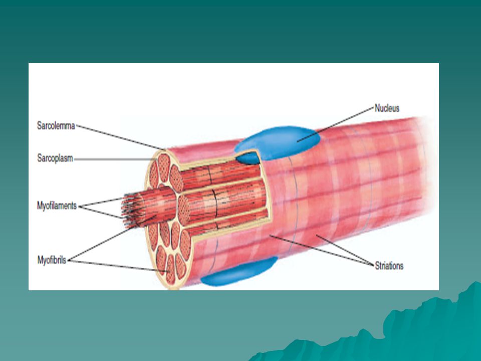
Physiologic Anatomy of Skeletal Muscle

4
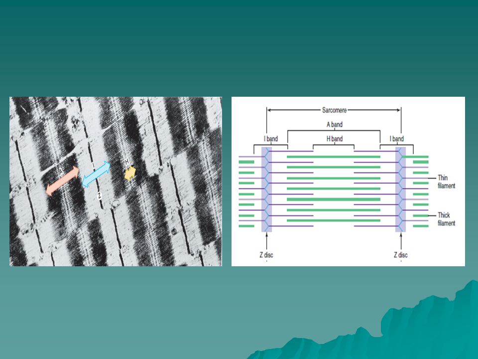
The striations of skeletal muscles are produced by thick and thin filaments.

7
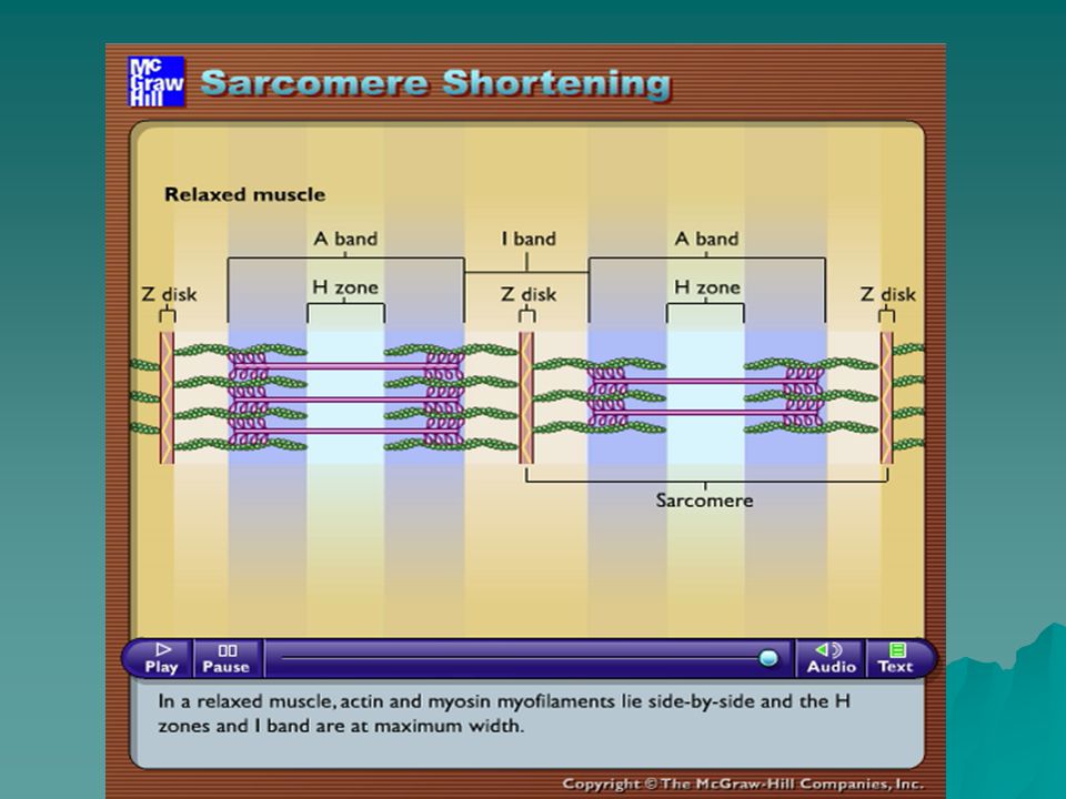
Molecular mechanisms of muscle contraction Huxley-Hanson Theory 1. Myofibrils are contracted due to contraction of a large number of sarcomeres 2. The length of actin and myosin filaments not change during contraction 3. The process of contraction is a result of sliding actin filaments along myosin filaments 4. The process of muscle contraction requires energy of ATP

8
Molecular Characteristics of the Myosin Filament myosin molecules The myosin filament is composed of multiple myosin molecules. The myosin molecule is composed of six polypeptide chains—two heavy chains, and four light chains. The two heavy chains wrap spirally around each other to form a double helix, which is called the tail of the myosin molecule. One end of each of these chains is folded bilaterally into a globular polypeptide structure called a myosin head. Thus, there are two free heads at one end of the double-helix myosin molecule. The four light chains are also part of the myosin head, two to each head. These light chains help control the function of the head during muscle contraction.

9
The myosin filament is made up of 200 or more individual myosin molecules. The central portion of one of these filaments, displaying the tails of the myosin molecules bundled together to form the body of the filament, while many heads of the molecules hang outward to the sides of the body. Part of the body of each myosin molecule hangs to the side along with the head, thus providing an arm that extends the head outward from the body. The protruding arms and heads together are called cross-bridges. Each cross-bridge is flexible at two points called hinges—one where the arm leaves the body of the myosin filament, and the other where the head attaches to the arm. The hinged arms allow the heads either to be extended far outward from the body of the myosin filament or to be brought close to the body. The hinged heads in turn participate in the actual contraction process, as discussed in the following sections.

10
Structure of myosin filament

11
When a muscle contracts, work is performed and energy is required. Large amounts of ATP are cleaved to form ADP during the contraction process; the greater the amount of work performed by the muscle, the greater the amount of ATP that is cleaved, which is called the Fenn effect. The following sequence of events is believed to be the means by which this occurs: 1. Before contraction begins, the heads of the cross-bridges bind with ATP. The ATP-ase activity of the myosin head immediately cleaves the ATP but leaves the cleavage products, ADP plus phosphate ion, bound to the head. In this state, the conformation of the head is such that it extends perpendicularly toward the actin filament but is not yet attached to the actin.

12
2. When the troponin-tropomyosin complex binds with calcium ions, active sites on the actin filament are uncovered, and the myosin heads then bind with these. 3. The bond between the head of the cross-bridge and the active site of the actin filament causes a conformational change in the head, prompting the head to tilt toward the arm of the cross- bridge. This provides the power stroke for pulling the actin filament. The energy that activates the power stroke is the energy already stored, like a “cocked” spring, by the conformational change that occurred in the head when the ATP molecule was cleaved earlier. 4. Once the head of the cross-bridge tilts, this allows release of the ADP and phosphate ion that were previously attached to the head. At the site of release of the ADP, a new molecule of ATP binds. This binding of new ATP causes detachment of the head from the actin.

13
5. After the head has detached from the actin, the new molecule of ATP is cleaved to begin the next cycle, leading to a new power stroke. That is, the energy again “cocks” the head back to its perpendicular condition, ready to begin the new power stroke cycle. 6. When the cocked head (with its stored energy derived from the cleaved ATP) binds with a new active site on the actin filament, it becomes unlocked and once again provides a new power stroke. Thus, the process proceeds again and again until the actin filaments pull the Z membrane up against the ends of the myosin filaments or until the load on the muscle becomes too great for further pulling to occur.
 binds with a new active site on the actin filament, it becomes unlocked and once again provides a new power stroke. Thus, the process proceeds again and again until the actin filaments pull the Z membrane up against the ends of the myosin filaments or until the load on the muscle becomes too great for further pulling to occur..")
14
Molecular Characteristics of the Actin Filament The actin filament is also complex. It is composed of three protein components: actin, tropomyosin, and troponin. The backbone of the actin filament is a double stranded F-actin protein molecule. The two strands are wound in a helix in the same manner as the myosin molecule. Each strand of the double F-actin helix is composed of polymerized G-actin molecules, each having a molecular weight of about 42,000. Attached to each one of the G- actin molecules is one molecule of ADP. It is believed that these ADP molecules are the active sites on the actin filaments with which the cross-bridges of the myosin filaments interact to cause muscle contraction. The active sites on the two F-actin strands of the double helix are staggered. The bases of the actin filaments are inserted strongly into the Z discs; the ends of the filaments protrude in both directions to lie in the spaces between the myosin molecules.

15
Molecular Characteristics of the Actin Filament

16
Tropomyosin Molecules. The actin filament also contains another protein, tropomyosin. These molecules are wrapped spirally around the sides of the F-actin helix. In the resting state, the tropomyosin molecules lie on top of the active sites of the actin strands, so that attraction cannot occur between the actin and myosin filaments to cause contraction. Troponin and Its Role in Muscle Contraction. Attached intermittently along the sides of the tropomyosin molecules are still other protein molecules called troponin. These are actually complexes of three loosely bound protein subunits, each of which plays a specific role in controlling muscle contraction. One of the subunits (troponin I) has a strong affinity for actin, another (troponin T) for tropomyosin, and a third (troponin C) for calcium ions. This complex is believed to attach the tropomyosin to the actin. The strong affinity of the troponin for calcium ions is believed to initiate the contraction process, as explained in the next section.
 has a strong affinity for actin, another (troponin T) for tropomyosin, and a third (troponin C) for calcium ions. This complex is believed to attach the tropomyosin to the actin. The strong affinity of the troponin for calcium ions is believed to initiate the contraction process, as explained in the next section..")
17
When calcium ions combine with troponin C, each molecule of which can bind strongly with up to four calcium ions, the troponin complex supposedly undergoes a conformational change that in some way tugs on the tropomyosin molecule and moves it deeper into the groove between the two actin strands. This “uncovers” the active sites of the actin, thus allowing these to attract the myosin cross-bridge heads and cause contraction to proceed.

18
“Walk-along” mechanism for contraction of the muscle Figure demonstrates this postulated walk-along mechanism for contraction. The figure shows the heads of two cross-bridges attaching to and disengaging from active sites of an actin filament. It is postulated that when a head attaches to an active site, this attachment simultaneously causes profound changes in the intramolecular forces between the head and arm of its cross-bridge. The new alignment of forces causes the head to tilt toward the arm and to drag the actin filament along with it. This tilt of the head is called the power stroke. Then, immediately after tilting, the head automatically breaks away from the active site.

19
Next, the head returns to its extended direction. In this position, it combines with a new active site farther down along the actin filament; then the head tilts again to cause a new power stroke, and the actin filament moves another step. Thus, the heads of the cross-bridges bend back and forth and step by step walk along the actin filament, pulling the ends of two successive actin filaments toward the center of the myosin filament. Each one of the cross-bridges is believed to operate independently of all others, each attaching and pulling in a continuous repeated cycle. Therefore, the greater the number of cross-bridges in contact with the actin filament at any given time, the greater, theoretically, the force of contraction.

20
Molecular Mechanism of Muscle Contraction

21
Stages of muscle contraction Stage 1 - Excitation of the membrane of muscle fibers Stage 2 - Electromechanical coupling Stage 3 - The actual reduction Stage 4 - Relaxation

23
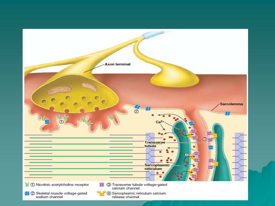
The initiation and execution of muscle contraction occur in the following sequential steps. 1.An action potential travels along a motor nerve to its endings on muscle fibers. 2. At each ending, the nerve secretes a small amount of the neurotransmitter substance acetylcholine. 3. The acetylcholine acts on a local area of the muscle fiber membrane to open multiple “acetylcholine gated” channels through protein molecules floating in the membrane. 4. Opening of the acetylcholine-gated channels allows large quantities of sodium ions to diffuse to the interior of the muscle fiber membrane. This initiates an action potential at the membrane.

24
5. The action potential travels along the muscle fiber membrane in the same way that action potentials travel along nerve fiber membranes. 6. The action potential depolarizes the muscle membrane, and fiber. Here it causes the sarcoplasmic reticulum to release large quantities of calcium ions that have been stored within this reticulum. 7. The calcium ions initiate attractive forces between the actin and myosin filaments, causing them to slide alongside each other, which is the contractile process.

25
8. Аfter a fraction of a second, the calcium ions are pumped back into the sarcoplasmic reticulum by a Ca++ membrane pump, and they remain stored in the reticulum until a new muscle action potential comes along; this removal of calcium ions from the myofibrils causes the muscle contraction to cease.

Similar presentations


, which.>")


 The Muscle Action Potential ( AP ) Muscle RMP = -90 mV ( same as in nerves.>")
