Download presentation
Presentation is loading. Please wait.

1
سبحانك لا علم لنا إلا ما علمتنا إنك أنت العليم الحكيم
بسم الله الرحمن الرحيم سبحانك لا علم لنا إلا ما علمتنا إنك أنت العليم الحكيم صدق الله العظيم

2
Phases of Embryonic Development
Abdelalim Gadallah (Ph.D.) Lecturer of Comparative Anatomy and Embryology Zoology Department, Faculty of Science, MANSOURA UNIVERSITY EGYPT
 Lecturer of Comparative Anatomy and Embryology. Zoology Department, Faculty of Science, MANSOURA UNIVERSITY. EGYPT.")
3
7 steps: 2. fertilization 3. cleavage 4. blastulation 5. gastrulation
1. gametogenesis 2. fertilization 3. cleavage 4. blastulation 5. gastrulation 6. neurulation 7. organogenesis

4
1.Gametogenesis is a process by which the diploid germ cells undergo a number of chromosomal and morphological changes to form mature haploid gametes. Animals produce gametes directly through meiosis in organs called gonads. Males and females of a species that reproduces sexually have different forms of gametogenesis: spermatogenesis (male) in testes produce sperms. oogenesis (female) in Ovary produce ova.
 in testes produce sperms. oogenesis (female) in Ovary produce ova.")
5
From this picture, let the student know a primardial germ cell can form 4 sperms. Don’t talk too much about the whole stages. The terminology will confuse them.

6
Structure of sperm Spermatozoon is just the sperm. It consists of head, midpiece and tail. The tail plays an important role in fertilization.

7
Structure of ovum of sea urchin

8
Common terms Animal Pole: the pole (end) of the egg where yolk is least concentrated. Animal hemisphere: the hemisphere of the egg where animal pole is located. Vegetal pole: the pole (end) of the egg where yolk is the most concentrated. Vegetal hemisphere: the hemisphere of the egg where vegetal pole is located.
 of the egg where yolk is the most concentrated. Vegetal hemisphere: the hemisphere of the egg where vegetal pole is located.")
9
2.Fertilization: is the process whereby two sex cells (gametes) fuse together to create a new individual with genetic potentials derived from both parents. Have two separate activity: Combining of genes derived from the two parents. Creation of new organisms. Thus The first function is: Transmit genes from parents to offspring. The second is : initiate reactions in the egg cytoplasm that proceed development. Also, Restoration of the diploid number of chromosomes reduced during meiosis.
 fuse together to create a new individual with genetic potentials derived from both parents. Have two separate activity: Combining of genes derived from the two parents. Creation of new organisms. Thus. The first function is: Transmit genes from parents to offspring. The second is : initiate reactions in the egg cytoplasm that proceed development. Also, Restoration of the diploid number of chromosomes reduced during meiosis.")
10
It have 4 major steps: Sperm contacts the egg
Sperm or its nucleus enters the egg, and regulation of sperm entry . Sperm and egg nuclei fuse Egg becomes activated and developmental changes begin

11
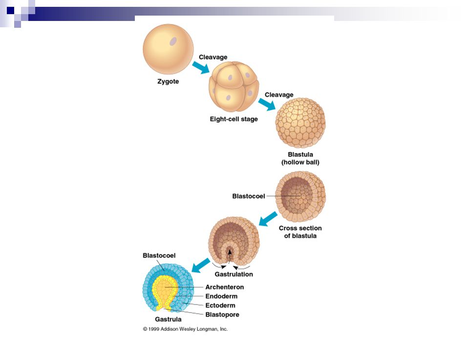
3. Cleavage Is the process of repeated rapid mitotic cell divisions of the zygote (unicellular structure) to form the Blastula (multicellular structure). The produced cells named Blastomeres. During this stage the size of the embryo does not change, the blastomeres become smaller with each division. The type & pattern of cleavage differ from species to species. continues divisions to form a ball of 32 cells called the morula. The morula continues divisions to form the hollow blastula with up to several hundred cells. The cavity of the blastula is the blastocoel.
 to form the Blastula (multicellular structure). The produced cells named Blastomeres. During this stage the size of the embryo does not change, the blastomeres become smaller with each division. The type & pattern of cleavage differ from species to species. continues divisions to form a ball of 32 cells called the morula. The morula continues divisions to form the hollow blastula with up to several hundred cells. The cavity of the blastula is the blastocoel.")
12
Figure 47.8x Cleavage in a frog embryo

13
4. Blastulation The result (end period) of cleavage.
The production of a multicellular blastula Blastula cells are called blastomeres. A cavity forms within the ball of the cells called the blastocoel.

14
Blastula of frog

15
Sea urchin blastula

16
Human blastula

17
morula Starfish development, unfertilized egg. 4 blastomeres.
Starfish development, nonmotile blastula. 16 blastomeres. 32 blastomeres. morula

18
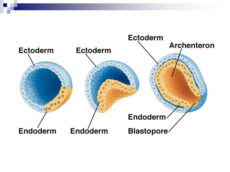
5. Gastrulation The morphogenetic process called gastrulation rearranges the cells of a blastula into a three-layered (triploblastic) embryo, called a gastrula, that has a primitive gut. It means rearrangement of blastula cells that transforms the blastula into a gastrula. The blastula develops a hole in one end and cells start to migrate into the hole; this forms the gastrula Characterized by cell movement. Blastocoel is gradually disappear and a new cavity is formed Gastrocoel.
 embryo, called a gastrula, that has a primitive gut. It means rearrangement of blastula cells that transforms the blastula into a gastrula. The blastula develops a hole in one end and cells start to migrate into the hole; this forms the gastrula. Characterized by cell movement. Blastocoel is gradually disappear and a new cavity is formed Gastrocoel.")
19
The gastrula is a three-layered embryo
The formation of three primary embryonic germ layers Endoderm (inner) Mesoderm (middle) Ectoderm (outer) The pattern of gastrulation is affected by the amount of yolk. The cells at the vegetal pole invaginate, initiating gastrulation.
 Mesoderm (middle) Ectoderm (outer) The pattern of gastrulation is affected by the amount of yolk. The cells at the vegetal pole invaginate, initiating gastrulation.")
22
Gastrulation in a Frog Embryo

23
Figure 47.10 Gastrulation in a frog embryo
The blastocoel of the frog blastula is off-center. Gastrulation begins when dorsal lip of the blastopore appears. Future endoderm and mesoderm cells roll inward over the dorsal lip and move away from the blastopore into the interior of the gastrula. Future ectoderm cells spread over the embryo’s outer surface. Externally, the lip of the blastopore starts becoming circular. Internally, the advancing endoderm, mesoderm, and the archenteron, lined by endoderm, are filling the space occupied by the blastocoel. Late in gastrulation, the circular blastopore surrounds a plug of yolk cells (the yolk plug). The three germ layers are now in place, ready for organogenesis.
. The three germ layers are now in place, ready for organogenesis.")
24
6. Neurulation Chordates Only
formation of a dorsal, hollow neural tube by ectodermal cells flatten into neural plate the center of the plate sinks forming neural groove edge of plate is elevated to form neural folds neural folds fuse and form neural tube anterior end develops into brain posterior end develops into spinal cord

26
Neurulation Dorsal view of the entire frog embryo, showing the ectodermal neural plate with edges elevated, forming the neural folds. Seen in transverse section, the neural folds meet and fuse, forming the neural tube.

27
The Neural Crest The neural crest is a critical structure that guides formation of several organ systems The neural crest forms on either side of the point of fusion Its cells migrate to form the dorsal root ganglia, the postganglionic sympathetic neurons, many sense organs and all pigment-forming cells

28
Organogenesis Endoderm Mesoderm
Organogenesis is the formation of the organs The layers are germ layers; they have specific fates in the developing embryo: Endoderm The innermost layer Goes on to form the gut Mesoderm The middle layer. Goes on to form the muscles, circulatory system, blood and many different organs Ectoderm The outermost Goes on to form the skin and nervous system

30
Organogenesis Begins With Development of the Nervous System
The nervous system is the first organ system to develop. The notochord grows and induces overlying ectoderm to form the neural plate. Cells of the neural plate fold to form the neural groove and the surrounding neural folds fuse to form the neural tube. The anterior portion forms the brain; the rest forms the spinal cord.

31
7. Organogenesis Ectoderm forms:
Development of organs from three primary germ layers Ectoderm forms: skin and associated glands, nervous system. Mesoderm forms: muscles, skeleton, gonads, excretory system, circulatory system. Endoderm forms: lining of digestive tract, liver, pancreas, lungs.

33
Figure 47.11 Organogenesis in a frog embryo

34
Somatic &Germ Cells Somatic Cells:
Found in all body tissues except gonads. Contain diploid numbers of chromosomes (2N). Replacement of dead cells Reproduce by mitotic division. Functions: Responsible for formation of different system and organs. Have other specific functions e.x.: muscular system have myoplast for contraction and relaxation . nervous system have neurons for transmission of impulses.
. Replacement of dead cells. Reproduce by mitotic division. Functions: Responsible for formation of different system and organs. Have other specific functions. e.x.: muscular system have myoplast for contraction and relaxation . nervous system have neurons for transmission of impulses.")
35
digestive system have secretory cells for secretion of enzymes for digestion.
bone have osteoplasts make hardening of bone. Lung have cells for respiration. Germ Cells: Found only in gonads (testes & ovary) Contain haploid number of chromosomes (1N) Reproduce by meiotic division (meiosis). Function : Formation of gametes (male & female)
 Contain haploid number of chromosomes (1N) Reproduce by meiotic division (meiosis). Function : Formation of gametes (male & female)")
36
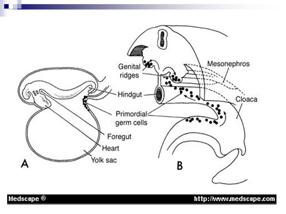
Primordial germ cells Appears in the wall of the endodermal layer of the yolk sac due to their large size and high content of alkaline phosphatase , and migrate by amoeboid movement toward the hind gut epithelium and then through dorsal mesentery reach to the primordia of the gonads ( primitive sex glands). Become recognizable at 24 days post-fertilization. Invading the genital ridges in the 6th week of development. The primordia of the gonads give a chemostatic attractant for the germ cells.
. Become recognizable at 24 days post-fertilization. Invading the genital ridges in the 6th week of development. The primordia of the gonads give a chemostatic attractant for the germ cells.")
37
Also may be the germ cells directed toward the gonads by the mature of cellular and non-cellular microenvironment that surround them. Or by tissue rearrangement that occur in the early embryos. If they fail to reach the ridges, the gonads will not develop. So, it have the inductive influence on the development of gonads into ovary and testes. Determine sex of the embryo. They will form different stages of spermatogenesis and oogenesis in the future. Found in adult in gonads (testes and ovary)
")
39
Basic Developmental Vocabulary
Fertilization – activates egg & brings together the nuclei of the egg and sperm. Cleavage partitions the zygote into many smaller cells. Gastrulation rearranges the blastula to form a three-layered embryo with a primitive gut, the archenteron. Organogenesis is the process by which the organs in the animal body form from the three embryonic germ layers.

40
Basic Developmental Vocabulary
Blastula – a hollow sphere of cells (128 cells) formed by cleavage of the morula. The blastula contains the blastocoel that is fluid-filled. The blastopore is the place where gastrulation begins. Gastrulation – the process leading to the creation of the primitive gut or archenteron. Invagination at the blastopore results in the gut. Gastrula – transformation of the blastula into an embryo possessing 3 germ layers, ectoderm, mesoderm, endoderm. Morula a solid mass of cells formed by cleavage.
 formed by cleavage of the morula. The blastula contains the blastocoel that is fluid-filled. The blastopore is the place where gastrulation begins. Gastrulation – the process leading to the creation of the primitive gut or archenteron. Invagination at the blastopore results in the gut. Gastrula – transformation of the blastula into an embryo possessing 3 germ layers, ectoderm, mesoderm, endoderm. Morula a solid mass of cells formed by cleavage.")
41
Questions? Dr. Abdelalim Gadallah
Thank you for your time. Questions? Dr. Abdelalim Gadallah

Similar presentations


 How do oogenesis and spermatogenesis differ? (Ch. 46) How do these hormones affect the menstrual cycle? LH FSH Estrogen Progesterone.>")


