Download presentation
Presentation is loading. Please wait.

1
고려대학교 구로병원 강 대 현 kangsono@naver.com

2
Goal of Ultrasound QA/QC Program To make sure a system is set up correctly and performs to specified standards. ( 장비의 올바른 설치와 기준에 맞는 작동을 보장 ) To maintain the consistency of the performance. ( 장비 성능의 일관성 유지 ) To reveal problems at its earliest stage before it severely interferes with the clinical practices. ( 임상업무를 심각하게 방해 하기 전에 초기에 문제점을 밝혀낼 수 있다 ) 장비의 정확하고 지속적인 성능 유지 초음파 영상 의 진단능력을 항상 최고로 유지시키기 위함
 To maintain the consistency of the performance. ( 장비 성능의 일관성 유지 ) To reveal problems at its earliest stage before it severely interferes with the clinical practices. ( 임상업무를 심각하게 방해 하기 전에 초기에 문제점을 밝혀낼 수 있다 ) 장비의 정확하고 지속적인 성능 유지 초음파 영상 의 진단능력을 항상 최고로 유지시키기 위함.")
3
Good Quality Assurance will ensure proper equipment operation detect gradual changes in performance minimize equipment down time reduce the number of repeat scans optimize operator and patient safety

5
When should quality assurance be performed? Routine preventative maintenance ( 기본예방정비 ) Performed by Sonographer on daily or weekly basis Routine performance testing ( 기본성능시험 ) Performed by Sonographer on weekly or monthly basis Acceptance testing ( 승인검사 ) Performed when new equipment arrives and is installed.
 Performed by Sonographer on daily or weekly basis Routine performance testing ( 기본성능시험 ) Performed by Sonographer on weekly or monthly basis Acceptance testing ( 승인검사 ) Performed when new equipment arrives and is installed..")
6
Three Levels of Ultrasound QC Testing Level 1 – Frequent QC testingLevel 1 – Frequent QC testing : Operator’s QC Level 2 – Semi-annual or annual QC testingLevel 2 – Semi-annual or annual QC testing : Physicist’s QC Level 3 – Acceptance testingLevel 3 – Acceptance testing / baseline set- up

7
Guideline By The Institute of Physical Sciences in Medicine (IPSM, 미국의학물리과학협회 ) Report No. 70 “Testing of Doppler Ultrasound Equipment” Edited by PR Hoskins, SB Sherriff and JA Evans York: ISPM, 1994

8
Guideline By The Institute of Physical Science in Medicine (IPSM, 미국 의학물리과학협회 ) Report No. 71 “Routine Quality Assurance of Ultrasound Imaging Systems” Edited by R. Price York: ISPM, 1995

9
Guideline By American Institute of Ultrasound in Medicine (AIUM, 미국 의학초음파 학회 ) “Quality Assurance Manual for Gray Scale Ultrasound Scanners (Stage 2)” Edited by E. Madsen AIUM, Laurel, MD, 1995

10
American Association of Physicists in Medicine (AAPM, 미국 의학물리협회 ) Ultrasound Task Group No. 1 “ Real-time B-mode ultrasound quality control test procedures” By MM Goodsitt et al Medical Physics. 25(8):1385-406, 1998 Aug. Guideline By
: , 1998 Aug. Guideline By.")
11
ACR Accreditation ( 인가 ) www.acr.org Breast Ultrasound Accreditation Program (including Ultrasound-guided Breast Biopsy) General Ultrasound Accreditation Program Obstetrical Gynecological General Vascular Combination of the above
 Breast Ultrasound Accreditation Program (including Ultrasound-guided Breast Biopsy) General Ultrasound Accreditation Program Obstetrical Gynecological General Vascular Combination of the above")
12
AIUM Accreditation www.aium.org Ultrasound practices in various fields: Breast Obstetrical and Trimester-Specific Obstetrical Gynecological Abdominal/General

13
ACR Accreditation QC Requirements A QA program should be in place; The QA program must be directed by a medical physicist or the supervising physician ; Routine QC testing must be done at least semi-annually ; Do the same tests, monitor changes over time and take corrective actions when necessary ; Testing results, corrective action, and the effects of corrective action must be documented and maintained on site. There is currently no ACR-preferred single phantom.

14
ACR Recommended Semi-Annual QC Tests for Breast Ultrasound Accreditation Maximum depth of visualization Distance accuracy (vertical and horizontal) Uniformity Electrical-mechanical cleanliness condition Anechoic void perception Ring down Lateral resolution Quality Control Checklist
 Uniformity Electrical-mechanical cleanliness condition Anechoic void perception Ring down Lateral resolution Quality Control Checklist")
15
ACR Recommended Technologist’s QC Tests for Breast Ultrasound Accreditation Adherence to universal infection control procedures for each biopsy All transducers should be cleaned between patients Distance calibration – quarterly Gray scale photography - quarterly

16
ACR Required semi-Annual QC Tests for General Ultrasound Accreditation System sensitivity and/or penetration capability Image uniformity Photography and other hard copy recording Low contrast object detectability (optional) Assurance of electrical and mechanical safety Vertical and horizontal distance accuracy (recommended only when the program is initiated for a scanner)
 Assurance of electrical and mechanical safety Vertical and horizontal distance accuracy (recommended only when the program is initiated for a scanner)")
17
Daily or weekly check lists General Machine Cleanliness Keyboard and knobs clean Monitors clean Air Filters cleans Mechanical and Electrical Wheels fastened securely and rotate easily? Wheels locks work well? Accessories fixed securely? Cords attached securely? ALI Workstation Contrast and Brightness between scanner and workstation Transducer Inspection (for each probe) Cables Transducer Cracks Matching layer delaminations
 Cables Transducer Cracks Matching layer delaminations.")
18
Assurance of Electrical and Mechanical Safety The equipment related physical checks are also worth attention. These include: Checking for any cracks or delamination in the transducers Noting for loose and frayed electric cables, loose handles or control arms Checking the working condition of the wheels and the wheel locks Making sure all the accessories (such as VCR, printer, etc.) are fastened securely to the system Making sure the image monitors are clean and the air filter are not dusty
 are fastened securely to the system Making sure the image monitors are clean and the air filter are not dusty.")
19
General Machine Cleanliness Philips General Machine Cleanliness / Philips System Power Switch Filter Main Power Switch ( 장비 뒷 편에 위치 ) Monitor Control Filter Power Switch IU 22 System Envisor System
 Monitor Control Filter Power Switch IU 22 System Envisor System")
20
HDI ® 5000 System System Power Switch Main Power Switch ( 장비 뒷 편에 위치 ) Filter Monitor Control Philips
 Filter Monitor Control Philips")
21
ACCUVIX System Track ball Medison

22
ACCUVIX Filer Medison ACCUVIX System Filter

23
제품 뒤쪽 화살표 부위를 청소한다. Medison SA8000 System Fan Accuvix

24
Acuson Sequoia 512 System Filter : 장비의 후면에 위치 System Power switch Main Power switch Key board

25
Acuson Antares System System Power switch Main Power switch Filter : 장비의 후면에 위치

26
Acuson G50/ G60S Both Filter : 본체 전면 하단 다리측 2 개 System Power switch : 장비 좌측하단

27
Mechanical and Electrical check

28
Transducer Inspection check Transducer Cleaner

29
Annually or semi-annually check lists Performance test Dead zone test Vertical Measurement accuracy test Horizontal Measurement Calibration System sensitivity test Uniformity test Grayscale photography test Axial resolution test Lateral resolution test Functional resolution, Definition and Fill-in Doppler QC tests Doppler signal sensitivity Doppler angle accuracy Color display and Gray-scale image congruency Range-gate accuracy Flow readout accuracy

30
Ultrasound QC Phantoms B-mode: 1.Multi-purpose or general purpose tissue/cyst phantom 2.Low contrast resolution phantom 3.Spherical lesion phantom 4.Beam profile and slice thickness phantom 5.Prostate QC phantom Doppler-mode: 1.Flow phantom 2.String phantom

31
Multipurpose Phantom Model 539

32
Specifications of Multipurpose Phantom Model 539 1.Tissue-mimicking material - Speed of sound: 1,540m/sec - Attenuation : 0.7 ㏈ / ㎝ / ㎒ - Freezing point: 0 ℃ (32 ℉ ) - Melting point: 78 ℃ (174 ℉ ) 2. Containers - Wall : 3/8"Acrylic - Scanning surface : 0.4mm (0.015")polyurethane(saran) 3. Targets - Cyst speed of sound : 1,540m/sec - Cyst attenuation : 0.05 ㏈ / ㎝ / ㎒ - Nylon lines: 0.3mm (0.012") diameter - Phantom size and weight: 15×18×5Cm, 2.5Kg
polyurethane(saran) 3. Targets - Cyst speed of sound : 1,540m/sec - Cyst attenuation : 0.05 ㏈ / ㎝ / ㎒ - Nylon lines: 0.3mm (0.012 ) diameter - Phantom size and weight: 15×18×5Cm, 2.5Kg.")
33
Multipurpose Phantom Model 539 ScanningTargetDiagram

34
측정항목 Dead zone test System sensitivity and/ or penetration capability test Image Uniformity test Grayscale photography and other hard copy recording test Axial and lateral resolution test Functional resolution, Definition and Fill-in Vertical and horizontal distance accuracy test (recommended only when the program is initiated for a scanner)
")
35
측정 시 주의사항 Scanning : vertical to phantom surface

36
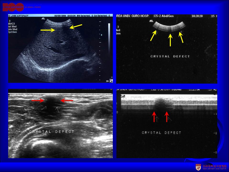
Dead Zone (Ring down) A group of 9 reflectors consisting of fibers are placed at different separations from the top of the phantom(~2-10mm). As the transducer scan across the top, the distance from the transducer to the first reflector completely imaged is equal to the dead zone (ring down) distance.
 distance..")
37
Dead Zone (Ring down) Reduced near field resolution by Transducer-skin interface
 Reduced near field resolution by Transducer-skin interface")
38
Dead Zone (Ring down) Dead Zone (Ring down) test Dead zone test 2mm ~ 10mm, 9 targets
 Dead Zone (Ring down) test Dead zone test 2mm ~ 10mm, 9 targets")
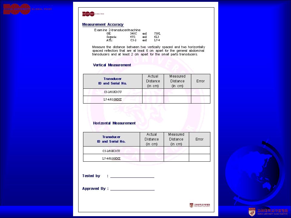
39
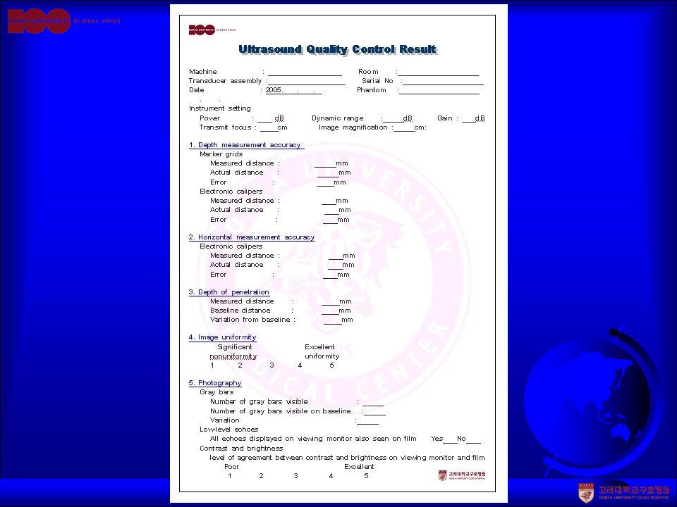
Distance Accuracy test Scan the phantom with vertical column and a horizontal row of reflectors; The digital caliper readout on screen is checked against the known distance between reflectors (10cm); Criteria Vertical : 1,5% of the actual distance or 2mm, whichever is greater. Horizontal : 3% of the actual distance or 3mm, whichever is greater.

40
Vertical Distance accuracy test

41
Horizontal Distance accuracy test

42
System Sensitivity/Penetration This test should be done with following setting: Maximum transmit power Proper receiver gain and TGC that allows echo texture to be visible in the deep region Transmit focus at the deepest depth The maximum depth of visualization is determined by comparing the gradually weakening echo texture to electronic noises near the bottom of the image. Do this test with the same setting and monitor the changes over time.

43
System Sensitivity/Penetration 3.5 MHz 12 MHz 7 MHz

44
Axial and Lateral Resolution Spatial resolution may be evaluated by either of the following ways: Count the number of the pins distinguished without overlap in lateral and axial direction; thus the minimum target spacing is documented Measure the size of the pin in lateral and axial direction

45
Axial resolution test Axial Resolution Array 3.0 mm 2.0 mm 1.0 mm 0.5 mm 0.25 mm 1.0 mm 0.25mm

46
Lateral resolution test Lateral Resolution Array 3.0 mm 2.0 mm 1.0 mm 0.5 mm 0.25 mm 3.0 mm 3 mm

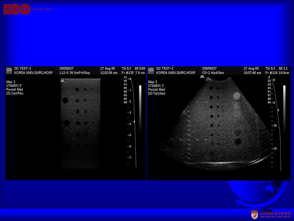
47
Image Uniformity Adjust the TGC controls and other sensitivity controls to obtain an image as uniform as possible Inspect the image to detect any kinds of Vertical or radially oriented streaks( 수직 또 는 방사형태로 출현하는 줄무늬 ) Dropouts Reduction of brightness near edges of the scan( 주사면 끝부분의 밝기 저하 ) Brightness transitions between focal zones ( 초점 영역 사이의 밝기 변화 )
 Dropouts Reduction of brightness near edges of the scan( 주사면 끝부분의 밝기 저하 ) Brightness transitions between focal zones ( 초점 영역 사이의 밝기 변화 )")
48
Image Uniformity

49
Functional resolution, Definition and Fill-in QA Phantom 에서 anechoic structure 의 spherical targets 를 명확하게 detection 할 수 있는 능력 2, 3, 4, 6, 8mm diameter spherical targets

50
Functional resolution, Definition and Fill-in 1 1/2D (Matrix) Transducer Conventional array Transducer 초음파 영상은 단면 data 가 아니라 volume data 이기 때문임. 초음파 영상은 단면 data 가 아니라 volume data 이기 때문임.

52
Soft and/or Hard Copy Recording Ⅰ The shades of gray, weak, and strong echo texture should be optimized and consistent between the image display on the ultrasound scanner and the photographic hard copies or soft copy displays on the workstation in the reading room. For quick follow-up testing, the Gray-scale bar pattern on the clinical image display can be used

53
Soft and/or Hard Copy Recording Ⅱ Use the SMPTE test pattern and other patterns if they are available on the ultrasound scanner. Workstation monitor display should be included in QC tests.

54
Soft and/or Hard Copy Recording Ⅲ Film processor QC needs to be done daily. Darkroom fog test needs to be done at least semi-annually. Sensitometry

55
Low Contrast Object Detectability Scan of a low contrast resolution phantom can reveal the low contrast object detectability which is an optional test on the ACR semi-annual QC test list for general ultrasound accreditation.

56
Ultrasound Doppler QC Testing Doppler QC tests include Doppler signal sensitivity Doppler angle accuracy Color display and Gray-scale image congruency Range-gate accuracy Flow readout accuracy

57
Doppler String Phantom

58
Doppler Velocity Accuracy: Variations among 6 clinical units, each with transducers of 3.5MHz, 5MHz and 7 MHz. Flow Rate (ml/min) Frequency 1002373986118001093AVG 3.5MHz 5.7%10.2%6.7%8.0%10.2%13.0%9.0% 5 MHz 17.4%7.7%10.6%6.9%13.9%13.0%11.6% 7 MHz 8.2% 4.1%9.2%7.1%12.5%8.2% Average 10.4%8.7%7.1%8.0%10.4%12.9%9.6%
 Frequency AVG 3.5MHz 5.7%10.2%6.7%8.0%10.2%13.0%9.0% 5 MHz 17.4%7.7%10.6%6.9%13.9%13.0%11.6% 7 MHz 8.2% 4.1%9.2%7.1%12.5%8.2% Average 10.4%8.7%7.1%8.0%10.4%12.9%9.6%.")
63
Discussion The guidelines for ultrasound QA/QC programs are descriptive. Maintaining an ultrasound QA/QC program is straightforward and testing consistency. However, we need to know that the tissue mimicking material can only mimic some of the properties of tissue in real clinical situations. Assessment of the efficacy and relevance of ultrasound QA procedures is rare. The guidelines need to be updated periodically as ultrasound technology develops.

Similar presentations


>")


