Download presentation
Presentation is loading. Please wait.

1
ACUTE APPENDICITIS

2
ACUTE APPENDICITIS Appendicitis is defined as an inflammation of the inner lining of the vermiform appendix that spreads to its other parts. This condition is a common and urgent surgical illness with protean manifestations, generous overlap with other clinical syndromes, and significant morbidity, which increases with diagnostic delay.

4
Anatomy Normal appendix; barium enema radiographic examination. A complete contrast-filled appendix is observed (arrows), which effectively excludes the diagnosis of appendicitis
, which effectively excludes the diagnosis of appendicitis.")
6
Variations in topographic position of the appendix
From its base at the cecum, the appendix may extend (A) upward, retrocecal and retrocolic; (B) downward, pelvic; (C) downward to the right, subcecal; or (D) upward to the left, ileocecal (may pass anterior or posterior to the ileum)
 upward, retrocecal and retrocolic; (B) downward, pelvic; (C) downward to the right, subcecal; or (D) upward to the left, ileocecal (may pass anterior or posterior to the ileum)")
7
Surgical Anatomy - Position

8
Blood supply to the appendix.
A and B. Usual type with a single appendicular artery. C. Paired appendicular arteries.

9
Incidence The lifetime rate of appendectomy is 12% for men and 25% for women, with approximately 7% of all people undergoing appendectomy for acute appendicitis during their lifetim Despite the increased use of ultrasonography, computed tomography (CT), and laparoscopy, the rate of misdiagnosis of appendicitis has remained constant (15.3%), as has the rate of appendiceal rupture. The percentage of misdiagnosed cases of appendicitis is significantly higher among women than among men
, and laparoscopy, the rate of misdiagnosis of appendicitis has remained constant (15.3%), as has the rate of appendiceal rupture. The percentage of misdiagnosed cases of appendicitis is significantly higher among women than among men.")
10
AGE

11
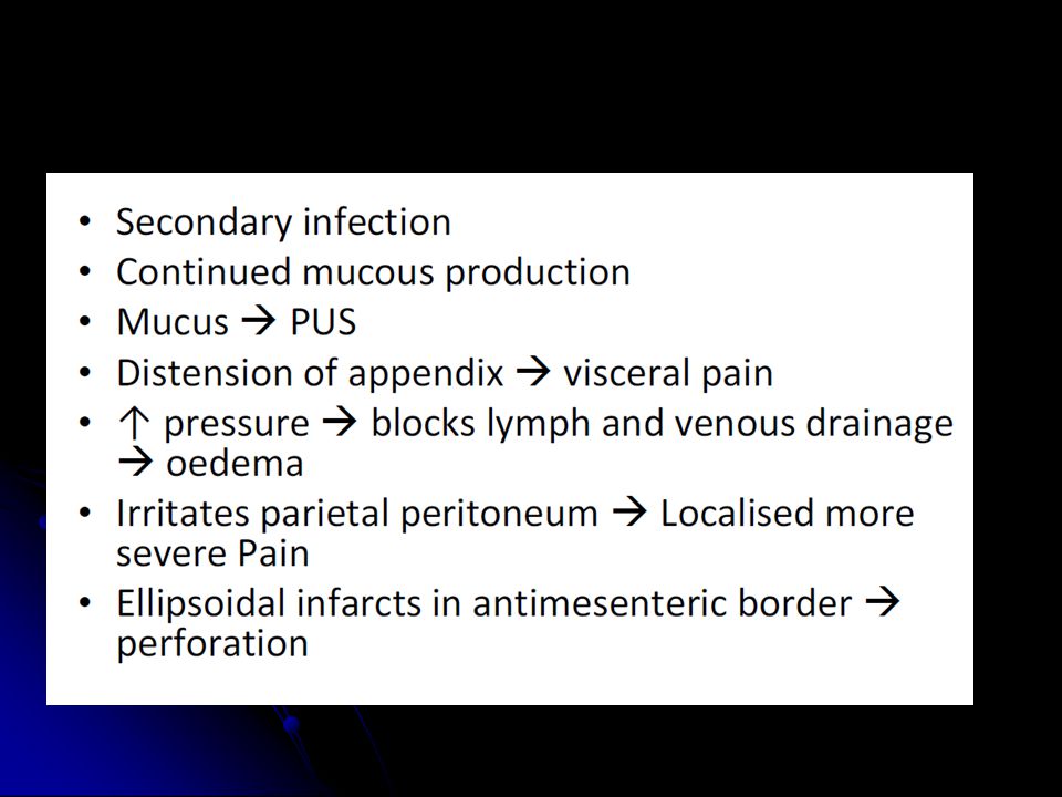
Etiology Obstruction of the lumen is the dominant etiologic factor in acute appendicitis. – Submucosal lymphoid hyperplasia – Faecolith / faecal stasis – Inspissated barium – Vegetable/fruit seeds – Worms (Entrobius vermicularis – Tumours of caecum/appendix

13
Common organisms seen in patients with acute appendicitis
Bacteriology Common organisms seen in patients with acute appendicitis

14
Classification (by V.I. Kolesnikov)
1. Appendiceal colic. 2. Simple superficial appendicitis. 3. Destructive appendicitis: а) phlegmonous; б) gangrenous; в) perforated. 4. Complicated appendicitis: а) appendicular infiltrate; б) appendicular abscess; в) diffuse purulent peritonitis. 5. Other complications of acute appendicitis (pylephlebitis, sepsis, retroperitoneal phlegmon, local abscesses of abdominal cavity).
 phlegmonous; б) gangrenous; в) perforated. 4. Complicated appendicitis: а) appendicular infiltrate; б) appendicular abscess; в) diffuse purulent peritonitis. 5. Other complications of acute appendicitis (pylephlebitis, sepsis, retroperitoneal phlegmon, local abscesses of abdominal cavity).")
15
Symptoms of simple appendicitis
1. Pain localized in a right iliac area. In 70 % of patients the pain arises in a epigastric area – it is an epigastric phase of acute appendicitis. In 2-4 hours it migrates to the area of appendix (the Kocher’s sign). 2. Single nausea and vomiting. 3. Fever to C. 4. Retention of stool or single diarrhea. 5. Muscular tension in a right iliac area. Rovsing's sign - pain in right lower quadrant during palpation of left lower quadrant Sitkovsky’s sign - increase of pain in a right iliac area when the patient lies on the left side Bartomier’s sign - the increase of pain intensity during the palpation of right iliac area when the patient lies on the left side. Dunphy's sign-increased pain with coughing
. 2. Single nausea and vomiting. 3. Fever to C. 4. Retention of stool or single diarrhea. 5. Muscular tension in a right iliac area. Rovsing s sign - pain in right lower quadrant during palpation of left lower quadrant. Sitkovsky’s sign - increase of pain in a right iliac area when the patient lies on the left side. Bartomier’s sign - the increase of pain intensity during the palpation of right iliac area when the patient lies on the left side. Dunphy s sign-increased pain with coughing.")
16
Symptoms of phlegmonous appendicitis
1. Expressed pain in a right iliac area. 2. Fever to C. 3. Muscular rigidity in a right iliac area. 4. Peritoneal signs Blumberg’s sign. After gradual pressing by fingers of anterior abdominal wall quick taking off the hand causes the sharp increase of pain. Voskresenky’s sign. The increase of pain during quick sliding movements by the tips of fingers from epigastric to right iliac area. Rozdolsky’s sign. Painfulness in a right iliac area during percussion.

17
Symptoms of gangrenous appendicitis
1. Pain in a right iliac area. 2. Grave condition of the patient. 3. Signs of local peritonitis. 4. Signs of intoxication

18
Symptoms of retrocaecal appendicitis
1. Non-expressive abdominal clinic. 2. Expressed pain in a right lumbar area. 3. Pain and muscular rigidity in a right iliac area during palpation. Yaure-Rozanov sign - Painfullness during palpation of Petit triangle Gabay’s sign - Blumberg’s sign in Petit triangle Pasternatsky’s sign - tapping of lumbar region cause the pain Psoas sign - pain on extension of right thigh

19
Symptoms of retrocaecal retroperitoneal appendicitis
1. Clinic of retroperitoneal phlegmon. 2. The signs of retrocaecal appendicitis. 3. Flank tenderness in right lower quadrant.

20
Symptoms of pelvic appendicitis
1. Clinic of irritation of pelvic organs (dysuria, pulling rectal pain, tenesmi). 2. Absence of muscular tenderness. 3. Painfullness of anterior rectal wall and posterior vaginal vault.
. 2. Absence of muscular tenderness. 3. Painfullness of anterior rectal wall and posterior vaginal vault.")
21
Complications 1. Appendicular infiltrate. 2. Appendicular abscess.
3. Diffuse peritonitis. 4. Pilephlebitis

22
Diagnostics 1. Anamnesis. 2. Objective examination.
3. General blood and urine analyses. 4. Vaginal examination for women. 5. Rectal examination for men.

23
Abdominal X-ray

24
Graded compression Ultrasound
Depends on the technique and experience Normal appendix – a blind-ended, tubular structure with a maximum wall thickness of 2 mm with an outer diameter of 6 mm, – No peristalsis – Originates from the base of the cecum

25
Graded compression Ultrasound

26
CT variable degree of distension (diameter 6–40 mm)
wall thickness of 1–3 mm. Wall - asymmetrically thickened enhances with intravenous contrast medium. periappendiceal inflammatory mass Thickening and enhancement with intravenous contrast - adjacent wall of the cecum or ileum

27
CT

28
Differential diagnostics
Gastrointestinal Cholecystitis Crohn's disease Duodenal ulcer Gastroenteritis Intestinal obstruction Meckel's diverticulitis Mesenteric lymphadenitis Necrotizing enterocolitis Neoplasm (carcinoid, carcinoma, lymphoma) Gynecologic Ectopic pregnancy Endometriosis Ovarian torsion Pelvic inflammatory disease Ruptured ovarian cyst Tubo-ovarian abscess
 Gynecologic. Ectopic pregnancy. Endometriosis. Ovarian torsion. Pelvic inflammatory disease. Ruptured ovarian cyst. Tubo-ovarian abscess.")
29
Differential diagnostics
Systemic Diabetic ketoacidosis Henoch-Schonlein purpura Pulmonary Pleuritis Pneumonia (basilar) Pulmonary infarction Genitourinary Kidney stone Pyelonephritis Wilms' tumor Other Parasitic infection Psoas abscess Rectus sheath hematoma
 Pulmonary infarction. Genitourinary. Kidney stone. Pyelonephritis. Wilms tumor. Other. Parasitic infection. Psoas abscess. Rectus sheath hematoma.")
30
Differential diagnostics of acute appendicitis with perforative peptic ulcer
Pain in the right iliac region Muscular tenderness in the right iliac region Single vomiting and diarrhea Sharp acute diffuse pain Ulcerative anamnesis Absence of hepatic dullness On X-ray of the abdomen air above the liver (air sickle) Rigidity of anterior abdominal wall
 Rigidity of anterior abdominal wall.")
31
Differential diagnostics of acute appendicitis with intestinal obstruction
Constant pain in the right iliac region Muscular tenderness in the right iliac region Single vomiting and diarrhea Periodic acute diffuse pain Constant vomiting and nausea without any relief Retention of stool and gases Abdominal distension On X-ray of the abdomen Kloiber's cups (air-fluid levels) Splashing sound, increased peristalsis
 Splashing sound, increased peristalsis.")
32
Management

33
Open Appendicectomy • Incission (transverse, Mc Burney’s point)
• Open in layers. (muscle split along its fibres) • Check for fluid (+/-c&S) • Identify caecum and exteriorized – follow taeniae to appendix • Mesoappendix divided + ligated • Clamp appendix 5mm above caecum and ligated • Cauterise residual mucosa +/- purse string (not req) • Return caecum, wash with warm saline • Close in layers
 • Check for fluid (+/-c&S) • Identify caecum and exteriorized – follow taeniae to appendix. • Mesoappendix divided + ligated. • Clamp appendix 5mm above caecum and ligated. • Cauterise residual mucosa +/- purse string (not req) • Return caecum, wash with warm saline. • Close in layers.")
34
Open Appendicectomy

35
Open Appendicectomy

36
Open Appendicectomy

37
Open Appendicectomy

38
Laparoscopic Appendicectomy

39
Laparoscopic Appendicectomy

40
Laparoscopic Appendicectomy

41
Laparoscopic Appendicectomy

42
Complications

Similar presentations