Download presentation
Presentation is loading. Please wait.

1
Chapter 18 Regulation of Gene Expression

2
Genetic Diversity in Asexual Bacteria? I. From Within A. High mutation rate B. Transposons II. Recombination A. Transformation B. Transduction C. Conjugation

3
I. Bacterial Regulation A.Advantage to expressing only the genes necessary? B. Metabolic control 1. Adjust enzyme activity 2. Adjust enzyme production Gene expression control

4
I. Bacterial Regulation C. The Operon Model 1. Discovery – Jacob and Monod, 1961 2. The Basic Concept Operon – A cluster of genes related to the same pathway See E.coli – Tryptophan example (352)
.")
5
I. Bacterial Regulation 2. The Basic Concept Operon a. Components; A Single promoter sequence A Single operator sequence Multiple genes (One for each protein in the pathway)
.")
6
I. Bacterial Regulation 2. The Basic Concept Operon b. Functions as a single transcription unit With a single “on-off” switch Allows coordination and control

7
I. Bacterial RegulationI. Bacterial Regulation 2. The Basic Concept Operon c. “On-off” switch = Operator Segment of DNA Upstream of the genes Within the promotor Contols access of RNA Polymerase to the genes

8
I. Bacterial RegulationI. Bacterial Regulation 2. The Basic Concept Operon d. Mechanism? (Example – Trp operon) Normally switched on Switched off when Active Repressor binds to the operator. This blocks RNA Polymerase from transcribing the genes
 Normally switched on Switched off when Active Repressor binds to the operator. This blocks RNA Polymerase from transcribing the genes.")
10
I. Bacterial Regulation 2. The Basic Concept Operon d. Mechanism? (Example – Trp operon) Repressors are specific to their operon Source of Repressor? Coded for by a Regulatory Gene Located a distance away Has its own promotor
 Repressors are specific to their operon Source of Repressor. Coded for by a Regulatory Gene Located a distance away Has its own promotor.")
11
2. The Basic Concept Operon d. Mechanism? (Example – Trp operon) Repressor Activity Regulatory genes continually expressed A Few repressors always present Binding to operator is reversible On or off depends number of active repressors around Repressor – allosteric protein Active state and inactive state Corepressor must bind to it to activate it
 Repressor Activity Regulatory genes continually expressed A Few repressors always present Binding to operator is reversible On or off depends number of active repressors around Repressor – allosteric protein Active state and inactive state Corepressor must bind to it to activate it.")
12
Corepressor can act as feedback control Example – Trp Operon As tryptophan is produced to high levels It binds to repressors activating them This blocks transcription, shutting down tryptophan production

14
I. Bacterial Regulation D. Negative Gene Regulation Involves a Repressor attaching to the gene operator Two types 1. Repressible operons 2. Inducible operons

15
I. Bacterial Regulation D. Negative Gene Regulation 1. Repressible Operons (anabolic) On = NormalTurn it off No CoRepessor+ CoRepressor Repressor inactiveRepr. Activated Operon NOT repressedOperon is repress. Protein Synthesis No synthesis
 On = NormalTurn it off No CoRepessor+ CoRepressor Repressor inactiveRepr. Activated Operon NOT repressedOperon is repress. Protein Synthesis No synthesis.")
16
Best Example; The Trp Operon

17
I. Bacterial Regulation D. Negative Gene Regulation 2. Inducible Operons (Catabolic) Normally OffTurn it on No InducerMake Inducer Repressor activeShuts off Repressor Operon blockedOperon active No synthesisSynthesis
 Normally OffTurn it on No InducerMake Inducer Repressor activeShuts off Repressor Operon blockedOperon active No synthesisSynthesis.")
18
Best Example; LAC Operon

19
I. Bacterial Regulation E. Positive Gene Regulation Involves an Activator attaching to genome (CAP for Glucose example)
.")
20
I. Bacterial Regulation E. Positive Gene Regulation Low GlucoseHigh Glucose cAMP upcAMP down Binds to CAP Act.CAP act. Looses CAP activatedCAP inactive Binds above promotorNo synthesis Synthesis occurs

21
II. Eukaryotic Regulation A.Gene regulation especially important for multicellular organisms Cell specialization What causes differences? Differential gene expression!

22
II. Eukaryotic Regulation B. Differential Gene Expression Expression of different genes by cells with the same genome Typical cell – 20% expression Less in more specialized cells How expressed? Transcription

24
II. Eukaryotic Regulation B. Regulation in Chromatin Structure

25
II. Eukaryotic Regulation C. Regulation by Chromatin Structure 1. Location of a Gene’s promotor relative to nucleosomes scaffold attachment nuclear lamina

26
II. Eukaryotic Regulation C. Regulation by Chromatin Structure 2. Location in Heterochromatin? Highly condensed Usually not expressed

27
II. Eukaryotic RegulationII. Eukaryotic Regulation C. Regulation by Chromatin Structure 3. Chemical modifications to histones and histone tails

28
II. Eukaryotic RegulationII. Eukaryotic Regulation 3. Chemical modifications Histone Acetylation Acetyl groups attached to lysines in the histone tails Effects + charges neutralized No longer bind to other nucleosomes Stays loosely packed Deacetylation - repacks

29
II. Eukaryotic RegulationII. Eukaryotic Regulation 3. Chemical modifications to histones and histone tails Methyl Groups – promotes condensing Phosphate Groups – reverses methyl effect Histone Code Hypothesis Combinations of chem modifications are the key

30
II. Eukaryotic Regulation D. DNA Methylation Direct methylation of DNA Bases (usually cytosine) 1. More methylation – DNA less active 2. Proteins can bind to methyl groups Recruit histone deacetylation enzymes Results in a dual mechanism to repress transcription 3. Critical in embryonic development Cell differentiation Methylation pattern passed down cell lines Genomic Imprinting!!

31
II. Eukaryotic Regulation E. Epigenetic Inheritance (Differential expression patterns are passed on) Passing Traits without code sequence changes These changes are reversible
 Passing Traits without code sequence changes These changes are reversible.")
32
F. Regulating Transcription Initiation Ultimately – Control the binding and action of RNA Polymerase. 1. Basic Organization of a Eukaryotic Gene Upstream Promotor Proteins assemble here = Transcription Init. Complex Control Elements Upstream DNA Sequences Bind control proteins

33
Eukaryotic Gene Organization

34
F. Regulating Transcription Initiation 2. Transcription Factors RNA Polymerase requires these a. General transcription factors required for all protein code genes Bind to promotor (TATA) and other proteins Make Transcription Initiation complex Then RNA Polymerase can function Leads to low level transcription
 and other proteins Make Transcription Initiation complex Then RNA Polymerase can function Leads to low level transcription.")
35
F. Regulating Transcription Initiation 2. Transcription Factors RNA Polymerase requires these b. Specific Transcription Factors Specific to a gene Interact with control factors leads to high level transcription

36
Eukaryotic Gene Organization

37
F. Regulating Transcription Initiation 3. Control Elements – The basics a. Proximal Control Elements Close to Promotor b. Distal Control Elements Called Enhancers 1000s of bases away from promotor A gene may - multiple enhancers Enhancers specific to a gene

38
Eukaryotic Gene Organization

39
F. Regulating Transcription Initiation 4. Regulation using Control Elements Key concept Protein-protein interactions work to assemble Transcr.Init.Complex on the promotor

41
F. Regulating Transcription Initiation 4. Regulation using Control Elements Mechanism Activators bind to enhancer Mediator proteins form link between activators and promotor DNA Bending protein bends enhancer to promotor Link builds Transc.Init.Complex

43
F. Regulating Transcription Initiation 4. Regulation using Control Elements Repression Repressors (methods) Bind to enhancers – Blocking Activators Bind to activators – blocking other protein binding Many other repression methods
 Bind to enhancers – Blocking Activators Bind to activators – blocking other protein binding Many other repression methods.")
45
F. Regulating Transcription Initiation 5. Enhancer Combination Effects Enhancer control element sequences repeat for many genes Each enhancer has around 10 control element sequences The combination of the control elements provides specificity for specific genes

47
F. Regulating Transcription Initiation 6. Coordinate Control of Related genes Prokaryotes – Operons! What about Eukaryotes???? a. Related genes may be clustered close together But – separate promotors

48
F. Regulating Transcription Initiation 6. Coordinate Control of Related genes b. Most commonly Co-expressed genes scattered! So how? - Coordination by specific control element sequences - often response to signal from outside of cell – binds to all enhancers with a specific control element sequence

49
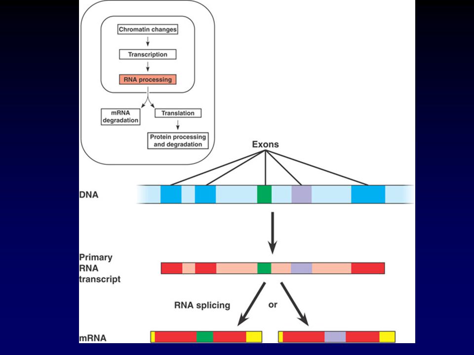
G. Post-Transcriptional Regulation 1.RNA Processing Alternative RNA Splicing – Different mRNAs are made from same primary transcript Cell types Regulatory proteins bind to determine intron / exon choices

51
G. Post-Transcriptional Regulation 2. mRNA Degradation Control of the life span of mRNA 3. Initiation of Translation a. Regulatory proteins attach at 5’ end of mRNA prevents binding of Ribosome b. Poly-A tail manipulation Necessary for Ribosome binding Some mRNAs lacking in Poly-A No translation until enzyme adds additional As

52
G. Post-Transcriptional Regulation 2. mRNA Degradation Control of the life span of mRNA 3. Initiation of Translation c. manipulation of the proteins that initiate translation

53
G. Post-Transcriptional Regulation 4. Protein processing Control of cleavage and reassembly Degradation of proteins = limit protein life span Tag proteins with ubiquitin Attracts proteasomes to degrade them

54
III. Impact of Non-Coding RNA A. Large part of the genome codes for Non-protein coding RNA (Non-Coding RNA) Many questions remain on what these do and how many types exist B. Role in regulation? Two places 1. mRNA translation (C,D, and E) 2. Chromatin configuration (F)
 Many questions remain on what these do and how many types exist B. Role in regulation. Two places 1. mRNA translation (C,D, and E) 2. Chromatin configuration (F).")
55
III. Impact of Non-Coding RNA C. Micro-RNA = miRNA Small and single stranded (ssRNA) 1. Formation – from large precurser RNA Trimmed by enzyme “Dicer”

56
miRNA Production

57
III. Impact of Non-Coding RNA C. Micro-RNA = miRNA Small and single stranded (ssRNA) 2. Function Degrades a target mRNA Blocks translation of a target mRNA

58
III. Impact of Non-Coding RNA D. siRNA – Small interfering RNA Turns off genes with same sequence Made by same machinery that makes miRNA Slight distinctions – see text pg365 E. Are these informational, catalytic, or structural?

59
III. Impact of Non-Coding RNA F. Impact on Chromatin Configuration siRNA – heterochromatin in yeast Associate with proteins to recruit enzymes – condensing chromatin Check experiments – 365-366

60
IV. Cell Differentiation A.Key Context – Embryonic Development B.The Genetic Programming of development 1. Three necessary components Cell Division Cell Differentiation Morphogenesis – Shaping 3D organization of cell types

61
IV. Cell Differentiation A.Key Context – Embryonic Development B.The Genetic Programming of development 2. Key Concepts Differential gene expression leads to specialization Different collections of activators Different patterns 3. BUT …… What sets this all up?????

62
IV. Cell Differentiation C. Cytoplasmic Determinants and Induction What tells a cell which collection of genes to express? Two sources of information 1. The Egg’s cytoplasm 2. Environment around a cell

63
IV. Cell Differentiation C. Cytoplasmic Determinants and Induction 1.The Egg’s cytoplasm Not homogeneous Cytoplasmic Determinants Maternal substances unevenly distributed mitotic divisions – new cells get differing concentrations

64
IV. Cell Differentiation C. Cytoplasmic Determinants and Induction 2. Surrounding Environment Signals from surrounding cells Contact with surrounding cell surfaces Growth factors from neighbors Changes from these sources called Induction Cell-surface receptors important Untilizes signal transduction pathways

65
IV. Cell Differentiation D. Sequential Regulation 1. Determination – the events that lead to observable differentiation of a cell Completed determination is irreversible Caused by differential gene expression for tissue specific proteins

66
IV. Cell Differentiation D. Sequential Regulation 2. Pathway to differentiation Signal mRNAs Specific protein Observable phenotype 3. Expression pattern is SEQUENTIAL 4. Key point of regulation - transcription

67
IV. Cell Differentiation D. Sequential Regulation 5. There is a hierarchy of differentiation 6. Study muscle cell example – pg368

68
Cell Differentiation

69
IV. Cell Differentiation E. Pattern Formation– the spatial arrangement of tissues and organs. Positional Information cues – establish the three key axes of the body Cytoplasmic determinants Inductive signals

70
IV. Cell Differentiation E. Pattern Formation 1. Information gleaned from D. melanogaster

71
IV. Cell Differentiation E. Pattern Formation 1. Information gleaned from D. melanogaster a. Modular construction - ordered series of segments Head Thorax Abdomen Sub-segments

72
IV. Cell Differentiation E. Pattern Formation 1. Information gleaned from D. melanogaster b. 3 Axes Anterior-posterior Dorsal- ventral Laterals (side-side)
.")
73
IV. Cell Differentiation E. Pattern Formation 1. Information gleaned from D. melanogaster c. Pattern formation? Axes from cytoplasmic determinants

74
IV. Cell Differentiation E. Pattern Formation 2. Drosophila sequence Edward B. Lewis (1940s) Genetic approach to embryology using D. melanogaster Born Wilkes-Barre PA Caltech professor Nobel prize 1995 Died 2004 (cancer)
 Genetic approach to embryology using D. melanogaster Born Wilkes-Barre PA Caltech professor Nobel prize 1995 Died 2004 (cancer).")
75
2. Drosophila sequence Initial discovery by Lewis Specific gene mutations led to extra legs or wings growing in the wrong places Homeotic Genes – Control pattern formation Identification of specifics? 30 years! Nusskein-Volhard and Wieschaus (Germany)
.")
76
2. Drosophila sequence Nusskein-Volhard and Wieschaus (Germany) Eventually – dicovered 1200 genes controlling pattern formation 1995 nobel prize for these three!
 Eventually – dicovered 1200 genes controlling pattern formation 1995 nobel prize for these three!.")
77
2. Drosophila sequence Genes discovered by tracking recessive mutations Embryonic Lethals

78
2. Drosophila sequence Axis establishment Maternal Effect Genes – build the cytoplasmic determinants in eggs Also called Egg-polarity Genes Example – the Bicoid Gene Mutant form leads to Two tail ends, no head Gene sets up the anterior-posterior axis

79
2. Drosophila sequence The Bicoid Gene This gene exemplifies the Morphogen Gradient Hypothesis Axes set up by concentration gradients of morphogens

80
2. Drosophila sequence The Bicoid Gene Bicoid process Bicoid mRNA concentrated in the anterior end of unfert. Egg Fertilization = zygote Bicoid mRNA translated to protein Bicoid protein diffuses through zygote Creates conc. Gradient High conc. = anterior Low conc. = posterior First gene-protein specifically linked to pattern formation

81
2. Drosophila sequence Beyond Bicoid example - Other gene linked gradients found for dorsal – ventral axis Later positional information Finer scale patterns correct orientation of each segment

82
V. Cancer and Gene Expression A. Types of Genes associated with Cancer Deal with cell growth and division 1. Growth factor genes receptor genes signaling pathway genes 2. Causes spontaneous mutations environmental influences Tumor viruses

83
V. Cancer and Gene Expression A. Types of Genes associated with Cancer 3. Tumor Viruses First - Payton Rous 1911 Epstein-Barr virus - Mono … Linked to lymphoma Papillomaviruses - cervical cancer HTLV-1 - Leukemia

84
V. Cancer and Gene Expression A. Types of Cancer Genes 4. Oncogenes and Proto-Oncogenes Proto-Oncogenes - normal versions Promote growth and division Oncogene - mutated - leads to cancer

85
V. Cancer and Gene Expression A. Types of Cancer Genes 4. Oncogoenes and Proto-Oncogenes How proto-oncogene to oncogene? Mutation effects protein product a. DNA moves in genome (protoOncogene move to active promotor) b. amplification of gene (increase copies of gene) c. point mutation
 b. amplification of gene (increase copies of gene) c. point mutation.")
86
V. Cancer and Gene Expression A. Types of Cancer Genes 4. Oncogoenes and Proto-Oncogenes How proto-oncogene to oncogene? Mutation effects protein product c. point mutation In promotor or enhancer increasing expression In gene itself protein more active or resistant

87
V. Cancer and Gene Expression A. Types of Cancer Genes 5. Tumor-Suppressor Genes Genes that inhibit growth or division How? Repair damaged DNA Control cell adhesion Cell signaling to control division Mutations can lead to tumors

88
V. Cancer and Gene Expression B. Interference with Cell-Signaling Two key genes studied ras proto-oncogene p53 tumor suppressor gene 1. Ras gene ----- ras protein (G-protein) Normal - growth factor growth factor receptor G-protein Kinase cascade Cell cycle stimulus
 Normal - growth factor growth factor receptor G-protein Kinase cascade Cell cycle stimulus.")
89
V. Cancer and Gene Expression B. Interference with Cell-Signaling 1. Ras gene ----- ras protein (G-protein) Mutated gene hyperactive ras protein triggers cascade without signal excessive cell division
 Mutated gene hyperactive ras protein triggers cascade without signal excessive cell division.")
90
V. Cancer and Gene Expression B. Interference with Cell-Signaling 2. P53 gene - activated by DNA damage encodes transription factor promotes synthesis of cycle inhibitors such as p21 - binds cdk’s activates DNA repair genes activates Apoptosis genes p53 = guardian angel of the genome

91
V. Cancer and Gene Expression C. Multistep Model for Cancer Multiple mutations necessary for cancer Cancer more common with age. Example Colorectal cancer Needs ras oncogene and mutated p53 gene Most cancers need At least one oncogene multiple damaged tumor suppressors

92
V. Cancer and Gene Expression C. Multistep Model for Cancer In many malignant tumors Telomerase gene is activated! Increasing life span of tumor

93
V. Cancer and Gene Expression D. Inherited Cancer Predisposition 1. Key - since many genetic problems are needed to make cancer, if one is inherited - one step closer! Best example - BRCA1 and BRCA2 mutations Increase Breast Cancer risk Mary-Claire King, 1990

Similar presentations


. Gene.>")


 Nucleosomes DNA & histones (proteins) DNA wrapped around 8-piece histone bead.>")


