Download presentation
Presentation is loading. Please wait.

1
Copyright © 2004 Pearson Education, Inc., publishing as Benjamin Cummings Spinal cord

2
Copyright © 2004 Pearson Education, Inc., publishing as Benjamin Cummings

3
The cord gray matter is the integrative area for the cord reflexes After entering the cord every sensory signal travels to two separate destination 1-one branch terminates in the gray matter and elicits local reflexes 2-another branch transmits signals to higher level Organization of spinal cord

4
Copyright © 2004 Pearson Education, Inc., publishing as Benjamin Cummings

5
Downloaded from: StudentConsult (on 11 July 2006 12:57 PM) © 2005 Elsevier
 © 2005 Elsevier")
6
Copyright © 2004 Pearson Education, Inc., publishing as Benjamin Cummings Sensory relay neurons Motor neurons Interneurons Each segment of spinal cord has :

7
Copyright © 2004 Pearson Education, Inc., publishing as Benjamin Cummings Alpha motor neurons These fibers branch many times after they enter the muscle and innervate large skeletal muscle fibers Stimulation of a single alpha nerve fiber excites from three to several hundred muscle fibers, which collectively called motor unit Anterior motor neurons

8
Next slide Motor unit = Motor neurone Motor axon + branches + terminals All muscle fibres to which it connects One motor neurone has exclusive Control of many muscle fibres eg 10 oculomotor, 1000 biceps One muscle fibre innervated by one motor nerve terminal Motor neurone Motor axon Skeletal muscle fibre

9
Motor Unit All the muscle cells controlled by one nerve cell

10
Copyright © 2004 Pearson Education, Inc., publishing as Benjamin Cummings Gamma motor neurons Transmit impulses through much smaller type A gamma Go to intrafusal fibers Anterior motor neurons

11
Downloaded from: StudentConsult (on 11 July 2006 12:57 PM) © 2005 Elsevier
 © 2005 Elsevier")
12
Copyright © 2004 Pearson Education, Inc., publishing as Benjamin Cummings Are present in all areas of spinal cord gray matter Are about 30 times as many as motor neurons Are very small and highly excitable, exhibit spontaneous activity Have many interconnections with one another and synapse with the anterior motor neuron Interneurons

13
Copyright © 2004 Pearson Education, Inc., publishing as Benjamin Cummings Only a few incoming sensory signals from spinal nerves or brain terminate directly on the anterior motor neurons Almost all signals are transmitted first through interneurons, where they appropriately processed Interneurons

14
Copyright © 2004 Pearson Education, Inc., publishing as Benjamin Cummings Corticospinal tract from the brain terminate almost entirely on interneurons The signals from this tract are combine with the signals from other spinal tracts or spinal nerves before converging on anterior motor neurons to control muscle function Interneurons

15
Copyright © 2004 Pearson Education, Inc., publishing as Benjamin Cummings Located in the anterior horn Almost immediately after the anterior motor neuron axon leaves the cell body, collatral branches from the axon pass to adjacent renshaw cells. Renshaw cells send inhibitory signals to surrounding motor neurons Lateral inhibition Renshaw cells

16
Copyright © 2004 Pearson Education, Inc., publishing as Benjamin Cummings Muscle spindles Muscle length and rate of change of length Golgi tendon organs Tendon tension and rate of change of tension Muscle sensory receptors

17
Copyright © 2004 Pearson Education, Inc., publishing as Benjamin Cummings They operate almost completely at a subconscious level They transmit tremendous amount of information to spinal cord, also to cerebellum and cerebral cortex Muscle sensory receptors

18
Copyright © 2004 Pearson Education, Inc., publishing as Benjamin Cummings Each spindle is 3-10 mm long Each contain 3-12 intrafusal fibers The midway of each fiber has few or no actin and myosin fillaments It functions as a sensory receptor Structure and motor innervation of muscle spindle

19
Copyright © 2004 Pearson Education, Inc., publishing as Benjamin Cummings Primary endings (annulaspiral): Ia fibers Secondary endings: II fibers Sensory innervation of muscle spindle
: Ia fibers Secondary endings: II fibers Sensory innervation of muscle spindle")
20
Downloaded from: StudentConsult (on 11 July 2006 12:57 PM) © 2005 Elsevier
 © 2005 Elsevier")
21
Copyright © 2004 Pearson Education, Inc., publishing as Benjamin Cummings Stretch reflex

22
Copyright © 2004 Pearson Education, Inc., publishing as Benjamin Cummings

23
Is used to asses the degree of facilitation of spinal cord centers Large lesions in the motor areas cause exaggerated muscle jerk Knee jerk reflex

24
Copyright © 2004 Pearson Education, Inc., publishing as Benjamin Cummings Golgi tendon reflex

25
Copyright © 2004 Pearson Education, Inc., publishing as Benjamin Cummings

26
Detect muscle tension as reflected by tension in itself Like primary receptor has both a dynamic response and static response Signals are transmitted through 1b Golgi tendon reflex

27
Copyright © 2004 Pearson Education, Inc., publishing as Benjamin Cummings Signals are transmitting to spinal cord, cerebellum and cerebral cortex The local cord signal excites a single inhibitory interneuron and inhibit anterior motor neuron Golgi tendon reflex

28
Copyright © 2004 Pearson Education, Inc., publishing as Benjamin Cummings Is a protective mechanism against tearing the muscle from its attachment to the bone Tendon reflex

29
Downloaded from: StudentConsult (on 11 July 2006 12:57 PM) © 2005 Elsevier
 © 2005 Elsevier")
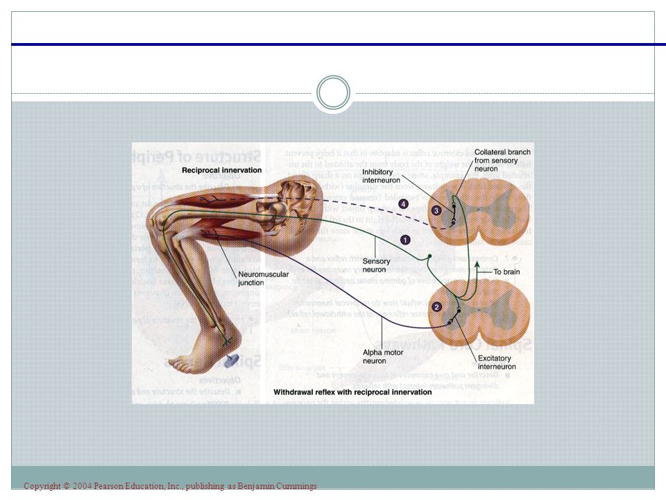
30
Copyright © 2004 Pearson Education, Inc., publishing as Benjamin Cummings The pathway is not pass directly to anterior neuron Pass into interneuronal pools and then to the motor neuron The shortest possible circuit is three of four Flexor reflex-withdrawal reflex

31
Copyright © 2004 Pearson Education, Inc., publishing as Benjamin Cummings

32
About 0.2 to 0.5 sec after a stimulus elicits a flexor reflex in one limb the opposite limb begins to extend Crossed extensor reflex

33
Copyright © 2004 Pearson Education, Inc., publishing as Benjamin Cummings

Similar presentations