Download presentation
Presentation is loading. Please wait.

1
Phase II Pediatric Block The Division of Pediatric Emergency Medicine Pediatric Emergency Medicine Jeffrey Bullard-Berent, MD Katherine Gnauck, MD Brian Moore, MD Grace Park, DO Robert Sapien, MD Sara Skarbek-Borowska,MD Bryan Upham, MD Julia Whitefield, MD PhD

2
PEM Critical Knowledge (in < 1 hour) Introduce the Pediatric Assessment Triangle Distinguishing PAT and ABCDEDistinguishing PAT and ABCDE Understand the Anatomic and Physiologic differences between adults and children in regard to: Respiratory DistressRespiratory Distress ShockShock TraumaTrauma
 Introduce the Pediatric Assessment Triangle Distinguishing PAT and ABCDEDistinguishing PAT and ABCDE Understand the Anatomic and Physiologic differences between adults and children in regard to: Respiratory DistressRespiratory Distress ShockShock TraumaTrauma")
3
Pediatric Assessment Triangle Appearance Breathing Circulation

4
Appearance Tone Interactiveness Consolability Look/Gaze Speech/Cry

5
Work of Breathing Abnormal airway sounds Abnormal positioning Retractions Nasal flaring Head bobbing

6
Circulation to Skin PallorMottlingCyanosis

7
General Approach Pediatric Assessment Triangle (PAT) Hands-on assessment of ABCDEs Pediatric differencesPediatric differences
 Hands-on assessment of ABCDEs Pediatric differencesPediatric differences")
8
Airway Airway opening maneuvers: Head tilt-chin lift, jaw thrust Suction: Often dramatic improvement in infants Age-specific obstructed airway support: <1 year: Back blow/chest thrust<1 year: Back blow/chest thrust >1 year: Abdominal thrust>1 year: Abdominal thrust Advanced airway techniques

9
Breathing: Respiratory Rate Varies with Age Slow or fast respirations are worrisome. Age Respiratory Rate Infant 30 to 60 Toddler 24 to 40 Preschooler 22 to 34 School-aged child 18 to 30 Adolescent 12 to 16

10
Breathing: Auscultation Midaxillary line, above sternal notch Stridor: Upper airway obstructionStridor: Upper airway obstruction Wheezing: Lower airway obstructionWheezing: Lower airway obstruction Grunting: Poor oxygenation; pneumonia, drowning, pulmonary contusionGrunting: Poor oxygenation; pneumonia, drowning, pulmonary contusion Crackles: Fluid, mucus, blood in airwayCrackles: Fluid, mucus, blood in airway Decreased /Decreased / absent breath absent breath sounds: Obstruction sounds: Obstruction

11
Circulation: Heart Rate varies by Age Age Normal Heart Rate Infant 100 to 160 Toddler 90 to 150 Preschooler 80 to 140 School-aged child 70 to 120 Adolescent 60 to 100

12
Circulation Pulse : Central, peripheral pulse quality Skin temp: “ Reverse thermometer” sign Capillary refill: ≤ 2 sec, warm finger, 5 sec B/P : Minimum = 70 + (2 X age in years)
")
13
Disability (and Dextrose) AVPU scale : AlertAlert Verbal: Responds to verbal commandsVerbal: Responds to verbal commands Painful: Responds to painful stimulusPainful: Responds to painful stimulus UnresponsiveUnresponsive (Pediatric) Glasgow Coma Scale Check Dextrose (glucose) if impaired
 AVPU scale : AlertAlert Verbal: Responds to verbal commandsVerbal: Responds to verbal commands Painful: Responds to painful stimulusPainful: Responds to painful stimulus UnresponsiveUnresponsive (Pediatric) Glasgow Coma Scale Check Dextrose (glucose) if impaired")
14
Exposure / Environment Full Exposure Necessary Evaluate physiologic functionEvaluate physiologic function Identify anatomic abnormalitiesIdentify anatomic abnormalities Maintain warm ambient environment Minimize heat loss Monitor temperature Warm IV fluids

15
Reassess General impression (PAT) ABCDE Continually reassess ABCs for response to therapy
 ABCDE Continually reassess ABCs for response to therapy")
16
The Bottom Line Begin with PAT, then ABCDEs. Form a general impression to guide priorities. Treat respiratory distress, failure, and shock as they are recognized. Focused history and detailed PE. Reassessment throughout ED stay.

17
Airway / Breathing

18
Objectives Compare anatomic, physiologic differences b/w adult & pediatric airway Distinguish respiratory distress from failure Describe clinical features of upper and lower airway obstruction and diseases of the lung

19
Respiratory arrest vs cardiac arrest intact survival rates in children

20
Anatomic Anatomic Physiologic Physiologic Why do children have more respiratory difficulties?

21
Large occiput – need shoulder roll Large tongue – obstruction Cephalad larynx – difficult to visualize Soft epiglottis – use Miller blades Smallest diameter below cords Small airways – high resistance Anatomy

22
Physiology: Pediatric vs Adult Higher Basic Metabolic Rate = Shorter time to Desaturation (6-8 mL/kg vs 3-4)(6-8 mL/kg vs 3-4) Smaller airways = Higher Airway Resistance (1/R 4 ) Prolonged respiratory distress -> failure
(6-8 mL/kg vs 3-4) Smaller airways = Higher Airway Resistance (1/R 4 ) Prolonged respiratory distress -> failure")
23
Physiology: Time to Desaturation

24
Signs of Respiratory Distress and Failure Respiratory Distress TachypneaTachypnea StridorStridor RetractionsRetractions Head bobbingHead bobbing Nasal FlaringNasal Flaring Respiratory Failure Altered mental status Poor resp effort Bradypnea Bradycardia Apnea Resp failure = inadequate oxygenation or ventilation

25
Mild Respiratory Distress: Accessory Muscle Usage

26
Severe Respiratory Distress: Accessory Muscle Usage

27
Airway Obstruction: Upper Vs. Lower StridorSturtor Wheeze ↓ breath sounds CoughRetractionsApnea Resp effort w/o air entry Upper Lower

28
Airway Obstruction Causes: Upper Vs. Lower Upper CroupCroup Foreign BodiesForeign Bodies Epiglotitis (rare)Epiglotitis (rare) Lower Bronchiolitis Asthma Foreign Bodies
Epiglotitis (rare) Lower Bronchiolitis Asthma Foreign Bodies.")
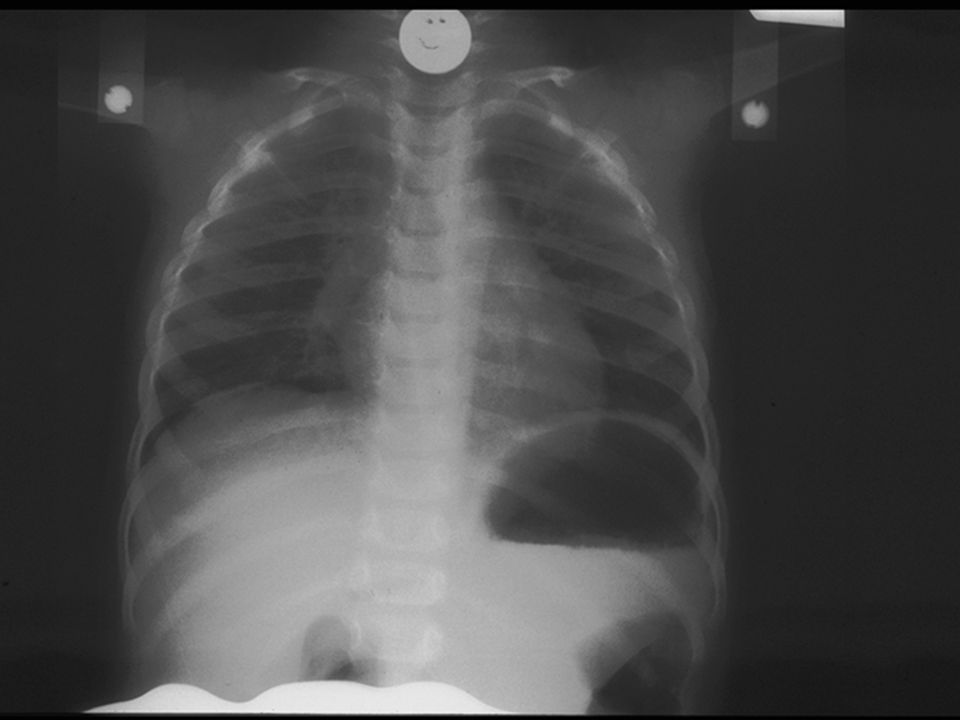
29
Upper Airway Obstruction: Croup

30
Upper Airway Obstruction: Treatment Reposition Airway Suction (infants, in particular) Partial obstruction – position of comfort Complete obstruction Back blows/ chest thrusts < 1 yoBack blows/ chest thrusts < 1 yo Abdominal thrusts > 1yearAbdominal thrusts > 1year N-P airway – sturtor; O-P airway (no gag) Advanced techniques
 Partial obstruction – position of comfort Complete obstruction Back blows/ chest thrusts < 1 yoBack blows/ chest thrusts < 1 yo Abdominal thrusts > 1yearAbdominal thrusts > 1year N-P airway – sturtor; O-P airway (no gag) Advanced techniques")
31
Lower Airway Obstruction: Treatment Oxygen / Supportive (bronchiolitis) Beta 2 agonist (albuterol in asthma) Bronchoscopy (foreign body)
 Beta 2 agonist (albuterol in asthma) Bronchoscopy (foreign body)")
32
Case 1 – Airway/Breathing cc: “Funny breathing” HPI: 14 month male with acute resp dist. No prior symptoms. Mom in other room, noted “funny breathing” while he was playing on the floor. ROS: No fevers, Otherwise well PE:AF, VSWNL except RR 50 Gen:Awake, alert, stridor at rest, mod resp dist Pulm:Retractions x3, transmitted airway sounds CV:RRR, no m/r/g, nl pulses, cap refill

33
Case 1 – Airway/Breathing Would you: a)Use a BVM, prepare to Intubate b)Give abdominal thrusts c)Give back blows/chest thrusts d)Place in position of comfort e)Start CPR f)Discharge home
Use a BVM, prepare to Intubate b)Give abdominal thrusts c)Give back blows/chest thrusts d)Place in position of comfort e)Start CPR f)Discharge home")
34
Case 1 – Airway/Breathing Would you: a)Use a BVM, prepare to Intubate b)Give abdominal thrusts c)Give back blows/chest thrusts d)Place in position of comfort e)Start CPR f)Discharge home
Use a BVM, prepare to Intubate b)Give abdominal thrusts c)Give back blows/chest thrusts d)Place in position of comfort e)Start CPR f)Discharge home")
37
Case 2 – Airway/Breathing cc: “Choked” HPI: 9 month male with difficulty breathing. Mom was feeding him grapes and peanuts. Had a choking episode, was making “funny sounds”, then during your exam stopped breathing and turned blue. ROS: No fevers, Otherwise well PE:AF, resp effort at 60, HR 190, SpO2 65% Gen:Limp, cyanotic. Pulm:Supraclavicular retractions, no air entry CV:Tachycardic, no m/r/g, 2+ pulses, ↓ CR

38
Case 2 – Airway/Breathing Would you: a)Use a BVM, prepare to Intubate b)Give abdominal thrusts c)Give back blows/chest thrusts d)Place in position of comfort e)Perform cricothyroidotomy f)Discharge home
Use a BVM, prepare to Intubate b)Give abdominal thrusts c)Give back blows/chest thrusts d)Place in position of comfort e)Perform cricothyroidotomy f)Discharge home")
39
Case 2 – Airway/Breathing Would you: a)Use a BVM, prepare to Intubate b)Give abdominal thrusts c)Give back blows/chest thrusts d)Place in position of comfort e)Perform cricothyroidotomy f)Discharge home
Use a BVM, prepare to Intubate b)Give abdominal thrusts c)Give back blows/chest thrusts d)Place in position of comfort e)Perform cricothyroidotomy f)Discharge home")
40
Age RR Newborn 35-50 Older infants/ toddlers 30-40 Elementary school age 20-30 Older child/ adolescent 12-20 Respiratory Rate Varies by Age

41
Case 3 – Airway/Breathing cc: “Trouble breathing” HPI: 9 month male with difficulty breathing. Cough, difficulty breathing x 4 days. Not taking fluids well, now “lethargic”. ROS: Fever to 102 F, decreased wet diapers. PE:AF, RR 12, HR 190, SpO2 78% Gen:Limp, shallow resp effort. Pulm:Coarse bs, poor air entry CV:Tachycardic, no m/r/g, central pulses only, ↓ CR 5-6 seconds

42
Case 3 – Airway/Breathing Would you: a)Use a BVM, prepare to Intubate b)Give abdominal thrusts c)Give back blows/chest thrusts d)Place in position of comfort e)Perform cricothyroidotomy f)Discharge home
Use a BVM, prepare to Intubate b)Give abdominal thrusts c)Give back blows/chest thrusts d)Place in position of comfort e)Perform cricothyroidotomy f)Discharge home")
43
Case 3 – Airway/Breathing Would you: a)Use a BVM, prepare to Intubate b)Give abdominal thrusts c)Give back blows/chest thrusts d)Place in position of comfort e)Perform cricothyroidotomy f)Discharge home
Use a BVM, prepare to Intubate b)Give abdominal thrusts c)Give back blows/chest thrusts d)Place in position of comfort e)Perform cricothyroidotomy f)Discharge home")
44
Circulation

45
Heart Rate Also Varies with Age Age Normal Heart Rate Infant 100 to 160 Toddler 90 to 150 Preschooler 80 to 140 School-aged child 70 to 120 Adolescent 60 to 100

46
CO = HR x Stroke Volume Cardiac Output Infants/children ↑ CO by ↑HR >> SV

47
Shock: Definition and Types Inadequate tissue perfusion (delivery of oxygen and nutrients) to meet the metabolic demands of the body. HypovolemicHypovolemic CardiogenicCardiogenic DistributiveDistributive SepticSeptic

48
Shock: Definitions Compensated: Vital organs perfused by compensatory mechanismsVital organs perfused by compensatory mechanisms B/P is normalB/P is normalDecompensated: Compensatory mechanisms overwhelmed, inadequateCompensatory mechanisms overwhelmed, inadequate Hypotension, high mortality riskHypotension, high mortality risk Aggressive treatment of early shock: Halts progression to decompensated shockHalts progression to decompensated shock

49
Shock Clinical Features: Your First Clues Abnormal mental status Apnea Tachycardia (hypotension NOT necessary) Grunting, respiratory distress Pale, cool skin; delayed capillary refill Warm shock CR will appear normal to briskWarm shock CR will appear normal to brisk
 Grunting, respiratory distress Pale, cool skin; delayed capillary refill Warm shock CR will appear normal to briskWarm shock CR will appear normal to brisk")
50
Clinical Features of Specific Shock Types Neurologic deficits (spinal cord injury) Urticaria, allergen trigger, wheezing (anaphylactic) Petechiae, erythroderma (septic)
 Urticaria, allergen trigger, wheezing (anaphylactic) Petechiae, erythroderma (septic)")
51
Hypovolemic Shock Fluid loss: Diarrhea, vomiting, anorexia, diuresisDiarrhea, vomiting, anorexia, diuresis HemorrhageHemorrhageResuscitation: Fluid replacementFluid replacement NS or LR 20 mL/kg bolus, reassess, repeat as neededNS or LR 20 mL/kg bolus, reassess, repeat as needed Blood transfusion for excessive hemorrhageBlood transfusion for excessive hemorrhage

52
Septic Shock Has elements of distributive shock and cardiogenic shock: Inappropriate vasodilation with a maldistribution of blood flowInappropriate vasodilation with a maldistribution of blood flow Myocardial depressionMyocardial depressionResuscitation: Fluid boluses: 20cc/kg x3 in the first 15 minutesFluid boluses: 20cc/kg x3 in the first 15 minutes Dopamine, Epinephrine or NorepinephrineDopamine, Epinephrine or Norepinephrine AntibioticsAntibiotics

53
Trauma

54
Trauma: Epidemiology Trauma is leading cause of death and disability in children worldwide. Most common injuries: Infants: Physical abuseInfants: Physical abuse Preschoolers: FallsPreschoolers: Falls School aged: Motor vehicle collision – pedestrian or bicyclistSchool aged: Motor vehicle collision – pedestrian or bicyclist Differences in anatomy, physiology, mechanism drive differences in injury pattern, response.

55
How Trauma Differs in Pediatrics (1 of 3) Anatomy/Physiology Proportionately larger head Large occiput and tongue Injury Response Higher frequency of head trauma, higher c-spine trauma More airway obstruction
 Anatomy/Physiology Proportionately larger head Large occiput and tongue Injury Response Higher frequency of head trauma, higher c-spine trauma More airway obstruction")
56
How Trauma Differs in Pediatrics (2 of 3) Anatomy/Physiology Thinner chest wall, more flexible ribs Horizontal ribs, weaker intercostals, more mobile mediastinum Abdominal organs more anterior and less subcutaneous fat Injury Response Higher frequency of pulmonary injury Tension pneumothorax poorly tolerated Higher risk of intra- abdominal injury and bleeding
 Anatomy/Physiology Thinner chest wall, more flexible ribs Horizontal ribs, weaker intercostals, more mobile mediastinum Abdominal organs more anterior and less subcutaneous fat Injury Response Higher frequency of pulmonary injury Tension pneumothorax poorly tolerated Higher risk of intra- abdominal injury and bleeding")
57
How Trauma Differs in Pediatrics (3 of 3) Anatomy/Physiology Softer bones, thicker periosteum Active, unfused bony growth plates Compensatory vasoconstriction Larger body surface area/mass ratio Injury Response Higher frequency of incomplete fractures Disrupted growth after growth plate fractures Normal blood pressure with early shock Greater heat loss from exposed body surfaces (head)
 Anatomy/Physiology Softer bones, thicker periosteum Active, unfused bony growth plates Compensatory vasoconstriction Larger body surface area/mass ratio Injury Response Higher frequency of incomplete fractures Disrupted growth after growth plate fractures Normal blood pressure with early shock Greater heat loss from exposed body surfaces (head)")
58
Trauma Assessment Primary survey Pediatric Assessment Triangle (PAT)Pediatric Assessment Triangle (PAT) ABCDEs (F- family)ABCDEs (F- family) Treat ABCDE in the order you find themTreat ABCDE in the order you find them Secondary survey Head to toe examHead to toe examReassess
Pediatric Assessment Triangle (PAT) ABCDEs (F- family)ABCDEs (F- family) Treat ABCDE in the order you find themTreat ABCDE in the order you find them Secondary survey Head to toe examHead to toe examReassess")
59
Summary PAT = General impression (Appearance, B, C) ABCDE = Airway, Breathing, Circulation, Disability / Dextrose, Extremities/Environment, F=Family Reassess response to treatment Respiratory Distress – Anat / Phys / Environmental Shock – Compensated /Decompensated, Tachycardia Trauma – Anatomic differences, logical approach
 ABCDE = Airway, Breathing, Circulation, Disability / Dextrose, Extremities/Environment, F=Family Reassess response to treatment Respiratory Distress – Anat / Phys / Environmental Shock – Compensated /Decompensated, Tachycardia Trauma – Anatomic differences, logical approach")
60
Thank you!

Similar presentations


: Jeff Holmes MD, Maine Medical Center License:>")


