Download presentation
Presentation is loading. Please wait.

2
Primary inflammatory myopathies are clinically, pathologically and therapeutically distinct entities Inflammatory changes are probably due to an immune mediated process rather than directly pathogenic These are acquired as opposed to the inherited dystrophies and are classified as follows : Polymyositis childhood Adult form INFLAMMATORY MYOPATHY

3
Dermatoyositis Inclusion body myositis Inflammatory myopathy associated with malignant disease Inflammatory myopathy associated with collagen vascular disorder – e.g lupus erythematosus, systemic sclerosis, rheumatoid arthritis Infective – viral e.g. coxsackie, echo. Parasitic, e.g. cysticerocosis, trichinosis, taenia solium, toxoplasma, toxocara Sarcoid myopathy – some with this multi-system disease have granulomas in skeletal muscle

4
Polymyositis / Dermatomyositis : There are two principal forms of inflammatory myopathy-polymyositis and dermatomyositis- which are separated clinically by the dermatological findings in the latter All age groups are affected. Annual incidence is 8 per 100 000. These disorders are sporadic though familial cases are described

5
All autoimmune basis for these disorders is supported by: -Response to immunosuppressive therapy. -Association with other known immunological disorders, e.g. collagen vascular disorders. -Elevated IgG in blood and presence of circulating auto antibodies, e.g. antinuclear antibody in some cases -An increased incidence of certain histocompatibility antigens (HLA antigens) – B8,DR3
 – B8,DR3.")
6
-The reproduction of a similar disorder in laboratory animals by injection of muscle extract with Freund’s adjuvant Humoral and cell mediated immune mechanism seem responsible for these disorders but the trigger factor's remain unknown

9
Inclusion Body Myositis (IBM) : Recognized now as the commonest inflammatory muscle disorder in the middle aged and elderly (women are less commonly affected and more likely to be younger) Unlike the other inflammatory myopathies symmetrical weakness is painless and distal including foot extensors and finger flexors Most patients have a protracted course unaffected by immunosuppressive therapies Occasionally it is associated with connective tissue disorders such as Sjogren’s syndrome
 : Recognized now as the commonest inflammatory muscle disorder in the middle aged and elderly (women are less commonly affected and more likely to be younger) Unlike the other inflammatory myopathies symmetrical weakness is painless and distal including foot extensors and finger flexors Most patients have a protracted course unaffected by immunosuppressive therapies Occasionally it is associated with connective tissue disorders such as Sjogren’s syndrome")
12
Outcome of inflammatory myopathies The natural history of these conditions is uncertain; mortality is low though perhaps only a minority recover completely Inclusion body myositis is slowly and steadily progressive Polymyositis and dermatomyositis respond in varying degrees to treatment and eventually become inactive Safe monitoring of treatments and protection against side effects (e.g. steroid induced bone disease) is critical
 is critical.")
13
Polymyositis and Dermatomyositis Associated with Malignant Diseases Approximately 10% of adults with inflammatory myopathy have underlying neoplasia usually carcinoma In dermatomyositis, of those over 40 years of age as many as 60% harbour neoplasia Neoplasia may present before or after the development of inflammatory myopathy

14
Polymyositis and dermatomyositis associated with collagen vascular diseases Approximately 15% of adults with inflammatory myopathy have symptoms and signs of an associated collagen vascular disorder In 5-10% of persons with these disorder (systemic lupus erythematosus etc.), inflammatory myopathy develops at some stage in their illness In the ‘overlap’ syndromes (mixed collagen vascular diseases) muscle involvement is more common
, inflammatory myopathy develops at some stage in their illness In the ‘overlap’ syndromes (mixed collagen vascular diseases) muscle involvement is more common")
15
Unlike inflammatory myopathy the weakness in these conditions is more chronic and is unassociated pathologically with inflammation Serum CK is usually normal and EMG and biopsy (if performed) show non-specific myopathic changes Correction of the underlying endocrine disturbance results in recovery Usually the other features of endocrine dysfunction are more problematical and myopathy is of secondary importance ENDOCRINE MYOPATHIES
 show non-specific myopathic changes Correction of the underlying endocrine disturbance results in recovery Usually the other features of endocrine dysfunction are more problematical and myopathy is of secondary importance ENDOCRINE MYOPATHIES")
16
Pituitary : Acromegaly Proximal weakness with fatigue Entrapment neuropathies, e.g. carpal tunnel syndrome may complicate the clinical picture of myopathy Other features of growth hormone excess are evident

17
Parathyroid : Hyperparathyroidism and osteomalacia Weakness of a proximal distribution with muscle tenderness occurs in 50% of patients wityh osteomalacia but is less common in primary hyperparathyroidism The legs are mainly affected and a waddling gait results Pathogenesis of hyperparathyroid myopathy is uncertain; hypercalcaemia, Vitamin D deficiency of chronic phosphate deficiency is implicated, tetany results and the CK may be elevated but weakness is uncommon

18
Adrenal : Hyperadrenalism and hypoadrenalism These may both be associated with proximal myopathy Muscle weakness, fatigue and cramping are frequent in Addison’s disease with attacks of severe episodic hypokalaemic weakness (periodic paralysis) requiring glycocorticoid and mineral corticoid replacement Hypokalaemic periodic paralysis is also frequent in hyperaldosteronism
 requiring glycocorticoid and mineral corticoid replacement Hypokalaemic periodic paralysis is also frequent in hyperaldosteronism")
19
Cushing’s syndrome and exposure to excessive exogenous glucocorticoids commonly results in insidious proximal weakness Reduction of steroid dosage results in improvement Thyroid : Hyperthyroidism Weakness occurs in 20% of thyrotoxic patients Shoulder girdle weakness is more marked than pelvic

20
Reflexes are brisk, fasciculation and atrophy may be present Distinction must be made from motor neuron disease There is always clinical evidence of thyrotoxicosis in these patients Diagnosis is confirmed by thyroid function studies Hypothyroidism : Hypothyroidism impairs muscle glycolysis and mitochondrial oxidative capacity Proximal weakness involves pelvic girdle more than shoulder

21
Painful cramps and muscle stiffness are common Muscle enlargement in limbs and tongue often occur (Hoffman’s syndrome) There is always clinical evidence of hypothyroidism in these patients Diagnosis is confirmed by thyroid function tests and response to thyroid hormone therapy is excellent In chronic proximal weakness, careful clinical history taking, examination and appropriate investigation will separate the various endocrine causes
 There is always clinical evidence of hypothyroidism in these patients Diagnosis is confirmed by thyroid function tests and response to thyroid hormone therapy is excellent In chronic proximal weakness, careful clinical history taking, examination and appropriate investigation will separate the various endocrine causes")
22
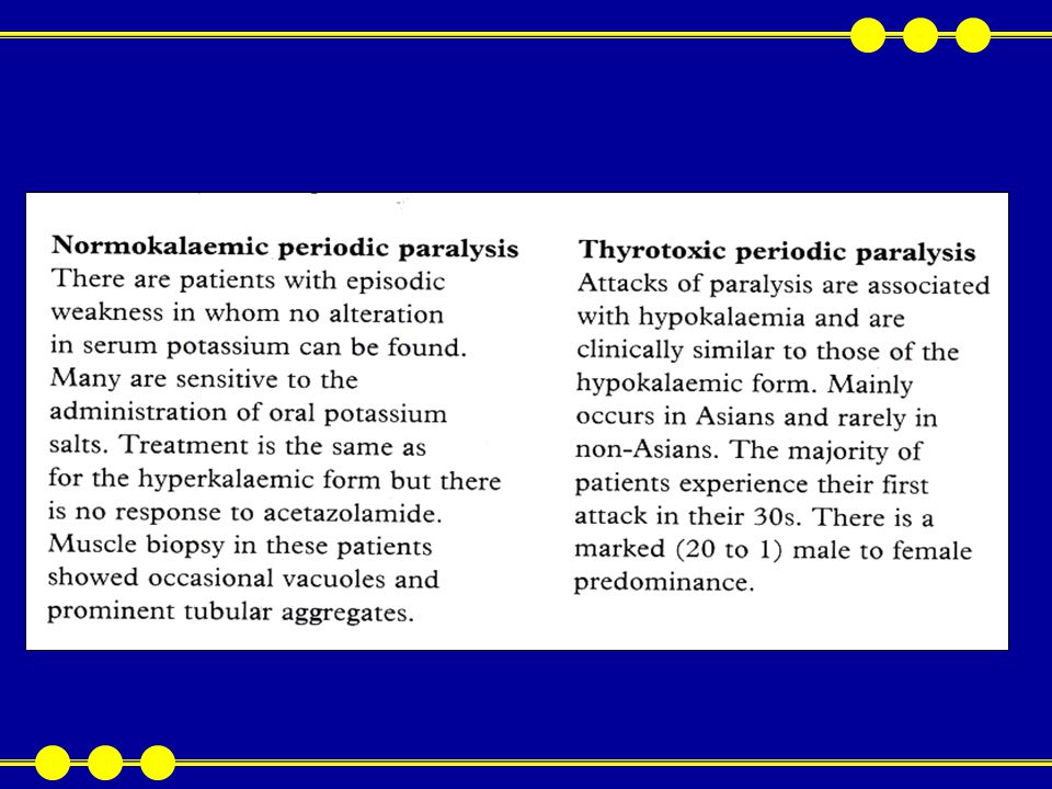
Periodic paralyses are associated with vchanges in serum potassium concentrations The primary periodic paralyses are classified into two categories: hypokalemic and hyperkalemic (or potassium-sensitive) Hypokelemic periodic paralysis shows the clearest relationship between epsodic weakness and alterations in potassiu Metabolic Myopathies : The Periodic Paralyses
 Hypokelemic periodic paralysis shows the clearest relationship between epsodic weakness and alterations in potassiu Metabolic Myopathies : The Periodic Paralyses")
23
Hyperkalemic periodic paralysis is more accurately a ‘potassium sensitive’ periodic paralysis as weakness can be provoked by potassium administration, whilst serum potassium may rise only marginally during spontaneous attacks Paramyotonia can be associated with either hypo or hyperkalaemic periodic paralysis Importantly episodes of weakness, with alterations in serum potassium, are most commonly secondary to drugs (e.g. diuretics and corticosteroids) or disorders such as alcoholism, renal and endocrine disease
 or disorders such as alcoholism, renal and endocrine disease.")
26
Metabolic and Toxic Myopathies

27
Carnitine palmitoyltranferase deficiency – Carnitine palmitoyltransferase (CPT) enzymes transfer fatty acids across the muscle mitochondrial membrane. CPT 1 attaches and CPT 2 detaches these fatty acids Infrequent episodes of myalgia and myoglobinuria following fasting or strenuous exercise. Onset is in adolescence, occasionally in adulthood Though an autosomal recessive disorder, males are more commonly symptomatice Neurological examination is normal. Serum CK, EMG and muscle biopsy (including histochemistry) are normal between attacks Patients are advised to take a low fat/high protein and carbohydrate diet and to avoid prolonged exercise of fasting
 are normal between attacks Patients are advised to take a low fat/high protein and carbohydrate diet and to avoid prolonged exercise of fasting.")
28
Acid maltase deficiency (ADM) - A lysosomal glycogen storage disease with infantile, childhood, and adult types The casual gene localizes to chromosome 17 with different mutations accounting for ages of onset Treatment Infantile AMD (Pompe’s disease)-progressive muscle weakness, cardiomegaly with congestive heart failure Death occurs before 1 year Glycogen accumulates in cardiac, skeletal muscle and in the CNS
 - A lysosomal glycogen storage disease with infantile, childhood, and adult types The casual gene localizes to chromosome 17 with different mutations accounting for ages of onset Treatment Infantile AMD (Pompe’s disease)-progressive muscle weakness, cardiomegaly with congestive heart failure Death occurs before 1 year Glycogen accumulates in cardiac, skeletal muscle and in the CNS")
29
Childhood AMD - Slower clinical course, with respiratory muscle weakness developing between 5 and 20 years. Histologically, muscle contains glycogen-filled vacuoles Adult AMD - proximal weakness in 3rd or 4th decade mimicking limb-gridle muscular dystrophy or polymyositis. Respiratory muscles are severely affected with risk of death from respiratory failure. Muscle biopsy again shows glycogen-filled vacuoles. Liver, heart and central nervous system are spared

30
Carnitine deficiency - Carnitine transports long-chain fatty acids into the mitochondria Deficiency results in systemic or myopathic features Systemic carnitine deficiency – Childhood onset weakness with hypoglycaemic encephalopathy, precipitated by fasting and resembling Reye’s syndrome. Serum and muscle carnitine levels are low Biopsy shows an excessive number of lipid droplets in type 1 fibres The liver, kidney, and heart contain excessive lipid

31
Cardiomyopathy is fatal Myopathic carnitine deficiency - Muscle weakness, exertional myalgias and myoglobinuria. Onset of symptoms is usually in childhood but can be delayed until adulthood. Some cases are complicated by cardiomyopathy. Muscle biopsy shows excessive lipid droplets, especially in type 1 fibres. Muscle and serum carnitine levels are low

32
Toxic myopathies Necrotising myopathy is the pathological consequence of toxic nuscle insult characterized bymuscle weakness, pain and tenderness Investigations – levated serum CK, myoglobinuria, myopathic motor units and fibrillation on EMG Muscle biopsy- necrosis and regeneration Numerous drugs have been incrimated. Examples are: lovastatin, when combined with other medications such as cyclosporin

33
Clofibrate in renal failure or hypoalbuminaemia Epsilon-aminocaproic acid, procainamide, zidovudine (AZT) and phencyclidine Focal muscle necrois can be caused by intramuscular injections
 and phencyclidine Focal muscle necrois can be caused by intramuscular injections")
34
The DNA of the mitochondria (mtDNA) is circular, and while mitochondria themselves reproduce by binary fission, mt DNA replication is controlled by the eukaryotic genome. Mitochondrial disorders are transmitted through the maternal line and not by affected males (nt DNA transmits through the ovum not sperm). The relative proprotion of normal to abnormal mt DNA determines the degree of expression for the mutation. As well as muscle involvement, characterized by ragged red muscle fibres on biopsy, many other clinical features are associated with mtDNa mutation syndromes and include: Mitochondrial Disorders
. The relative proprotion of normal to abnormal mt DNA determines the degree of expression for the mutation. As well as muscle involvement, characterized by ragged red muscle fibres on biopsy, many other clinical features are associated with mtDNa mutation syndromes and include: Mitochondrial Disorders.")
35
-Seizures, respiratory insufficiency, weakness and vomiting and failure to thrive in the neonate -Developmental delay, ataxia, optic atrophy, progressive external ophtalmoplegia, sensorineual deafness, stroke-like episodes, dementia, exercise intolerance, and short stature -Renal failure, diabetes mellitus, cataract and cardiomyopathy Certain specific syndromes are recognised though overlap and diversity of phenotype is common

Similar presentations


 Unlike: neuronopathies: secondary.>")


BB>")


 Ulcerative colitis is an inflammatory bowel disease (IBD) that causes chronic inflammation of the digestive tract It is.>")
