Download presentation
Presentation is loading. Please wait.

1
Management of Supraventricular Tachycardias
May 31, 2008 Sandeep K. Jain, M.D. Cardiac Electrophysiology

2
History Pattern of symptoms / palpitations Triggers Syncope
Caffeine, tobacco, alcohol Nasal decongestants Emotional events Syncope

3
Physical Exam AV dissociation Split S2
More than one arrhythmia can yield similar exam findings so not as useful for making a diagnosis

4
SVT mechanisms Sinus tachycardia Atrial flutter Atrial fibrillation
Junctional tachycardia Atrial tachycardia AV node reentry tachycardia (AVNRT) Accessory pathway mediated tachycardia (AVRT)
 Accessory pathway mediated tachycardia (AVRT)")
5
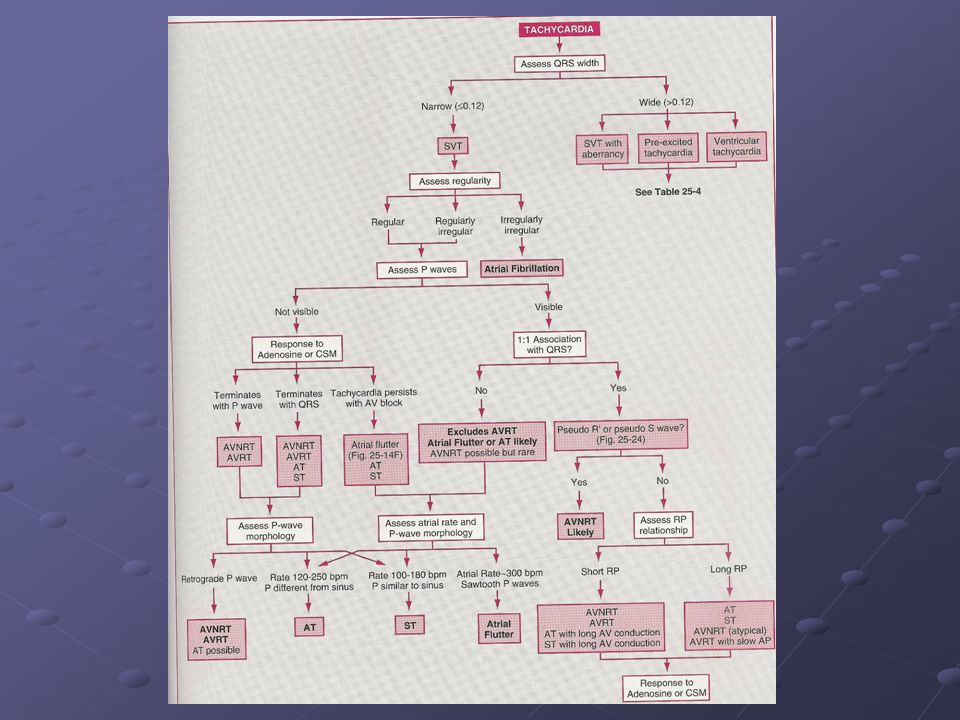
ECG Are P-waves present?
What are atrial and ventricular rates and are they the same ? Regular or Irregular ? Is the PR or RP constant ?

7
Carotid Sinus Massage Increases vagal tone
Sinus slowing and prolongs AV nodal refractoriness Sinus tach – gradually slows then returns to rate prior to massage AVNRT or AVRT can terminate AT, AF and AFL ventricular response slows

8
Sinus Tachycardia Gradual change in heart rate Normal P-wave contour
Constant PR interval before each QRS unless there is AV block (long RP) Carotid massage or Valsalva will gradually slow rate and then resume to original rate
 Carotid massage or Valsalva will gradually slow rate and then resume to original rate.")
9
Almost always secondary
Management is to treat underlying cause Rarely, inappropriate sinus tachycardia in which case beta-blockers, Ca-blockers can be utilized Sinus node reentry tachycardia is abrupt in onset, anxiety related and can be treated with RF ablation

10
Atrial Flutter Macro-reentrant rhythm in the atria
Usually associated with underlying heart disease Typical flutter is in the right atrium and can travel in counterclockwise and clockwise directions Incisional scars from prior cardiac surgery / congenital abnormalities Atrial rate is typically beats/min Ventricular rate of 150 – look out for 2:1 flutter

11
Counterclockwise Clockwise

13
Diagnostic maneuvers - Adenosine

14
Atrial Flutter - Management
Rate control / anticoagulation Beta-blocker, Ca-blocker, Digoxin Stroke risk likely not much different than atrial fibrillation Cardioversion DCCV with low energies possible (50-100J) Ibutilide successful in 60-90% Prolongs QT and need to be monitored 4-6 hours post administration Overdrive atrial pacing if available
 Ibutilide successful in 60-90% Prolongs QT and need to be monitored 4-6 hours. post administration. Overdrive atrial pacing if available.")
15
Atrial Flutter - Management
Antiarrhythmics Class Ia and Ic agents – convert and maintain sinus rhythm Class III – amiodarone, sotalol, dofetilide Need to have AV nodal agent on board to prevent 1:1 flutter Flutter rate slows and allows 1:1 conduction Class IA agents also have a vagolytic effect

16
Flecainide administration

17
Atrial Flutter - Management
Radiofrequency Ablation High success rates for typical flutter Atypical circuits can be approached percutaneously with advanced mapping systems Significant proportion of patients eventually develop atrial fibrillation indicating underlying substrate is the issue

18
Atrial Fibrillation Multiple wavelets propagating in different directions No effective atrial contraction Most common arrhythmia 1% of those older than 60 5% of those older than 69 Mutliple causes – consider mechanical such as from an RA catheter

19
Atrial Fibrillation Rate vs Rhythm Control Anticoagulation
No benefit of either if asymptomatic Anticoagulation Congestive Heart Failure Hypertension Age > 65 Diabetes Stroke Maintenance of sinus rhythm does not necessarily eliminate stroke risk

20
Atrial Fibrillation Rate Control Conversion
Ca-blocker, Beta-blocker, digoxin Conversion DCCV Class I : Procainamide, Flecainide, Propafenone Class III: Amiodarone, Sotalol (both poor as converting agents), ibutilide Maintenance of sinus rhythm Same as above + dofetilide 50-70% efficacy at 1 year RF ablation is an emerging tool
, ibutilide. Maintenance of sinus rhythm. Same as above + dofetilide % efficacy at 1 year. RF ablation is an emerging tool.")
21
Atrial Tachycardia Rapid, focal discharge in the atrium
Rate generally bpm P-wave contour different from sinus P-waves generally found in second half of tachycardia cycle (long RP / short PR) Most commonly in those with structural heart disease but also seen in those without any cardiac abnormality
 Most commonly in those with structural heart disease but also seen in those without any cardiac abnormality.")
22
Atrial Tachycardia Adenosine Response
Terminates tachycardia with an ‘R’ wave OR Tachycardia persists with AV Block

24
Atrial Tachycardia - Management
Beta- and Ca- blockers Class I antiarrhythmics in normal hearts Class III antiarrhythmics in abnormal hearts Radiofrequency ablation for those who are refractory / intolerant to medications

25
AV Node Reentry Tachycardia
Regular, sudden onset and termination at rates between bpm QRS usually normal unless aberrancy is present Reentry within the AV node P-wave seen just prior to or just after the QRS complex (Short RP tachycardia) Usually occurs without any structural heart disease
 Usually occurs without any structural heart disease.")
26
AV Node Reentry Tachycardia
Antegrade limb is the slow pathway Retrograde limb is the fast pathway

28
Baseline

29
AV Node Reentry Tachycardia

30
AVNRT 2:1

31
AV Node Reentry - Management
IV adenosine, vagal maneuvers, carotid massage Ca, Beta-blockers, rarely antiarrhythmics Overdrive atrial or ventricular pacing RF ablation is usually treatment of choice for chronic management – cost-effective and high success rate (~1% incidence of AV block requiring permanent pacemaker)
")
32
Accessory Pathways Muscular connections outside of the specialized conduction system connecting the atrium and ventricle while bypassing the AV node Can be manifest (WPW) or concealed (retrograde only conduction) Concealed pathways not apparent on ECG Manifest pathways have a delta wave representing pre-excitation of ventricular tissue prior to activation via the His-purkinje system
 or concealed (retrograde only conduction) Concealed pathways not apparent on ECG. Manifest pathways have a delta wave representing pre-excitation of ventricular tissue prior to activation via the His-purkinje system.")
33
Concealed Accessory Pathways
30% of people with SVT referred for EP evaluation Can present with syncope Normal baseline 12-lead ECG Orthodromic atrioventricular reentry tachycardia is the mechanism of SVT (down the AV node and up the accessory pathway) Will often see a retrograde P-wave during SVT – short RP unless slowly conducting pathway
 Will often see a retrograde P-wave during SVT – short RP unless slowly conducting pathway.")
34
Orthodromic AV reentry tachycardia
Most common arrhythmia with presence of an accessory pathway Macro-reentrant circuit in which the impulse travels down the AV node and up the accessory pathway Narrow QRS interval

35
Accessory Pathways

36
Accessory Pathways

37
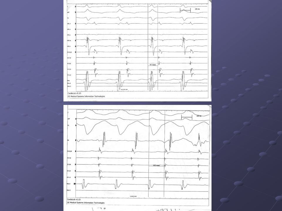
Response to BBB Aberration
Does VA interval increase with development of BBB? An increase in VA interval indicates the presence and participation of a bypass tract on the side of the blocked bundle

39
Concealed Accessory Pathways
Same management as for AV node reentry RF ablation should be considered early in symptomatic patients Atrial fibrillation in conjunction with a concealed pathway does not pose a risk of sudden death and IV Ca-blocker not contraindicated as the pathway does not conduct antegrade

40
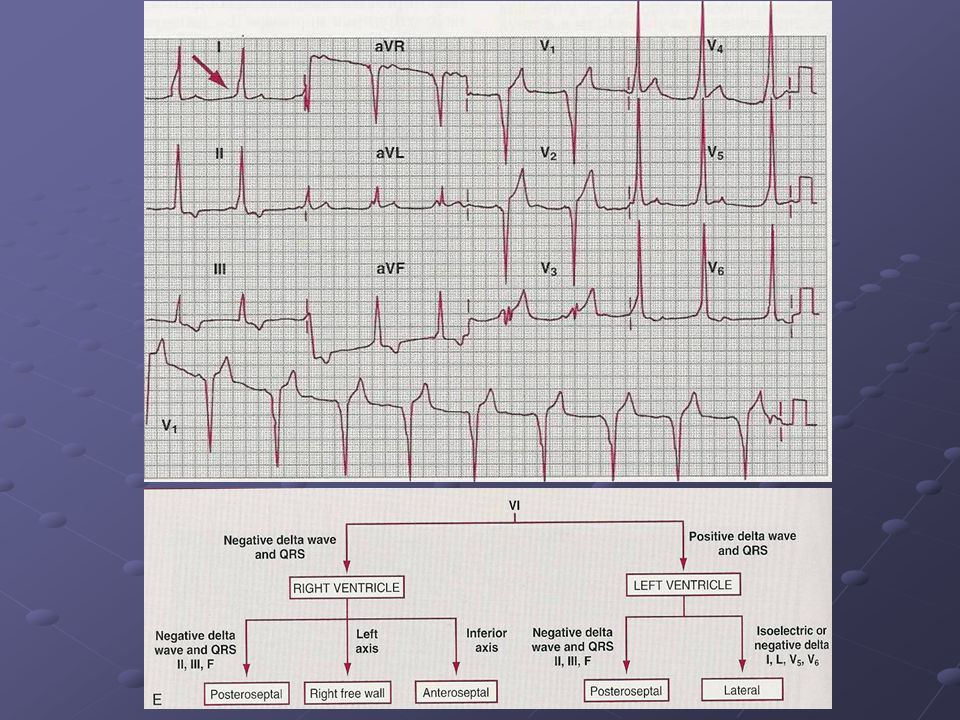
Pre-excitation syndrome - WPW
WPW syndrome is when tachycardia is associated with the finding of a delta wave on the ECG Incidence is 1.5 per thousand Ebstein’s anomaly associated with multiple bypass tracts Baseline ECG findings PR interval < 120ms QRS > 120ms with a slurred, slowly rising onset (delta wave) and usually normal terminal portion of QRS Secondary ST-T changes in opposite direction of delta wave
 and usually normal terminal portion of QRS. Secondary ST-T changes in opposite direction of delta wave.")
41
Accessory pathways Left free wall most common, then posteroseptal, right free wall and anteroseptal

42
Pre-excitation syndromes

44
Pre-excitation syndrome - WPW
Most common tachycardia is Orthodromic AVRT as seen in concealed bypass tracts Major difference is the capacity for anterograde conduction over the pathway during atrial fibrillation (15-30%) or atrial flutter (5%) and thus the rare incidence of sudden cardiac death Some children lose conduction in the pathway but usually persists if present at age 5
 or atrial flutter (5%) and thus the rare incidence of sudden cardiac death. Some children lose conduction in the pathway but usually persists if present at age 5.")
46
Pre-excitation syndromes

47
Pre-excitation syndromes

48
WPW - Treatment ECG abnormality only without arrhythmias may not require EP evaluation Symptomatic patients should receive treatment Ablation – Electrical or Surgical Good success rates but with procedural risks Pharmacologic Drugs prolong conduction and/or refractoriness in the AV node, accessory pathway, or both Some agents can suppress premature atrial contractions which can induce arrhythmias

49
WPW - Treatment Prolong conduction time and refractoriness in AV node:
Adenosine, verapamil, beta-blockers, digoxin Prolong refractory period in accessory pathway: Class IA and IC drugs Affect both AP and AV node: Class IC drugs, amiodarone, and sotalol

50
WPW – Acute Episode Normal QRS, regular R-R intervals, retrograde p-waves Approach same as AVNRT – vagal maneuvers, adenosine, IV Ca-blocker Note, atrial fibrillation can occur after drug administration, so external defibrillator back-up should be ready Atrial Fibrillation or Flutter with irregular R-R intervals and abnormal QRS complexes Agents which affect AV node and pathway (procainamide with beta blocker) Any signs of hemodynamic impairment – electrical cardioversion is initial treatment of choice
 Any signs of hemodynamic impairment – electrical cardioversion is initial treatment of choice.")
52
WPW - Treatment DRUGS NOT TO USE in AF/AFL with WPW:
Digoxin has varying effects on the accessory pathway and can shorten refractoriness and speed ventricular response to atrial fibrillation IV Lidocaine can also increase ventricular response rate IV Verapamil can precipitate ventricular fibrillation in this circumstance (may not happen with oral) Catecholamines
 Catecholamines.")
53
Supraventricular Tachycardias
Short RP / Long PR AV node reentry AV reentry Long RP / Short PR Atrial tachycardia Sinus node reentry Atypical AV node reentry AV reentry with a slowly conducting pathway

Similar presentations