Download presentation
Presentation is loading. Please wait.

1
Chap 32 Animal Evolution

2
( 1) Animals are multicellular, heterotrophic eukaryotes. –They must take in preformed organic molecules through ingestion, eating other organisms or organic material that is decomposing. 1. Structure, nutrition and life history define animals (2) Animal cells lack cell walls that provide structural supports for plants and fungi. The multicellular bodies of animals are held together with the extracellular proteins, especially collagen. In addition, other structural proteins create several types of intercellular junctions, including tight junctions, desmosomes, and gap junctions, that hold tissues together. (3) Animals have two unique types of tissues: nervous tissue for impulse conduction and muscle tissue for movement.
 Animal cells lack cell walls that provide structural supports for plants and fungi. The multicellular bodies of animals are held together with the extracellular proteins, especially collagen. In addition, other structural proteins create several types of intercellular junctions, including tight junctions, desmosomes, and gap junctions, that hold tissues together. (3) Animals have two unique types of tissues: nervous tissue for impulse conduction and muscle tissue for movement..")
3
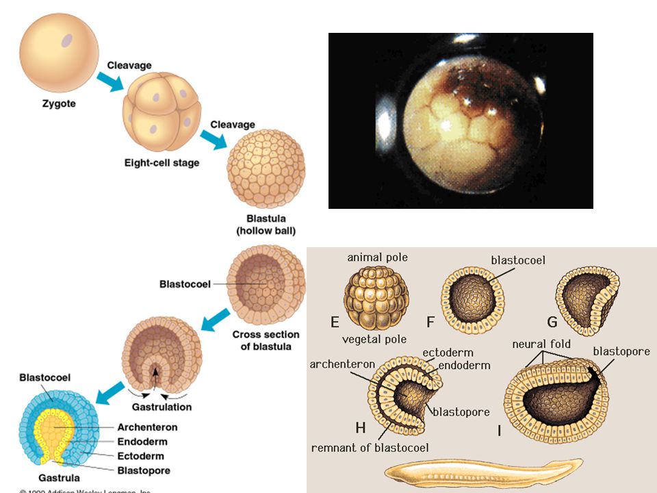
(4) Most animals reproduce sexually, with the diploid stage usually dominating the life cycle. –In most species, a small flagellated sperm fertilizes a larger, nonmotile eggs. –The zygote undergoes cleavage, a succession mitotic cell divisions, leading to the formation of a multicellular, hollow ball of cells called the blastula. Copyright © 2002 Pearson Education, Inc., publishing as Benjamin Cummings Fig. 32.1

5
This protist was probably related to choanoflagellates, a group that arose about a billion years ago. –Modern choanoflagellates are tiny, stalked organisms inhabiting shallow ponds, lakes, and marine environments. Fig. 32.2

6
One hypothesis for origin of animals from a flagellated protist suggests that a colony of identical cells evolved into a hollow sphere. The cells of this sphere then specialized, creating two or more layers of cells. Copyright © 2002 Pearson Education, Inc., publishing as Benjamin Cummings Fig. 32.3

7
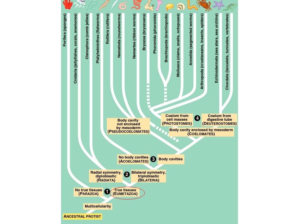
four deep branches. (1) The first branch point splits the Parazoa which lack true tissues from the Eumetazoa which have true tissues. –The parazoans, phylum Porifera or sponges, represent an early branch of the animal kingdom. –Sponges have unique development and a structural simplicity. Copyright © 2002 Pearson Education, Inc., publishing as Benjamin Cummings
 The first branch point splits the Parazoa which lack true tissues from the Eumetazoa which have true tissues. –The parazoans, phylum Porifera or sponges, represent an early branch of the animal kingdom. –Sponges have unique development and a structural simplicity. Copyright © 2002 Pearson Education, Inc., publishing as Benjamin Cummings.")
9
(2) The eumetazoans are divided into two major branches, partly based on body symmetry. –Members of the phylum Cnidaria (hydras, jellies, sea anemones and their relatives) and phylum Ctenophora (comb jellies) have radial symmetry and are known collectively as the Radiata. –The other major branch, the Bilateria, has bilateral symmetry with a dorsal and ventral side, an anterior and posterior end, and a left and right side. Copyright © 2002 Pearson Education, Inc., publishing as Benjamin Cummings
 and phylum Ctenophora (comb jellies) have radial symmetry and are known collectively as the Radiata. –The other major branch, the Bilateria, has bilateral symmetry with a dorsal and ventral side, an anterior and posterior end, and a left and right side. Copyright © 2002 Pearson Education, Inc., publishing as Benjamin Cummings.")
10
Fig. 32.5

11
Linked with bilateral symmetry is cephalization, an evolutionary trend toward the concentration of sensory equipment on the anterior end. –Cephalization also includes the development of a central nervous system concentrated in the head and extending toward the tail as a longitudinal nerve cord. The symmetry of an animal generally fits its lifestyle. –Many radial animals are sessile or planktonic and need to meet the environment equally well from all sides. –Animals that move actively are bilateral, such that the head end is usually first to encounter food, danger, and other stimuli.

13
The basic organization of germ layers, concentric layers of embryonic tissue that form various tissues and organs, differs between radiata and bilateria. The radiata are said to be diploblastic because they have two germ layers. –The ectoderm, covering the surface of the embryo, give rise to the outer covering and, in some phyla, the central nervous system. –The endoderm, the innermost layer, lines the developing digestive tube, or archenteron, and gives rise to the lining of the digestive tract and the organs derived from it, such as the liver and lungs of vertebrates. Copyright © 2002 Pearson Education, Inc., publishing as Benjamin Cummings

14
The bilateria are triploblastic. –The third germ layer, the mesoderm lies between the endoderm and ectoderm. –The mesoderm develops into the muscles and most other organs between the digestive tube and the outer covering of the animal. Copyright © 2002 Pearson Education, Inc., publishing as Benjamin Cummings

15
(3) The Bilateria can be divided by the presence or absence of a body cavity (a fluid-filled space separating the digestive tract from the outer body wall) and by the structure the body cavity. Acoelomates (the phylum Platyhelminthes) have a solid body and lack a body cavity. Copyright © 2002 Pearson Education, Inc., publishing as Benjamin Cummings Fig. 32.6a
 have a solid body and lack a body cavity. Copyright © 2002 Pearson Education, Inc., publishing as Benjamin Cummings Fig. 32.6a.")
16
In some organisms, there is a body cavity, but it is not completely lined by mesoderm. –This is termed a pseudocoelom. –These pseudocoelomates include the rotifers (phylum Rotifera) and the roundworms (phylum Nematoda). Copyright © 2002 Pearson Education, Inc., publishing as Benjamin Cummings Fig. 32.6b
 and the roundworms (phylum Nematoda). Copyright © 2002 Pearson Education, Inc., publishing as Benjamin Cummings Fig. 32.6b.")
17
Coelomates are organisms with a true coelom, a fluid-filled body cavity completely lined by mesoderm. –The inner and outer layers of tissue that surround the cavity connect dorsally and ventrally to form mesenteries, which suspend the internal organs. Copyright © 2002 Pearson Education, Inc., publishing as Benjamin Cummings Fig. 32.6b

18
(4) The coelomate phyla are divided into two grades based on differences in their development. –The mollusks, annelids, arthropods, and several other phyla belong to the protostomes, while echinoderms, chordates, and some other phyla belong to the deuterostomes. –These differences center on cleavage pattern, coelom formation, and blastopore fate. Copyright © 2002 Pearson Education, Inc., publishing as Benjamin Cummings

19
3 Differences between Protostomes and Deuterostomes

20
Many protostomes undergo spiral cleavage, in which planes of cell division are diagonal to the vertical axis of the embryo. –Some protostomes also show determinate cleavage where the fate of each embryonic cell is determined early in development. The zygotes of many deuterostomes undergo radial cleavage in which the cleavage planes are parallel or perpendicular to the vertical egg axis. –Most deuterostomes show indeterminate cleavage whereby each cell in the early embryo retains the capacity to develop into a complete embryo. Copyright © 2002 Pearson Education, Inc., publishing as Benjamin Cummings

21
Coelom formation begins in the gastrula stage. –As the archenteron forms in a protostome, solid masses of mesoderm split to form the coelomic cavities, called schizocoelous development. –In deuterostomes, mesoderm buds off from the wall of the archenteron and hollows to become the coelomic cavities, called enterocoelous development. Copyright © 2002 Pearson Education, Inc., publishing as Benjamin Cummings

22
The third difference centers on the fate of the blastopore, the opening of the archenteron. –In many protosomes, the blastopore develops into the mouth and a second opening at the opposite end of the gastrula develops into the anus. –In deuterostomes, the blastopore usually develops into the anus and the mouth is derived from the secondary opening. Copyright © 2002 Pearson Education, Inc., publishing as Benjamin Cummings

23
Nearly all the major animal body plans appear in Cambrian rocks from 543 to 525 million years ago. During this relatively short time, a burst of animal origins, the Cambrian explosion, left a rich fossil assemblage. –It includes the first animals with hard, mineralized skeletons Copyright © 2002 Pearson Education, Inc., publishing as Benjamin Cummings

Similar presentations