Download presentation
Presentation is loading. Please wait.

1
OMM in the Treatment of Spring Sports Injuries
Richard Ogden, DO, FACOFP, FAAFP Rance McClain, DO, FACOFP Kansas City University of Medicine and Biosciences College of Osteopathic Medicine

2
OMM in Spring Sport Injuries: Learning Objectives
Identify those sports injuries frequently encountered in spring activities. Develop and demonstrate skills utilizing the Still technique as a part of the treatment of these sports injuries.

3
The Still Technique “I want to make it plain that there are many ways of adjusting bones. And, when one operator does not use the same method as another, it does not show criminal ignorance on the part of either, but simply the getting of results in a different manner. Each operator should use his {her}own method of adjusting all bones of the body. It is not a matter of imitation and doing just as some successful operator does, but bringing of the bone from the abnormal to the normal.” Osteopathy: Research and Practice, Still.

4
Rotator Cuff Tendinitis
Rule out a complete tear of one of the four muscles: SITS, Supraspinatus: Abduction of the shoulder Infraspinatus: External rotation of shoulder Teres Minor: “ “ “ “ Subscapularis Internal rotation of shoulder

5
Supraspinatus: OMTreatment
Physician’s sensing finger on the supraspinatus muscle, arm abducted to 90°, elbow flexed. Physician’s operating hand cupping the elbow, applying axial compression to the sensing finger. Continue compression and move the arm inferior and medially until the elbow rests near the umbilicus. Reassess.

6
Teres Minor and Infraspinatus Tendinitis: OMTreatment
Physician’s hand stabilizing the acromio-clavicular joint and the other hand cupping the flexed elbow, externally rotating the forearm and bringing the upper arm to 90° abduction. Axial compression is made and maintained as the operating hand internally rotates the upper arm until the forearm is inferior and posterior to the torso. Reassess.

7
Subscapularis Tendinitis: OMTreatment
Physician’s sensing finger on the subscapularis (in axilla). Patient’s shoulder abducted to 90°, elbow flexed to 90°, fingers pointed toward floor. Physician operating hand grasps proximal forearm, applies continuous compression toward sensing hand and externally rotates humerus so that patient’s fingers point toward ceiling. Reassess.
. Patient’s shoulder abducted to 90°, elbow flexed to 90°, fingers pointed toward floor. Physician operating hand grasps proximal forearm, applies continuous compression toward sensing hand and externally rotates humerus so that patient’s fingers point toward ceiling. Reassess.")
8
Lateral Epicondylitis (Tennis Elbow) Diagnosis
Tenderness around the radial head. Tenderness increases with extension of the wrist or supination of the forearm against the physician’s resistance.

9
Common Extensor Tendon

10
Common Extensor Tendon
Lateral Epicondyle Common Extensor Tendon Radius Ulna

11
Lateral Epicondylitis: OMTreatment
The affected elbow is fully extended held in supination, is supported by the physician’s sensing hand, finger on the lateral epicondyle. The patient’s hand fingers are extended, physician feeling relaxation of the tissues at the lateral epicondyle.

12
Lateral Epicondylitis: OMTreatment
Axial compression is introduced from the hand to the lateral epicondyle. The elbow is then flexed, the arm adducted, the wrist and fingers are fully flexed and the upper arm comes to rest anterior to the chest. Reassess

13
Wrist Injuries: Carpal Tunnel Syndrome Diagnosis
Phalen’s test positive Tinel’s test positive Distribution of pain, paresthesias including the thenar eminence, thumb, index, long and radial edge of ring fingers. X ray to rule out fracture (esp. scaphoid).
.")
14
Carpal Tunnel Syndrome: OMTreatment
Patient’s supinated hand, physician’s thumbs grasp thenar and hypothenar eminences. Fold eminences toward the midline and hold until tissue release is palpated. Physician’s thumbs compress toward the carpal tunnel and externally rotates the eminences toward the radius and ulna respectively. Retest

15
Piriformis Syndrome Diagnosis
Subjective complaints of burning, aching, paresthesias down the back of the thigh/leg. Tender points midway along muscle. Seated or recumbent straight leg raising test to rule out a central origin of the symptoms.

16
Sciatica: Piriformis Syndrome

17
Piriformis OMTreatment
Patient supine, physician standing on the side of the dysfunction. Physician cephalic sensing finger on the piriformis, caudal hand on the ankle. Knee is fully flexed, hip is fully flexed and abducted. Compression is maintained as first the foot then the knee are fully adducted across the other thigh. Continuing compression, the foot followed by the knee are brought back over the midline and the knee and hip are fully extended. Reassess.

18
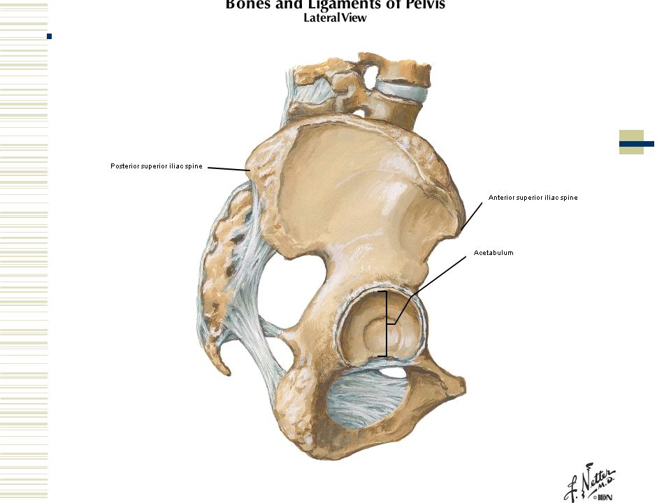
Innominates: Diagnosis
Standing flexion positive same side. Anterior Rotation: PSIS superior, ASIS inferior, medial malleolus inferior same side. Posterior Rotation: PSIS inferior, ASIS superior, medial malleolus superior same side.

21
Anterior Innominate Rotation: OMTreatment
The patient is supine and the physician is standing on the side of the dysfunction. Physician’s monitoring hand is placed on the SI joint and the hip and knee are slightly (60°) flexed to induce softening of the SI joint. The hip is slightly abducted and externally rotated, compression is maintained from the knee to the sensing finger as the hip is adducted in an arc across the midline. The hip and knees are then extended Reassess.
 flexed to induce softening of the SI joint. The hip is slightly abducted and externally rotated, compression is maintained from the knee to the sensing finger as the hip is adducted in an arc across the midline. The hip and knees are then extended. Reassess.")
22
Posterior Innominate Rotation: OMTreatment
The patient is supine and the physician is standing on the side of the dysfunction. Physician’s monitoring hand is placed on the SI joint and the hip and knee are fully (greater than 90°) flexed to induce softening of the SI joint. The hip is slightly adducted and internally rotated and compression is maintained from the knee to the sensing finger as the hip is abducted and externally rotated in an arc away from the midline. The hip and knee are then extended. Reassess.
 flexed to induce softening of the SI joint. The hip is slightly adducted and internally rotated and compression is maintained from the knee to the sensing finger as the hip is abducted and externally rotated in an arc away from the midline. The hip and knee are then extended. Reassess.")
23
Sacrum: Different Diagnosis for Still technique
Seated flexion test: Fingers on the SACRAL SULCUS, NOT INFERIOR SURFACE OF THE PSIS. Inferior lateral angle (ILA) is posterior and inferior and resists movement anterior.
 is posterior and inferior and resists movement anterior.")
24
Sacral Sulcus Inferior Lateral Angle Sacrotuberous ligament

25
Sacral Diagnosis: Dx; Seated Flexion Test; Inferior Lateral Angle
DX SFT. ILA Diag R R L+ Diag L L R+ Unilat. R R R+ Unilat. L L L+

27
Sacrum OMTreatment: Right Diagonal
This is a technique without a sensing hand. Patient must be reassessed after the treatment. Patient supine, physician standing at the side of the table. Knees flexed and hips are flexed to at least 90° and physician’s hand moves the knees to the left followed by the feet. Compression is initiated and maintained as the knees followed by the feet are moved past the midline toward the right. At approximately 45° past the midline, the hips and knees are extended to neutral. Reassess.

28
Sacrum OMTreatment: Left Unilateral
Patient is supine, physician standing on the ipsilateral side. Physician’s sensing fingers are on the left SI joint and the physician’s operating hand grasps the RIGHT leg just below the knee. The right lower extremity is abducted until softening of the SI tissues occurs. Compression is applied toward the SI joint as the right lower extremity is brought well past the midline, bringing the right pelvis off the table. Extend the knee and hip and reassess.

29
Groin Muscle Injuries: Pectineus, Adductors Brevis, Longus, Magnus
In general the positioning is similar with the palpable tender points for the muscles at differing locations. Pectineus: just lateral to pubic tubercle Adductor Brevis: ¼ distance down thigh Adductor Longus: ½ - 1/3 distance down thigh Adductor Magnus: just above anterior medial condyle.

30
Pectineus Adductor Longus Adductor Brevis Adductor Magnus

31
Pectineus Adductor Longus Gracilis

32
Pectineus Adductor Longus

33
Pectineus: OMTreatment
Patient is supine and physician is standing on ipsilateral side. Sensing finger is just lateral to the pubic insertion of the inguinal ligament. The hip is flexed and internally rotated to put the pectineus into a position of ease. Compression is applied and maintained as the hip is abducted and the foot brought toward the midline and the thigh and knee are then extended. Reassess.

34
Adductor Brevis: OMTreatment
Patient is supine, physician on ipsilateral side, sensing finger on tender point, approximately ¼ distance down medial thigh and operating hand grasping ankle. Adduct leg to sense relaxation of tender point. Apply compression and abduct leg (30°) off the side of the table, flexing the knee (30°) toward the floor. Reassess
 off the side of the table, flexing the knee (30°) toward the floor. Reassess.")
35
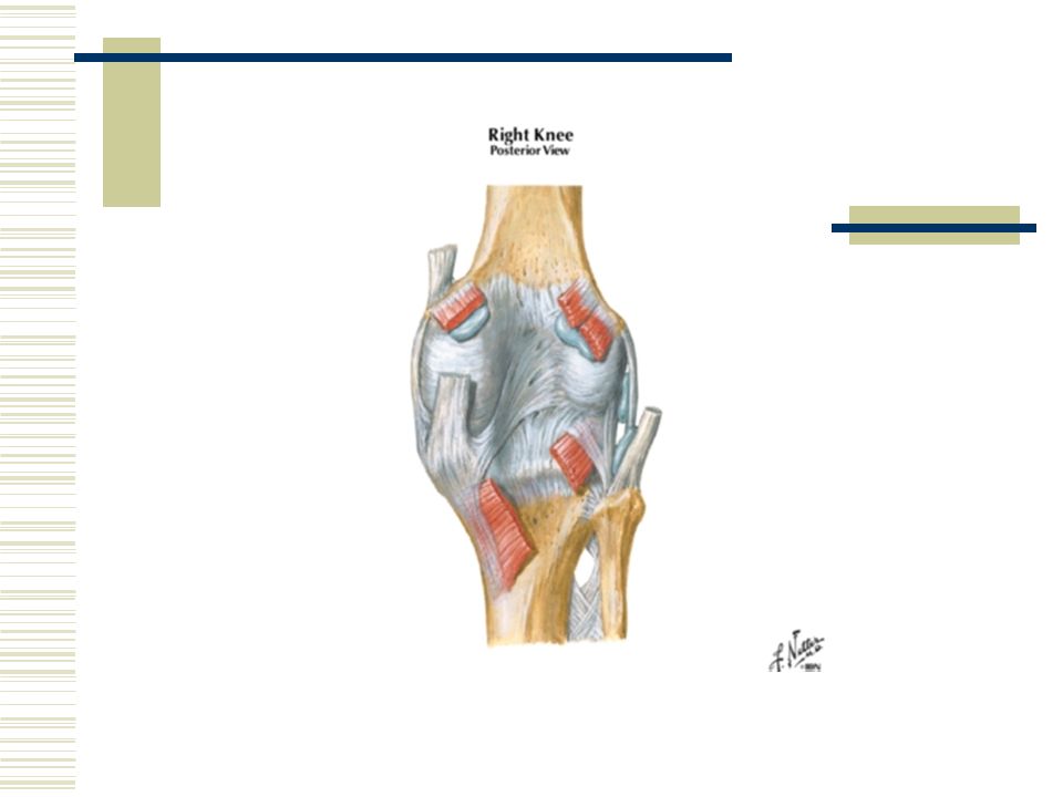
Knee: Fibular Head: Motion mechanics
If the knee is flexed, then with ankle dorsiflexion, the fibular head moves anterior and with plantar flexion, the fibular head moves posterior. BUT If the knee is extended, then with ankle plantar flexion, the fibular head moves anterior and with ankle dorsiflexion the fibular head moves posterior.

38
Fibular Head: Posterior OMTreatment
Patient is supine, physician standing at the ipsilateral side. Physician grasps the foot and gently dorsiflexes and externally rotates the leg, with sensing hand on the fibular head. Compression is applied and the foot is gently plantar flexed and internally rotated (supinated). Reassess
. Reassess.")
Similar presentations


 ABduction ◦ Move away from the midline of the body (lateral) >")


>")


