Download presentation
Presentation is loading. Please wait.

1
Innate Immunity of the oral cavity Dr. Aaron Weinberg DMD, PhD Department of Biological Sciences

2
Outline of Lecture Innate vs adaptive immunity Oral mucosal strategy Mucin Lysozyme Lactoferrin Salivary peroxidase Histatins PRPs Statherin Cystatins Epithelial cell derived antimicrobial peptides –“good bug” vs “bad bug”

3
Oral Mucosal Wounds

4
Innate Immunity Body’s first line of defense against infection Mechanical barriers at body surfaces Nonspecific/ no memory Rapid response Antibacterial substances in secretions -lysozyme & lactoferrin -low pH of stomach contents Evolutionarily ancient and conserved Alternate Complement Pathway

5
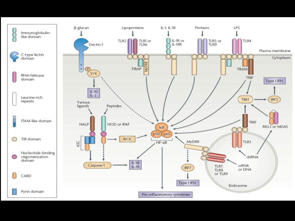
Epithelial Innate Immunity Recognition of bacteria –PAMPs Lipopolysaccharide, peptidoglycan, lipoteichoic acid, flagella, mannans, bacterial DNA, glucans –Toll-like Receptors (TLRs) TLR-2: peptidoglycan TLR-4: lipopolysaccharide TLR-9: bacterial DNA TLR-5: bacterial flagella Homodimers/Heterodimers –Intracellular signaling NF-kB signal Inler and Hoffman, Trends Cell Biol, 2001
 TLR-2: peptidoglycan TLR-4: lipopolysaccharide TLR-9: bacterial DNA TLR-5: bacterial flagella Homodimers/Heterodimers –Intracellular signaling NF-kB signal Inler and Hoffman, Trends Cell Biol, 2001")
6
kdo sugar alcohol phosphate polymers NAM-NAG sugar alcohol phosphate polymers

8
Toll mutant Christiane Nüsslein-Volhard, Nobel Laureate (Medicine, 1995 )
")
9
Lemaitre, B. et.al. 1996. The dorsoventral regulatory gene cassette spatzle/Toll/cactus controls the potent antifungal response in Drosophila adults. Cell. 86:973.

12
Host responses to infection

13
Innate vs Adaptive Immunity

14
Oral Mucosal Strategy fluid phase defense static phase defense recruitable defense EAPs Parotid Submandibular Sublingual

15
Salivary constituents and their functions defensins Mucins

16
SALIVARY FACTORS WITH ANTI-MICROBIAL ACTIVITY FACTOREFFECT AntibodiesBind bacteria, Neutralize and inactivate viral particles Cystatins Perform general antimicrobial activity; inhibit cysteine proteases DefensinsPerform general antimicrobial activity; carry out charge mediated disruption of bacterial membranes Histatinsantifungal LactoferrinBinds iron to inhibit bacterial growth LysozymeLyses bacteria MucinsEntrap and aggregate microbial particles Proline-Rich PeptidesBind bacteria StatherinBinds bacteria

17
Mucins Important in formation of lubricating biofilm to protect underlying mucosa and tooth surfaces from chemical and physical harm Important for physiological processes: speech, swallowing, clearance of microbes Glycoproteins, 30-90% carbohydrate w/serine or threonine-galactosylamine glycopeptide linkage Two major salivary mucins: –MG1 (from mucous acini of seromucous salivary glands) –MG2 (from serous cells of seromucous salivary glands + parotid gland) –Distinguished by differences in size, carbohydrate content, sulfation (and charge), sialic acid, subunit structure (see Table) – size: MG1 (MUC5B) > MG2 (MUC7) – carbohydrates, sulfation: MG1 > MG2 – MG1 bears ABH and Lewis blood group antigens for microbial adherence
 –MG2 (from serous cells of seromucous salivary glands + parotid gland) –Distinguished by differences in size, carbohydrate content, sulfation (and charge), sialic acid, subunit structure (see Table) – size: MG1 (MUC5B) > MG2 (MUC7) – carbohydrates, sulfation: MG1 > MG2 – MG1 bears ABH and Lewis blood group antigens for microbial adherence")
19
Mucins Characteristic/FunctionMG1MG2 Molecular weight (daltons)>10 6 2-2.5x10 5 Protein (composition)14.9%30.4% Carbohydrate (composition)78%68% Size of oligosaccharides4-17 residues2-7 residues Number of oligosaccharides46170 Number of sialic acid units1467 Sulfate7%1.6% Fatty acidsYesnegligible SubunitsMultiple SS-linked1 Conc. in submand/submax saliva6-8 mg/L16-18 mg/L Proposed protective functionPellicleBacterial clearance

20
Mucins and mucosal surfaces MG1 is tightly associated with mucosa Serves as barrier against toxins, hydrolytic enzymes, acids, carcinogens Traps various host defense factors, providing high concentrations of these factors near surface sIgA concentrated w/i mucin layer overlying epithelium In solution, MG1 forms complexes with various proteins: amylase, PRPs, statherin, histatins

21
Mucins and dental surfaces Mucins play important role in generation of the dental pellicle “Pellicle” 1-2 m layer containing lipids + salivary proteins/glycoproteins: albumin, lysozyme, PRPs, lactoferrin, statherin, bacterial debri etc.. MG1 covers outer layers of pellicle while MG2 more inside ( Kajisa et al, 1990) Pellicle important for colonization of first line of bacteria; “good guys”
 Pellicle important for colonization of first line of bacteria; good guys .")
22
Mucins and fluid phase defenses Mucins exert several antimicrobial activities in fluid phase. MG2 prevents bacterial colonization of the pellicle coated-tooth by presenting identical surface carbohydrates in the fluid phase (Levine et al, 1985: Reddy et al, 1993) MG1 believed to protect mucosa by preventing viral infections; ex. HIV and herpes simplex (Mandel and Ellison, 1985)
 MG1 believed to protect mucosa by preventing viral infections; ex. HIV and herpes simplex (Mandel and Ellison, 1985).")
23
Serous secretions Provided by the parotid gland Contain innate immune factors –Lysozyme, histatins, salivary peroxidase, lactoferrin, cystatins, PRPs, calprotectin Contain adaptive immune factors –sIgA

24
Lysozyme Muramidase, N-acetylmuramide glycanohydrolase Encoded on chromosome 12 Cationic protein Mol wt. 14.3 kD Produced by myeloid cells and glandular epithelium Parotid saliva: up to 10 mg/L (unstimulated); down to 1 mg/L (stimulated) Activity: cleaves -1,4 linkage between NAM-NAG in bacterial cell wall peptidoglycan. Protein contains deep groove capable of binding 6 sugar units of the NAM-NAG structure causing the backbone structure to snap. Due to its charge, has some non-enzymatic microbicidal activity against bacteria and oral fungi (Laible and Germaine, 1985; Tobji et al, 1988)
; down to 1 mg/L (stimulated) Activity: cleaves -1,4 linkage between NAM-NAG in bacterial cell wall peptidoglycan. Protein contains deep groove capable of binding 6 sugar units of the NAM-NAG structure causing the backbone structure to snap. Due to its charge, has some non-enzymatic microbicidal activity against bacteria and oral fungi (Laible and Germaine, 1985; Tobji et al, 1988).")
25
Lactoferrin (lactotransferrin) LF iron chelating glycoprotein 10-20 mg/L saliva; 1 g/L in milk Produced by neutrophils (not other myeloid cells) and glandular epithelium Encoded on chromosome 3 LF single polypeptide; MW 80 kD; 2 homologous domains that each binds one Fe +2 ion Activity: blocks growth of iron dependent organisms (Ca, Pg) Apolactoferrin (iron-less) can kill certain oral bacteria (S. mutans, A.a.) by binding to metal and destabilizing outer membrane (conjecture; Ellison et al, 1988) “Lactoferricin” microbicidal peptide domain released from LF by pepsin (gastric hydrolase) (Yamauchi et al, 1993)
 by binding to metal and destabilizing outer membrane (conjecture; Ellison et al, 1988) Lactoferricin microbicidal peptide domain released from LF by pepsin (gastric hydrolase) (Yamauchi et al, 1993).")
26
Salivary peroxidase 78 kD enzyme Produced by salivary gland epithelium Catalyzes reduction of H 2 O 2 to H 2 O and oxidation of electron donor Main donor is thiocyanate (SCN - ), halide ion, 1-2 mM in saliva; H 2 O 2 + SCN - H 2 O + OSCN - (hypothiocyanite) Activity: –potentiates antimicrobial activity of fluoride against S. mutans (Lenander-Lumikari et al, 1997) –potentiates activities of lysozyme and lactoferrin –may function against H 2 O 2 generating bacteria (Thomas et al, 1983) –neutralizes H 2 O 2 along the mucosa (antioxidant effect) H 2 O 2 oral releasing bacteria can induce ulceration of the mucosa (conjecture) Acatalasemia (deficiency in catalase; enzyme catalyzes H 2 O 2 ) has been associated with extensive ulceration of oral tissues Strep. H 2 O 2 release is strain dependent; 0-165 nmol/min/mg bacterial protein in presence of glucose (Miyasaki et al, 1988) Tissue destructive effects of H 2 O 2 is probably indirect, requiring further reduction of H 2 O 2 to OH ? aphthous ulcerations respond to salivary peroxidase enhancement therapy; clinical study, 45 of 64 aphthous patients reported symptomatic relief; ? Oral strep connection?? (Hoogendoorn and Piessens, 1987)
 –potentiates activities of lysozyme and lactoferrin –may function against H 2 O 2 generating bacteria (Thomas et al, 1983) –neutralizes H 2 O 2 along the mucosa (antioxidant effect) H 2 O 2 oral releasing bacteria can induce ulceration of the mucosa (conjecture) Acatalasemia (deficiency in catalase; enzyme catalyzes H 2 O 2 ) has been associated with extensive ulceration of oral tissues Strep. H 2 O 2 release is strain dependent; nmol/min/mg bacterial protein in presence of glucose (Miyasaki et al, 1988) Tissue destructive effects of H 2 O 2 is probably indirect, requiring further reduction of H 2 O 2 to OH . aphthous ulcerations respond to salivary peroxidase enhancement therapy; clinical study, 45 of 64 aphthous patients reported symptomatic relief; . Oral strep connection . (Hoogendoorn and Piessens, 1987).")
27
Histatins (HRPs) Basic, histidine rich, -helical peptides (7-38 aa) Up to 12 different HRPs Produced by salivary gland epithelium Found in parotid and submandibular secretions 50-425 g/ml saliva (Edgerton et al, 1998) Strong anticandidal peptides; some anti- S. mutans and anti-P. gingivalis activity 2 families of HRPs based on sequence analysis

28
Antifungal activity of Histatins Most important antifungal agents in saliva Anti-histatin immunoaffinity adsorption of saliva removes candidacidal activity Decrease in salivary histatins associated with increased incidence of candidiasis secondary to HIV (Mandel et al, 1992) Topical histatins have been shown to prevent denture stomatitis (DS) Dentures cover palate and prevent access of parotid saliva DS presents as a superficial fungal problem; little fungus is found in mucosa Clinical observations reveal the significance of histatins in preventing superficial oral candidiasis.
 Topical histatins have been shown to prevent denture stomatitis (DS) Dentures cover palate and prevent access of parotid saliva DS presents as a superficial fungal problem; little fungus is found in mucosa Clinical observations reveal the significance of histatins in preventing superficial oral candidiasis.")
29
Mouse model of oral candidiasis Normal mouse tongue Candida/hyphae

30
Proline-rich proteins (PRPs) Acidic, with 25-40% proline content 150-170 aa Multigene complex on chromosome 12 Multifunctional –In solution PRPs maintain salivary calcium phosphate in a supersaturated state (Gibbons and Hay, 1988) –Are a significant fraction of the acquired pellicle; important in dictating microbial attachment. –Pleomorphism Some variants may be associated with greater susceptibility to dental caries –Bacteroides thetaiotomicron in mice (Jeff Gordon, et al)
.")
31
Statherin 43 Kd phosphoprotein Encoded on chromosome 4 Like PRPs, maintains salivary calcium phosphate in a supersaturated state Found in acquired pellicle Involved in microbial attachment May be evolutionarily related to the histatins

32
Paul Kolenbrander

33
Cystatins Inhibitors of cysteine proteinases; widely distributed in tissues MW ~14 kD, 120 aa, chromosome 20 (family 2), chromosome 3 (family 1, 3); derived from submandibular secretions Most common in saliva are family 2 cystatins: S, (pI, 4.7), SA (pI, 4.3), SN (neutral), and C (pI, 7.5) Myeloid cells are source of cystatin C Cystatin C appears to increase in saliva from periodontitis and gingivitis cases Importance: –neutralizing against microbial-derived cysteine proteinases –cystatin SN may exert an anti-adhesive effect by binding to bacterial pili (Reddy, 1998)
, chromosome 3 (family 1, 3); derived from submandibular secretions Most common in saliva are family 2 cystatins: S, (pI, 4.7), SA (pI, 4.3), SN (neutral), and C (pI, 7.5) Myeloid cells are source of cystatin C Cystatin C appears to increase in saliva from periodontitis and gingivitis cases Importance: –neutralizing against microbial-derived cysteine proteinases –cystatin SN may exert an anti-adhesive effect by binding to bacterial pili (Reddy, 1998)")
34
Question *Why is the mouth so healthy in spite of constant trauma occurring in a very septic environment? Michael Zasloff Magainin Host Defense Peptides

35
Oral Mucosal Strategy fluid phase defense static phase defense recruitable defense EAPs S. sanguis antagonizes A.a. and competes with C.a. for biotin

36
Host defense peptides in humans Adrenomedullin: 52 aa vasoactive, cationic antibacterial peptide (Allakar RP, Kapas S, ’99, ’01, ’03) Calprotectin: Two-subunit protein ( , MRP8; , MRP18), anionic (Eversole, ’93; Ross and Herzberg, ’01) SLPI: 12kDa non-glycosylated protein (Wahl, ’97; Shugars, ’97) LL37: cathelicidin; PMNs, lymphocytes, macrophages, some epithelial cells (Lehrer and Ganz, ’02) Human -defensins: beta sheeted; PMNS, Paneth cells (Kagan et al, 1994; Ouellette and Selsted, ‘96) Human -defensins: beta sheeted; epithelial cells (Weinberg et al, ’98; Zasloff, ’02; Krisanaprakornkit et al, ’98, ’00; Quinones et al, ’03; Feng et al, ’05; Feng et al, ’06)
 Calprotectin: Two-subunit protein ( , MRP8; , MRP18), anionic (Eversole, ’93; Ross and Herzberg, ’01) SLPI: 12kDa non-glycosylated protein (Wahl, ’97; Shugars, ’97) LL37: cathelicidin; PMNs, lymphocytes, macrophages, some epithelial cells (Lehrer and Ganz, ’02) Human -defensins: beta sheeted; PMNS, Paneth cells (Kagan et al, 1994; Ouellette and Selsted, ‘96) Human -defensins: beta sheeted; epithelial cells (Weinberg et al, ’98; Zasloff, ’02; Krisanaprakornkit et al, ’98, ’00; Quinones et al, ’03; Feng et al, ’05; Feng et al, ’06)")
37
Human -Defensins Produced by epithelial cells Cationic, amphipathic peptides - hBD1, constitutive - hBD2, inducible - hBD3, inducible - hBD4, inducible ? - antibacterial, antifungal, antiviral Mechanism of action -anionic targets: LPS, LTA, phospholipids (phosphatidylglycerol) -form pores in bacterial membrane Cross-talk with adaptive immunity (Hancock, Lancet, 1997) + + C C C C C C
 -form pores in bacterial membrane Cross-talk with adaptive immunity (Hancock, Lancet, 1997) + + C C C C C C.")
38
Gram positive rod and hBD-3

39
Defensins in innate and adaptive immunity ___________________________________ ____ Ganz, Science 286:420, 1999 Yang et al, Science 286:525, 1999

40
- defensins in innate and adaptive immunity - defensins recruit iDCs and T cells via CCR6 (Yang et al, 1999) -defensins promote maturation of dendritic cells via TLR4 (Biragyn et al, 2002) Recruitment of monocytes is hBD3 isoform dependent (Wu et al, 2003) hBD2 is chemotactic towards human neutrophils via CCR6 (Niyonsaba et a, 2004) hBD2 and -3 interact with CXCR4 (Quinones-Mateu et al, 2003; Feng et al, 2006) hBD3 induces co-stimulatory molecule expression in human monocytes/mDCs via TLR1/2 (Funderberg et al, 2007)
 -defensins promote maturation of dendritic cells via TLR4 (Biragyn et al, 2002) Recruitment of monocytes is hBD3 isoform dependent (Wu et al, 2003) hBD2 is chemotactic towards human neutrophils via CCR6 (Niyonsaba et a, 2004) hBD2 and -3 interact with CXCR4 (Quinones-Mateu et al, 2003; Feng et al, 2006) hBD3 induces co-stimulatory molecule expression in human monocytes/mDCs via TLR1/2 (Funderberg et al, 2007)")
41
IMF of hBD-2 and hBD-3 in human oral epithelium Ge Jin, CWRU

42
carcinoma in situ normal H&E staining hBD-2 hBD-3 nucleus hBD-2/hBD-3

43
B. Dale-Crunk, U. Washington Gingiva Skin

44
Questions *Why is the mouth so healthy in spite of constant trauma occurring in a very septic environment? *The answers may lie with some of the ubiquitous bacteria of the mouth that induce epithelial cell derived antimicrobial peptides. *Why are hBDs constitutively expressed in oral mucosa?

45
Homo sapiens or Homo bacteriens? 10 13 eukaryotic cells 10 14 bacteria Henderson and Wilson, JDR, 1998

46
found in healthy sites involved in dental plaque formation Porphyromonas gingivalis black pigmented gram negative rod found in diseased sites associated with periodontal disease gram negative fusiform Fusobacterium nucleatum

47
F. nucleatum vs P. gingivalis induction of defensins hBD-2 - FnPg RT-PCR microbial challenge

48
F. nucleatum induction of hBD-3 in NHOECs

49
Can the “good guy” protect us from the “bad guy”

50
Are these “good guy” bacteria sensitive to defensins

51
F. nucleatum and P. gingivalis sensitivity to recombinant hBDs 2 X 10 5 bacteria incubated with rhBD-1, -2, or -3, anaerobically, 3 h, followed by serial dilutions and plating on sheep red blood agar plates.

52
1.1. Immunogold TEM of P. gingivalis after rhBD-2 incubation

53
2. Immunogold TEM of F. nucleatum after rhBD-2 incubation

54
3. Immunogold TEM of F. nucleatum after rhBD-2 incubation

55
F. nucleatum vs P. gingivalis F. nucleatum induces hBD2 and hBD3 in NHOECs and protects cells from P. gingivalis invasion F. nucleatum factor(s) isolated from the cell wall promotes defensin expression; 4 candidate peptides; 12-13 kDa, pI 4-5 F. nucleatum is resistant to -defensins ; P. gingivalis is not F. nucleatum resistance may be due to “fimbrial" extensions that sequester the hBD away from the outer membrane P. gingivalis does not induce hBD expression in NHOECs
 isolated from the cell wall promotes defensin expression; 4 candidate peptides; kDa, pI 4-5 F. nucleatum is resistant to -defensins ; P. gingivalis is not F. nucleatum resistance may be due to fimbrial extensions that sequester the hBD away from the outer membrane P. gingivalis does not induce hBD expression in NHOECs.")
Similar presentations


 BIOS 486A/586A>")


 BY. Dr. Shahzadi Tayyaba Hashmi DNT 353.>")


>")


 Lecture 18 Bacterial Pathogenesis (Based on other textbooks such as Madigan’s)>")

>")


