Download presentation
Presentation is loading. Please wait.

1
DNA The Genetic Material Replication
Chapter 13. DNA The Genetic Material Replication Biology 114

2
Scientific History The march to understanding that DNA is the genetic material T.H. Morgan (1908) Frederick Griffith (1928) Avery, McCarty & MacLeod (1944) Hershey & Chase (1952) Watson & Crick (1953) Meselson & Stahl (1958)
 Avery, McCarty & MacLeod (1944) Hershey & Chase (1952) Watson & Crick (1953) Meselson & Stahl (1958)")
3
Genes are on chromosomes
1908 | 1933 Genes are on chromosomes T.H. Morgan working with Drosophila (fruit flies) genes are on chromosomes but is it the protein or the DNA of the chromosomes that are the genes? through 1940 proteins were thought to be genetic material… Why? What’s so impressive about proteins?!
 genes are on chromosomes. but is it the protein or the DNA of the chromosomes that are the genes through 1940 proteins were thought to be genetic material… Why What’s so impressive about proteins !")
4
The “Transforming Factor”
1928 The “Transforming Factor” Frederick Griffith Streptococcus pneumonia bacteria was working to find cure for pneumonia harmless live bacteria mixed with heat-killed infectious bacteria causes disease in mice substance passed from dead bacteria to live bacteria = “Transforming Factor” Fred Griffith, English microbiologist, dies in the Blitz in London in 1941

5
The “Transforming Factor”
mix heat-killed pathogenic & non-pathogenic bacteria live pathogenic strain of bacteria live non-pathogenic strain of bacteria heat-killed pathogenic bacteria A. B. C. D. mice die mice live mice live mice die Transformation? something in heat-killed bacteria could still transmit disease-causing properties

6
DNA is the “Transforming Factor”
1944 DNA is the “Transforming Factor” Avery, McCarty & MacLeod purified both DNA & proteins from Streptococcus pneumonia bacteria which will transform non-pathogenic bacteria? injected protein into bacteria no effect injected DNA into bacteria transformed harmless bacteria into virulent bacteria What’s the conclusion?

7
Avery, McCarty & MacLeod
Oswald Avery Colin MacLeod Maclyn McCarty

8
Why use Sulfur vs. Phosphorus?
1952 | 1969 Confirmation of DNA Hershey & Chase classic “blender” experiment worked with bacteriophage viruses that infect bacteria grew phage viruses in 2 media, radioactively labeled with either 35S in their proteins 32P in their DNA infected bacteria with labeled phages Why use Sulfur vs. Phosphorus?

9
Hershey & Chase Martha Chase Alfred Hershey

10
Hershey & Chase Which radioactive marker is found inside the cell?
Protein coat labeled with 35S DNA labeled with 32P Hershey & Chase T2 bacteriophages are labeled with radioactive isotopes S vs. P bacteriophages infect bacterial cells bacterial cells are agitated to remove viral protein coats Which radioactive marker is found inside the cell? Which molecule carries viral genetic info? 35S radioactivity found in the medium 32P radioactivity found in the bacterial cells

12
Blender experiment Radioactive phage & bacteria in blender 35S phage
radioactive proteins stayed in supernatant therefore protein did NOT enter bacteria 32P phage radioactive DNA stayed in pellet therefore DNA did enter bacteria Confirmed DNA is “transforming factor” Taaa-Daaa!

13
Chargaff 1947 DNA composition: “Chargaff’s rules”
varies from species to species all 4 bases not in equal quantity bases present in characteristic ratio humans: A = 30.9% T = 29.4% G = 19.9% C = 19.8%

14
Structure of DNA 1953 | 1962 Watson & Crick
developed double helix model of DNA other scientists working on question: Rosalind Franklin Maurice Wilkins Linus Pauling Watson & Crick’s model was inspired by 3 recent discoveries: Chargaff’s rules Pauling’s alpha helical structure of a protein X-ray crystallography data from Franklin & Wilkins Franklin Wilkins Pauling

15
1953 article in Nature Watson and Crick

16
Rosalind Franklin ( ) A chemist by training, Franklin had made original and essential contributions to the understanding of the structure of graphite and other carbon compounds even before her appointment to King's College. Unfortunately, her reputation did not precede her. James Watson's unflattering portrayal of Franklin in his account of the discovery of DNA's structure, entitled "The Double Helix," depicts Franklin as an underling of Maurice Wilkins, when in fact Wilkins and Franklin were peers in the Randall laboratory. And it was Franklin alone whom Randall had given the task of elucidating DNA's structure. The technique with which Rosalind Franklin set out to do this is called X-ray crystallography. With this technique, the locations of atoms in any crystal can be precisely mapped by looking at the image of the crystal under an X-ray beam. By the early 1950s, scientists were just learning how to use this technique to study biological molecules. Rosalind Franklin applied her chemist's expertise to the unwieldy DNA molecule. After complicated analysis, she discovered (and was the first to state) that the sugar-phosphate backbone of DNA lies on the outside of the molecule. She also elucidated the basic helical structure of the molecule. After Randall presented Franklin's data and her unpublished conclusions at a routine seminar, her work was provided - without Randall's knowledge - to her competitors at Cambridge University, Watson and Crick. The scientists used her data and that of other scientists to build their ultimately correct and detailed description of DNA's structure in Franklin was not bitter, but pleased, and set out to publish a corroborating report of the Watson-Crick model. Her career was eventually cut short by illness. It is a tremendous shame that Franklin did not receive due credit for her essential role in this discovery, either during her lifetime or after her untimely death at age 37 due to cancer.
 that the sugar-phosphate backbone of DNA lies on the outside of the molecule. She also elucidated the basic helical structure of the molecule. After Randall presented Franklin s data and her unpublished conclusions at a routine seminar, her work was provided - without Randall s knowledge - to her competitors at Cambridge University, Watson and Crick. The scientists used her data and that of other scientists to build their ultimately correct and detailed description of DNA s structure in Franklin was not bitter, but pleased, and set out to publish a corroborating report of the Watson-Crick model. Her career was eventually cut short by illness. It is a tremendous shame that Franklin did not receive due credit for her essential role in this discovery, either during her lifetime or after her untimely death at age 37 due to cancer.")
17
Double helix structure of DNA
the structure of DNA suggested a mechanism for how DNA is copied by the cell

18
Directionality of DNA You need to number the carbons! nucleotide
it matters! nucleotide PO4 N base 5 CH2 O This will be IMPORTANT!! 4 1 ribose 3 2 OH

19
I mean it… This will be IMPORTANT!!
5 The DNA backbone PO4 Putting the DNA backbone together refer to the 3 and 5 ends of the DNA the last trailing carbon base CH2 O C O –O P O O base I mean it… This will be IMPORTANT!! CH2 O OH 3

20
Anti-parallel strands
Phosphate to sugar bond involves carbons in 3 & 5 positions DNA molecule has “direction” complementary strand runs in opposite direction “It has not escaped our notice that the specific pairing we have postulated immediately suggests a possible copying mechanism for the genetic material.” Watson & Crick

21
Bonding in DNA 5’ 3’ 3’ 5’ hydrogen bonds phosphodiester bonds
….strong or weak bonds? How do the bonds fit the mechanism for copying DNA?

22
Base pairing in DNA Purines Pyrimidines Pairing adenine (A)
guanine (G) Pyrimidines thymine (T) cytosine (C) Pairing A : T C : G
 Pyrimidines. thymine (T) cytosine (C) Pairing. A : T. C : G.")
23
Copying DNA Replication of DNA
base pairing allows each strand to serve as a pattern for a new strand

24
Models of DNA Replication
verify through experiments… Models of DNA Replication Alternative models so how is DNA copied?

25
Semi-conservative replication
1958 Semi-conservative replication Meselson & Stahl label nucleotides of “parent” DNA strands with heavy nitrogen = 15N label new nucleotides with lighter isotope = 14N “The Most Beautiful Experiment in Biology” parent replication make predictions…

26
Semi-conservative replication
1958 Semi-conservative replication Make predictions… 15N strands replicated in 14N medium 1st round of replication? 2nd round?

27
DNA Replication Large team of enzymes coordinates replication
let’s meet the team… DNA Replication Large team of enzymes coordinates replication Enzymes more than a dozen enzymes & other proteins participate in DNA replication

28
single-stranded binding proteins
Replication: 1st step Unwind DNA helicase enzyme unwinds part of DNA helix stabilized by single-stranded binding proteins single-stranded binding proteins

29
Where’s the ENERGY for the bonding! We’re missing something!
Replication: 2nd step Bring in new nucleotides to match up to template strands Where’s the ENERGY for the bonding! But… We’re missing something! What?

30
Is that the only energy molecule?
Energy of Replication Where does the energy for the bonding come from? energy You remember ATP! Is that the only energy molecule? CTP GTP TTP ATP CMP TMP AMP ADP GMP

31
Energy of Replication ATP GTP TTP CTP
The nucleotides arrive as nucleosides DNA bases with P–P–P DNA bases arrive with their own energy source for bonding bonded by DNA polymerase III ATP GTP TTP CTP

32
Replication Adding bases
5' 3' Replication energy DNA P III Adding bases can only add nucleotides to 3 end of a growing DNA strand strand grow 5'3’ energy energy The energy rules the process. energy B.Y.O. ENERGY 3' 5' leading strand

33
5' 3' 5' 3' ligase energy 3' 3' leading strand lagging strand 5' 5'

34
Leading & Lagging strands
Leading strand - continuous synthesis Okazaki Lagging strand - Okazaki fragments - joined by ligase - “spot welder” enzyme

35
Okazaki fragments

36
Priming DNA synthesis DNA polymerase III can only extend an existing DNA molecule cannot start new one cannot place first base short RNA primer is built first by primase starter sequences DNA polymerase III can now add nucleotides to RNA primer

37
Cleaning up primers DNA polymerase I removes sections of RNA primer and replaces with DNA nucleotides

38
direction of replication
Draw this in your notes and label each of the structures and their function Replication fork DNA polymerase III lagging strand DNA polymerase I 3’ Okazaki fragments primase 5’ 5’ ligase SSB 3’ 5’ 3’ helicase DNA polymerase III 5’ leading strand 3’ direction of replication

39
And in the end… Ends of chromosomes are eroded with each replication
an issue in aging? ends of chromosomes are protected by telomeres

40
Telomeres Expendable, non-coding sequences at ends of DNA
short sequence of bases repeated 1000s times TTAGGG in humans Telomerase enzyme in certain cells enzyme extends telomeres prevalent in cancers Why?

41
Replication bubble Adds 1000 bases/second!
Which direction does DNA build? List the enzymes & their role

42
Replication enzymes helicase DNA polymerase III primase
ligase single-stranded binding proteins

43
DNA polymerases DNA polymerase III DNA polymerase I 1000 bases/second
main DNA building enzyme DNA polymerase I 20 bases/second editing, repair & primer removal DNA polymerase III enzyme

44
Editing & proofreading DNA
1000 bases/second = lots of typos! DNA polymerase I proofreads & corrects typos repairs mismatched bases excises abnormal bases repairs damage throughout life reduces error rate from 1 in 10,000 to 1 in 100 million bases

45
Fast & accurate! It takes E. coli <1 hour to copy 5 million base pairs in its single chromosome divide to form 2 identical daughter cells Human cell copies its 6 billion bases & divide into daughter cells in only few hours remarkably accurate only ~1 error per 100 million bases ~30 errors per cell cycle

46
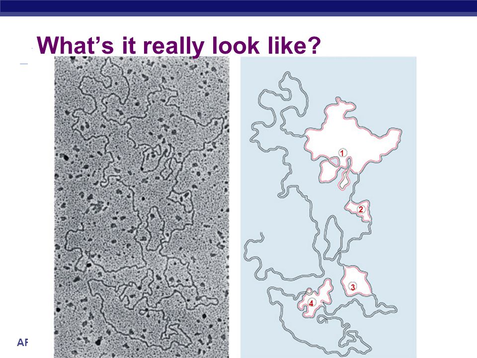
What’s it really look like?
1 2 3 4
47
DNA RNA protein The “Central Dogma”
flow of genetic information within a cell transcription translation DNA RNA protein replication

48
Any Questions?? Biology 114

Similar presentations


. Brief History Many people contributed to our understanding of DNA –T.H. Morgan (1908) –Frederick Griffith (1928) –Avery, McCarty & MacLeod.>")


 Brief History Many people contributed to our understanding of DNA – T.H. Morgan (1908) – Frederick Griffith (1928) – Avery, McCarty & MacLeod.>")


 for growth & development From.>")


